Signalment:
70-week-old, male, Wistar-Han, rat (
Rattus norvegicus).Tissue from the thoracic cavity of a control group male rat on a 2-year carcinogenicity study.
Gross Description:
The thoracic cavity contained a 3 x 3 x 2 cm, tan, multilobular mediastinal mass.
Histopathologic Description:
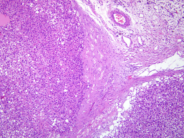
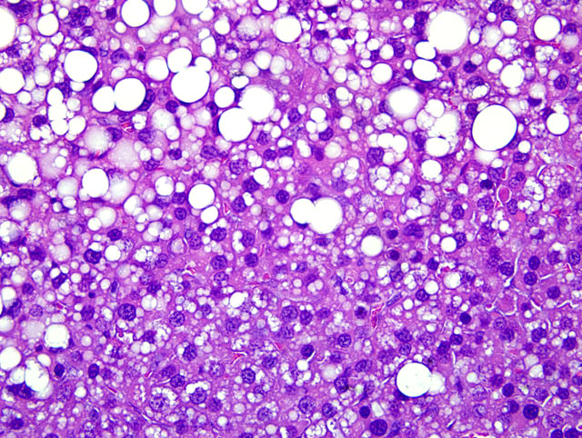
Fibroadipose tissue: Tissue consists of lobules of neoplastic cells separated by
irregular thin to broad trabeculae of fibrous connective tissue. Neoplastic cells are polyhedral with homogeneous
eosinophilic to microvacuolated cytoplasm and nuclei are round to oval with stippled chromatin and 0-2 nucleoli.
Mitoses are 0-1/HPF. These cells are frequently admixed with unilocular adipocytes that have eccentric nuclei.
There is marked anisocytosis and anisokaryosis, and there are scattered karyomegalic cells. There are multiple foci
of coagulative necrosis sometimes surrounded by foamy macrophages and multinucleate giant cells, and occasional
lakes of proteinaceous fluid containing foamy macrophages. In some sections, connective tissue trabeculae contain
hemorrhage and loose aggregates of pigmented macrophages (hemosiderophages), plasma cells, and lymphocytes.
Morphologic Diagnosis:
Hibernoma, malignant.
Lab Results:
Immunohistochemical stain: immunolabeling for UCP-1 (uncoupling protein 1).
Transmission electron microscopy: numerous mitochondria with transverse cristae.
Condition:
Malignant hibernoma
Contributor Comment:
Hibernomas are rare, well-characterized neoplasms of brown adipose tissue (BAT) in
humans and animals. Historically, the background incidence has been low in rodent carcinogenicity studies. This
tissue was from a twoyear study, in group-housed male and female Wistar-Han rats, with an unusually high
incidence of non-test article-related hibernomas (up to 12% in control animals).(3) For an unknown reason, the
incidence was higher in males. For males and females, the majority of hibernomas were noted in the thoracic cavity
as tan to red lobulated masses. Larger tumors had necrotic foci bordered by granulomatous inflammation. Tumor
emboli were present in vascular lumina as well as in the lungs. Characteristic ultrastructural features were abundant
mitochondria with parallel lamellar cristae and variably sized lipid droplets. Immunohistochemically, neoplastic
cells stained positively for UCP1 (uncoupling protein 1), generally considered a specific marker of brown
adipocytes. Although stimulation and regression of BAT secondary to changes in the ambient environment as well
as chronic disease states is reported,(7) a high incidence of spontaneous hibernomas occurred in this study.
Hibernomas were not observed in the concurrent mouse carcinogenicity study with the same test article conducted at
the same laboratory.(3) Although hyperplasia and/or neoplasia of BAT have been observed with several classes of
unrelated pharmaceutical compounds,(1,6,7) the historical incidence of spontaneous hibernomas in rats has been very
low. Increased incidences of spontaneous hibernomas unrelated to the administration of test articles were also
observed in Sprague-Dawley rats in three different carcinogenesis bioassays conducted during approximately the
same time period.(2)
The primary function of brown adipose tissue is to provide nonshivering thermogenesis during periods of coldinduced
stress. Brown adipose tissue is considered especially critical in neonates, in which deposits are within
abdominal and thoracic cavities as well as in subcutaneous tissue of the interscapular region. In rodents, BAT
deposits normally persist in multiple locations throughout life. Brown adipose cells, packed with mitochondria, are
specialized for thermogenesis.(4) Cold stress induces the release of norepinephrine, which binds to β-adrenergic
receptors on brown adipocytes.(2) This binding activates lipoprotein lipase to liberate free fatty acids, and initiates the
adenylcyclase-cAMP-lipase activation signal that stimulates βoxidation of fatty acids in mitochondria.(4) Free fatty
acids act as ionophore uncouplers in BAT mitochondria. UCP-1, localized to the inner mitochondrial membrane,
uncouples fatty acid oxidation from ADP phosphorylation and promotes the dissipation of the energy generated by
oxidation as heat.(2)
JPC Diagnosis:
Fibroadipose tissue: Hibernoma.
Conference Comment:
Participants generally agreed with the diagnosis of hibernoma for the lesion. With no
discernable normal tissue, features of malignancy, such as stromal invasion, evidence of metastasis or tumor emboli
within lymphatics or vasculature, cannot be adequately assessed. After the moderator provided additional
information from the contributor during the conference, a diagnosis of malignant hibernoma is appropriate.
Historically, the literature refers to benign tumors of brown adipose tissue as hibernomas while classifying
malignant tumors of brown adipose tissue liposarcomas.(5) Utilizing immunohistochemical and ultrastructural
examination, closer investigation of tumors of brown adipose tissue supports a diagnosis of malignant hibernorma
rather than liposarcoma.(2,3) This raises the possibility that malignant hibernomas previously have been
misdiagnosed/misclassified and are therefore more common than previous data suggest. This presents a problem
from a toxicologic pathology standpoint in that historical data may be skewed. It is therefore difficult to discern
whether malignant hibernomas are test-article related or are spontaneous occurrences in certain strains of rats.
The contributor provides an excellent overview of the function and physiology of brown adipose tissue.
References:
1. Brees DJ, Elwell MR, Tingley FD, et al. Pharmacological effects of nicotine on norepinephrine metabolism in rat
brown adipose tissue: relevance to nicotinic therapies for smoking cessation.
Toxicol Pathol. 2008;36:568-575.
2. Bruner RH, Novilla MN, Picut CA, et al. Spontaneous hibernomas in Sprague-Dawley rats.
Toxicol Pathol.
2009;37:547-552.
3. Chandra S, Dochterman W, Ploch S, et al. A carcinogenicity study with unusually high incidence of spontaneous
hibernomas in Wistar-Han rats.
Toxicol Pathol. 2009;37:127(P14).
4. Cheville NF. An introduction to interpretation In:
Ultrastructural Pathology. Ames, IA: Iowa State University
Press; 1994:14.
5. Greaves P, Faccini JM, Courtney CL. Proliferative lesions of soft tissues and skeletal muscles in rats, MST-1. In:
Guides for Toxicologic Pathology. Washington, DC: STP/ARP/AFIP; 1992:3.
6. Herman JR, Dethloff LA, McGuire EJ, et al. Rodent carcinogenicity with the thiazolidinedione antidiabetic agent
troglitazone.
Toxicol Sci. 2002;68:226-236.
7. Poulet FM, Berardi MR, Halliwell W, Hartman B, Auletta C, Bolte H. Development of hibernomas in rats dosed
with phentolamine mesylate during the 24-month carcinogenicity study.
Toxicol Pathol. 2004;32:558-566.