Signalment:
Gross Description:
Histopathologic Description:
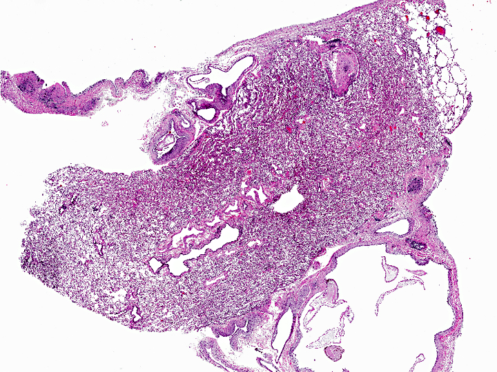
Small nodular areas of lymphoplasmacytic inflammation are noted on the serosa of the intestine and on the diaphragm.
Morphologic Diagnosis:
Condition:
Contributor Comment:
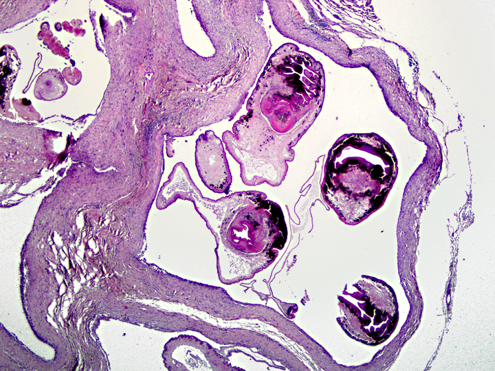
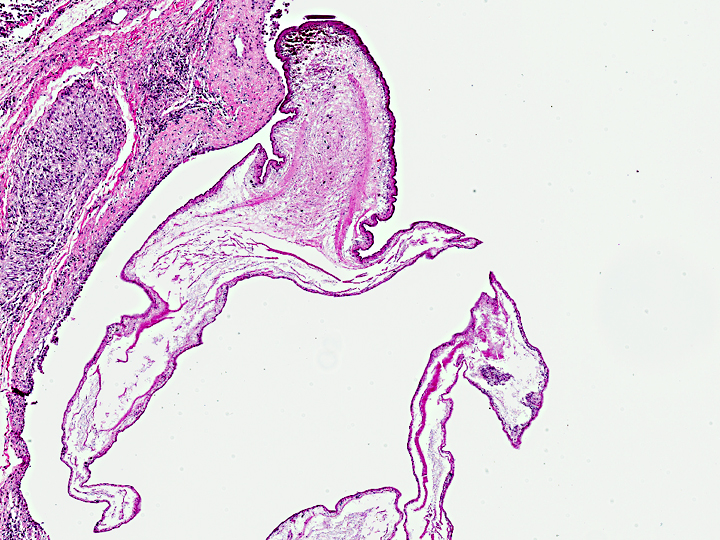
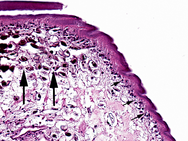
Pulmonary, pleural, or peritoneal infection of lemurs by cysticerci has been well documented anecdotally and in the literature.(2,4) Based on these reports, one of which included molecular confirmation, and the similar morphology including rare budding seen in this case, the most likely etiology is Cysticercus longicollis, the larval form of Taenia crassiceps, though other Taenia sp. are possible.
Exposure to feces of the definitive host (typically wild canids, but also domestic cats and dogs) or consumption of infected secondary rodent hosts are possible modes of transmission to the lemur in this case. After ingestion by an aberrant host, it is not well-documented as to how the cysticerci migrate to the peritoneal or pleural cavities, but once there, these cases can be remarkably fulminating since the organism can proliferate by budding both endogenously and exogenously. Infection and rapid proliferation is associated particularly with immunosuppression. This disease is zoonotic, most recently associated with AIDS patients. There was no history or clinical pathology data to suggest this lemur is immunocompromised, but the perceived high frequency of cysticercosis in lemurs might indicate a predisposition in this exotic species which has presumably never been exposed to canid or felid parasites in their evolutionary history until very recently.
JPC Diagnosis:
Conference Comment:
Other important cysticerci in veterinary species are as follows(1):
| ADULT TAPEWORM | DEFINITIVE HOST | LARVAL FORM | INTERMEDIATE HOST | SITE |
| Taenia saginata | man | Cysticercus bovis | cattle | muscle |
| Taenia solium | man | Cysticercus cellulosae | pig, man | muscle |
| Taenia(Multiceps) multiceps | dog | Coenurus cerebralis | sheep, cattle | CNS |
| Taenia hydatigena | dog | Cysticercus tenuicollis | sheep, cattle, pig | peritoneum |
| Taenia ovis | dog | Cysticercus ovis | sheep | muscle |
| Taenia pisiformis | dog | Cysticercus pisiformis | rabbit | peritoneum, liver |
| Taenia serialis | dog | Coenurus serialis | rabbit | connective tissue |
| Taenia taeniaeformis | cat | Cysticercus fasciolaris (strobilocercus) | mouse, rat | liver |
| Taenia krabbei | dog | Cysticercus tarandi | reindeer | muscle |
| Taenia mustelae | wild felids | rodents | liver | |
| Diphyllobothrium latum | bear, man | fish | muscle | |
| Diphyllobothrium pacificum | seal, sea lion | marine birds | muscle |
References:
2. Dyer NW, Greve JH: Severe Cysticercus longicollis cysticercosis in a black lemur (Eulemur macaco macaco). J Vet Diagn Invest 10: 362-364, 1998
3. Gardiner CH, Poynton SL: An atlas of metazoan parasites in animal tissues Armed Forces Institute of Pathology, Washington, D.C., 2006
4. Luzon M, de la Fuente-Lopez C, Martinez-Nevado E, Fernandez-Moran J, Ponce-Gordo F: Taenia crassiceps cysticercosis in a ring-tailed lemur (Lemur cata). J Zoo Wildl Med 41: 327-330, 2010