Signalment:
Fifteen-week-old mixed breed pig (
Sus scrofa domesticus)Diarrhea was reported in which there were flecks of undigested blood in a group of pigs. Approximately 10% of the pigs had diarrhea and 5% were markedly emaciated.
Gross Description:
The carcass of the euthanized pig was gaunt and the hair coat roughened. Stomach and small intestine contained scant amounts of ingesta. There was a moderate amount of semi-liquid feces in the spiral colon. Fresh blood and fibrin was mixed with the colon contents. Colonic mucosa was glistening and rough in appearance.
Histopathologic Description:
These sections of large intestine are characterized by multiple large mucosal erosions. Luminal surface is covered by a thick layer of neutrophilic debris mixed with mucus and occasionally extravasated erythrocytes. Rare large protozoa with a morphology consistent with
Balantidium coli are seen associated with the eroded areas. Mixed bacteria (including myriad long slender spirochetes) are seen in the luminal debris, eroded areas, and in superficial areas of mucosal crypts. Superficial mucosal crypts are mildly dilated with mucous plugs. Crypt epithelium is mildly hyperplastic and there are reduced numbers of goblet cells.
Morphologic Diagnosis:
Mild to moderate acute focally extensive erosive and catarrhal colitis
Lab Results:
Polymerase chain reaction testing for
Brachyspira hyodysenteriae on bacterial colonies from anaerobic cultures were positive.
Condition:
Brachyspira hyodysenteriae
Contributor Comment:
This is a classic case of swine dysentery (SD) caused by
Brachyspira hyodysenteriae.(3) Along with diarrhea, other signs seen with SD include fever, anorexia and dehydration. Diarrhea is the result of malabsorption of fluids and electrolytes in the large intestine.(1) Death is usually the result of dehydration, acidosis, and hyperkalemia.(3) Pathogenesis of SD is poorly understood, but in gnotobiotic pigs, there is a synergistic effect with other anaerobic bacteria.(5)
B. hyodysenteriae colonizes the mucus on the surface of the mucosa and in crypts and invades surface epithelial cells.(1) The main virulence factor shown to be associated with severity of disease is a hemolysin.(4)
JPC Diagnosis:
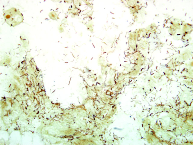
Colon: Colitis, erosive, multifocal, moderate, with necrosis, luminal mucin accumulation and argyrophilic spiral bacteria (
Fig. 4-1)
Conference Comment:
Swine dysentery (SD) generally affects pigs during the grow-finish phase of production. It causes a mucohemorrhagic diarrhea of the colon, and it can cause serious economic setbacks in a herd of pigs. SD is caused by
Brachyspira hyodysenteriae, a gram-negative, beta-hemolytic anaerobic spirochete. This organism was also formerly known as
Treponema hyodysenteriae, and then was reclassified as
Serpulina hyodysenteriae, and it is now known as
Brachyspira hyodysenteriae.(2) At least 4 other intestinal spirochetes have been identified in swine with
Brachyspira pilosicoli being the only other one to cause clinical disease.Â
B. pilosicoli can cause a mild colitis in pigs.(2)
Gross lesions for SD are normally present only in the large intestine. The ileocecal junction is normally the boundary for lesions caused by SD. Lesions often begin as hyperemic and edematous mucosal lesions and progress to a mucosa covered by copious amounts of mucus and fibrin admixed with blood. These lesions often form a pseudomembrane. There also may be multifocal erosions of the surface mucosa, and ulcerative lesions are not generally seen.(2)
Microscopically acute lesions consist of mucosal congestion, edema, and neutrophilic infiltrates in superficial lamina propria near blood vessels. Spirochetes may be seen in colonic crypts, and hyperplasia of goblet cells is common. These lesions progress with the development of superficial mucosal erosions and a characteristic mat of fibrin, mucous, hemorrhage, and cellular debris forms on the mucosal surface. Silver stains can be used to identify these spirochetes in tissue section.(2)
References:
1. Duhamel GE: The alimentary system.Â
In: Jubb, Kennedy, and Palmers Pathology of Domestic Animals, 5th edition; Maxie MG ed., Academic Press, Inc., Vol 2, pp. 210-213, 2007
2. Hampson DJ, Fellstrom C, Thomson JR: Swine dysentery.Â
In: Disease of Swine, eds. Straw BE, Zimmerman JJ, DAllaire S, Taylor DJ, 9th ed, pp. 785-805. Blackwell Publishing, Ames, Iowa, 2006
3. Harris DL, Hampson DJ, Glock RD: Swine Dysentery.Â
In: Disease of swine, eds. Straw BE, DAllaire S, Mengeling WL, Taylor DJ, 89th ed., pp.579-600. Iowa State University Press, Ames, Iowa, 1999
4. Muir S, Koopman MB, Libby SJ, Joens LA, Heffron F, Kusters JG: Cloning and expression of a Serpula (Treponema) hyodysenteriae hemolysin gene. Infect Immun
60:529-35, 1992
5. Whipp SC, Robinson IM, Harris DL, Glock RD, Matthews PJ, Alexander TJ: Pathogenic synergism between Treponema hyodysenteriae and other selected anaerobes in gnotobiotic pigs. Infect Immun
26:1042-7, 1979