Signalment:
Gross Description:
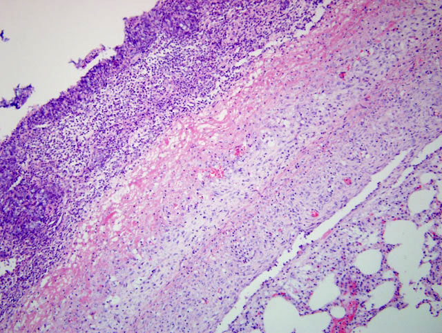
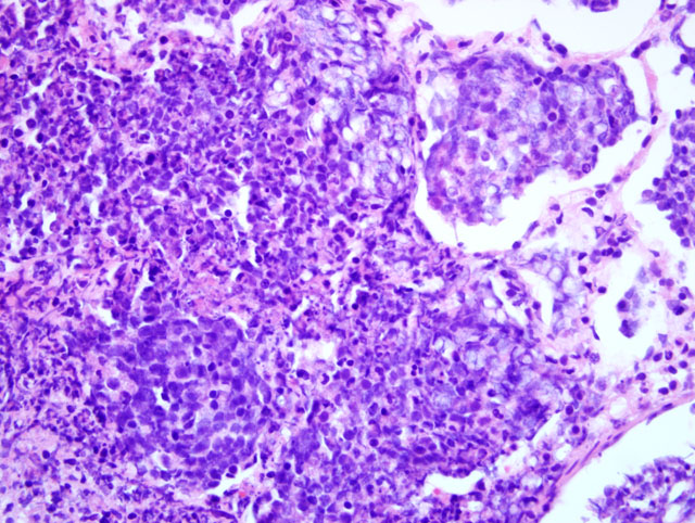
Histopathologic Description:
Morphologic Diagnosis:
Lab Results:
Virology: Swine influenza was not detected (RT-PCR).
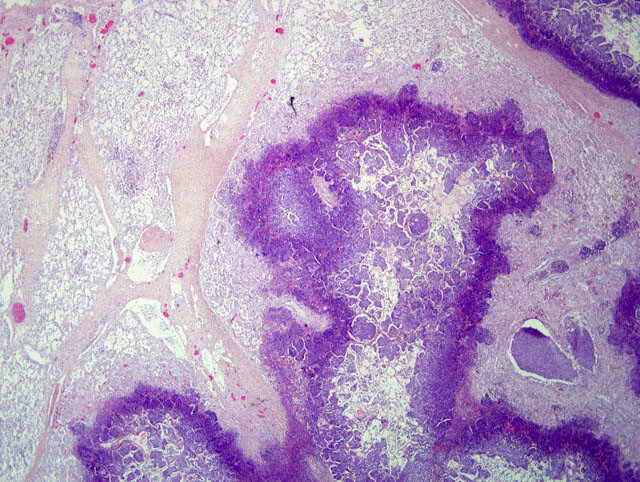
Immunohistochemistry: Porcine circovirus-2 antigen was demonstrated in lymph nodes.
Condition:
Contributor Comment:
In peracute cases, pigs can be found dead before clinical signs have been observed. In acute cases, pigs have high fever, apathy and anorexia, occasionally dyspnea and diarrhea or vomiting, cyanosis of skin and blood-tinged discharge from the nostrils is often seen. Gross lesions can be detected anywhere in the lungs, but often also one or both of the caudal lung lobes is affected. Blood stained froth fills the trachea. Fibrinous pleuritis and pleural adhesions are characteristic. Histologically there is a lobar fibrinosuppurative hemorrhagic and necrotizing pneumonia and fibrinous pleuritis.
Vasculitis with thrombosis is often seen. Differential diagnoses in acute cases include Salmonella Choleraesuis and Actinobacillus suis infections.
The disease is caused by a gram negative, encapsulated coccobacillus, Actinobacillus pleuropneumoniae. The bacterial agent is divided into two biovars according to the requirement of NAD (nicotinamide adenine dinucleotide). Biovar 1 strains are NAD- dependent and biovar 2 strains are NAD- independent. There are 15 serotypes based on capsular polysaccharides.
Clinical signs and pathological lesions are associated with several bacterial virulence factors combined with the host reaction. In addition to capsular components and cellular lipopolysaccharides, A. pleuropneumoniae produces several cytotoxins and proteolytic enzymes and can resist macrophage and complement killing. Acute disease is often followed by chronic encapsulated necrotic lung lesions and local pleuritis with adhesions. Chronic pleuritis seen at slaughter is common in herds with endemic infection. Asymptomatic carrier pigs spread the disease by direct contact or by aerosols.
A. pleuropneumoniae is capable of causing severe disease without predisposing factors, but the severity of the disease is affected by the immune status of the pigs and environmental factors such as crowding, ambient temperature or moving and mixing of animals. There are also differences in the virulence within and between the strains. Disease problems can be enhanced by other concurrent pathogens, for example Mycoplasma hyopneumoniae, swine influenza virus, PRRS (porcine respiratory and reproduction syndrome virus) or PMWS (porcine circovirus-2).
Other uncommon lesions associated with A. pleuropneumoniae include endocarditis, pericarditis, arthritis, osteomyelitis, meningitis, otitis media, granulomatous hepatitis and granulomatous pneumonia.(3) A. pleuropneumoniae was cultured from lung lesions of all the four examined pigs.
In this case, Pasteurella multocida was also isolated from the lungs. Pasteurella multocida can cause suppurative pneumonia in pigs, but it is often a secondary invader. The lymph nodes exhibited depletion of lymphocytes and loss of the follicular architecture. Porcine circovirus-2 antigen was also detected by immunohistochemistry in lymph nodes of this pig. These lesions were indicative of PMWS (postweaning multisystemic wasting syndrome). However, in this pig the lesions associated with PCV2 were considered to be an additional finding.
JPC Diagnosis:
Conference Comment:
| Pneumonia | Route of exposure | Anatomic Distribution | Lung Texture | Anatomic Location of Injury | Example | Common Pulmonary Sequelae |
| Broncho-pneumonia | Aerogenous | Cranioventral | Firm, hard | Bronchiolar-alveolar junction | Enzootic pneumonia; Pneumonic mannheimiosis | Abscesses, pleural adhesions |
| Interstitial | Aerogenous or hematogenous | Diffuse | Elastic, rubbery | Alveolar or interlobular septae | PRRS, PCV2 | Edema, type II pneumocyte hypertrophy, alveolar fibrosis |
| Bronchointerstitial | Aerogenous | Multifocal | Firm to rubbery | Bronchioloar and alveolar epithelium | BRSV, swine influenza | Mix of bronchoand interstitialpneumonias |
| Granulomatous | Aerogenous or hematogenous | Multifocal | Nodular | Non-specific | Tuberculosis, blastomycosis | Dissemination of infection |
| Embolic | Hematogenous | Multifocal | Nodular | Pulmonary vasculature | Vegetative endocarditis | Random abscesses |
The moderator discussed additional patterns of lung injury. Bronchitis and bronchiolitis result from direct damage to and subsequent necrosis of airway epithelium leading to airway inflammation. Potential etiologic agents or disease syndromes resulting in this pattern include feline asthma, viral infection, inhalation of toxic gases, toxins metabolized by P450 cytochrome oxidase of Clara cells, and inhaled irritants.(1)
As noted by the contributor, Actinobacillus pleuropneumoniaehas several virulence factors which play a unique role in the pathogenesis of porcine pleuropneumonia. The organism produces three RTX toxins: Apx I, II, and III which are cytolytic for neutrophils, erythrocytes, alveolar macrophages, and epithelial cells - once lysed, the contents of neutrophils and macrophages damage host tissue. Macrophages activated by lipopolysaccharide secrete neutrophil chemoattractants, activate complement, and promote coagulation. In addition to resisting macrophage phagocytosis, the bacterial capsule prevents complement activation. In order to survive in the milieu of cytotoxins and reactive oxygen species from lysed leukocytes, Actinobacillus pleuropneumoniae produces a variety of antioxidant enzymes including superoxide dismutase, catalase and hydrogen peroxide reductase.(1)
References:
2. Lopez A. Respiratory system. In: McGavin MD, Zachary JF, eds. Pathologic Basis of Veterinary Disease. 4th ed. St. Louis, MO: Elsevier; 2007:508-517.
3. Ohba T, Shibahara T, Kobayashi H,et al. Prevalence of granulomatous pleuropneumonia associated with Actinobacillus pleuropneumoniae serotype 2 in slaughter pigs. J Vet Med Sci. 2009;71:1089-1092.