Signalment:
Adult, female, red-footed tortoise (
Geochelone carbonaria).According to the provided history, an adult female red-footed tortoise (
Geochelone carbonaria) from a
privately-owned Midwestern zoological garden presented with progressive lethargy. As the animal was nonresponsive
to treatment, necropsy was performed on site following euthanasia, and tissues were submitted to the
Diagnostic Center for Population and Animal Health, Lansing, MI, for histopathologic examination.
Histopathologic Description:
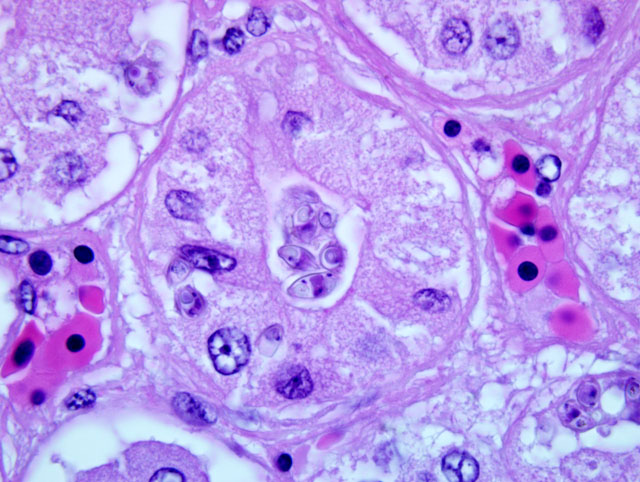
Kidney: Diffusely in a section of kidney, the renal tubular epithelium is variably
degenerate and necrotic. Within affected tubules, epithelial cells are often dissociated, variably swollen, with
eosinophilic finely vacuolated to floccular cytoplasm, and pyknotic to karyolytic to lost nuclei (degeneration and
necrosis). Few large, irregular, and mildly atypical cells with prominent vesicular nuclei are also present
(regeneration). Multifocally, tubules are filled with small to moderate amounts of eosinophilic smooth to floccular
material (proteinosis), sloughed-off epithelial cells, and few heterophils. Occasional tubular epithelial cells contain
up to 10, 10-15 μm, intracytoplasmic, retractile, amphophilic ellipsoidal organisms obscuring the cytoplasm and
nucleus. These organisms contain two apical polar capsules, approximately 2 μm in diameter, and basophilic
nuclear material in between polar capsules (morphology suggestive of myxozoans). Similar organisms are also
present in tubular lumina, within sloughed-off epithelial cells or individually amidst intraluminal debris. Low
numbers of lymphocytes and heterophils expand the renal interstitium, together with rare pigment-laden
macrophages (hemosiderin). Diffusely within the section, glomerular tufts are segmentally to globally, mildly to
moderately thickened. Multifocally, there is rare segmental synechiation and mild expansion of the urinary space,
which is mostly devoid of contents (clear space). Rare intratubular basophilic casts (mineral) are also identified.
Degenerate sloughed-off epithelial cells, heterophils, and myxozoan organisms are present in the renal pelvic lumen.
Multifocally within the subepithelial stroma of the pelvis are occasional foci of coarse deeply basophilic salt
deposition (dystrophic mineralization).
The above described organisms were not highlighted by PAS, acid-fast, or Giemsa histochemistries.
Additional findings in other organs (not included): multifocal myocardial mineralization and severe heterophilic
hepatitis. No organisms were observed within any other examined tissues, including but not limited to liver, biliary
ducts, and urinary bladder
Morphologic Diagnosis:
1. Kidney: Moderate diffuse tubular degeneration, and necrosis, with
intralesional, intraepithelial and intratubular myxozoa-type parasites (
Myxidium spp.).
2. Kidney: Moderate diffuse membranous glomerulonephritis, with mild tubular proteinosis.
3. Kidney: Minimal multifocal dystrophic mineralization.
Lab Results:
After a diagnosis was made, water from the pond where this tortoise was kept was submitted
for parasitological analysis but yielded no significant results.
Condition:
Myxidium sp.
Contributor Comment:
Microorganisms of the phylum Myxozoa are spore-forming, metazoan parasites that
infect the biliary, urinary, and gastrointestinal tracts of cold-blooded aquatic vertebrates, especially fishes but rarely
also reptiles, amphibians, and birds, with alternate life cycle stages in invertebrates. (2,4,5) Infection is believed to be
of little clinical importance in most species, although in the recent years, life-threatening disease has been
documented in a variety of species.(2) In the reported cases, the kidneys, biliary tract, and liver are the most
commonly compromised organs. Affected kidneys are often grossly pale and swollen, while histologically the
infection is characterized by degeneration and necrosis of the tubular epithelium infected with intralesional spores.(5)
All myxozoans known to infect reptiles are in the genus
Myxidium, and all reports involve aquatic turtles.(5)
In an attempt to elucidate the species involved in this case, additional ancillary tests such as ultrastructural analysis,
phase contrast microscopy, and molecular analysis of the samples were performed.
Ultrastructural analysis on infected kidney tissue revealed 60x20 μm spores, with a mean polar capsule dimension of
15x10 μm. Phase contrast microscopy and transmission electron microscopy demonstrated only mature spores, with
two asymmetrical valve cells and a binucleated sporoplasm between two opposing polar capsules. Valve cells were
longitudinally striated, with two overlapping sigmoidal capsule sutures. Polar capsules contained a single polar
filament, coiled 5 to 7 times and surrounded by a double-layered wall. Based on spore morphology, these myxozoa
were classified in the genus
Myxidium.
Macerated formalin-fixed tissue yielded plentiful spores from which DNA extraction was attempted. At least two
distinct extraction methods were used (using
Myxozoan and
Myxydium generic primers) with no success, likely as a
result of formalin-fixation nucleic acid damage to the samples (additional fresh samples were not readily available).
To the best of the contributors knowledge, renal myxozoanosis has not been documented in terrestrial chelonids.
JPC Diagnosis:
1. Kidney: Tubular degeneration and proteinosis, multifocal, mild with rare tubular epithelial
necrosis and intraepithelial and intratubular myxozoan parasites.
2. Adipose tissue, perirenal: Fat atrophy, diffuse moderate.
Conference Comment:
Several participants included some form of glomerulonephritis in the histomorphologic
diagnosis. The moderator pointed out that early studies and descriptions of the histomorphology of the chelonian
kidney mistakenly identified aging changes as glomerulonephritis, and he assessed the glomeruli in this tortoise as
essentially normal. Tortoises and other reptiles possess smaller glomeruli with reduced vascularity as compared to
amphibians, birds and mammals.(3) This morphologic feature acts to conserve water for these animals that often live
in an arid, dehydrating environment. The reduced glomerular size and vascularity results in an ostensibly thicker
mesangium with pronounced mesangial cells, and these normal histoanatomical features may be mistaken for
glomerulonephritis by pathologists lacking extensive experience in evaluating reptile tissues. Additionally, the
moderator believed there was little corroborative evidence to support a diagnosis of glomerulonephritis, e.g. lack
of interstitial inflammatory infiltrates.
A subtle, but important, histologic finding in this case is the depletion of perirenal adipose tissue. The moderator
indicated this was the most striking lesion, as it indicates negative energy balance. When conducting gross and
histologic examination of reptiles and amphibians, assessment of adipose tissue provides valuable insight into the
metabolic state of the animal.
Participants discussed the various animal species affected by myxozoan parasites. In addition to cold-blooded
vertebrates, the moderator indicated that several ducks, both native wild and captive exotic species, have been
infected with
Myxidium anatidum n. sp.(1) Myxozoan parasites which infect ducts preferentially infect biliary
epithelium.
References:
1. Bartholomew JL, Atkinson SD, Hallett SL, et al. Myxozoan parasitism in waterfowl.Â
Int J Parasitol. 2008;38(10):
1199-1207.
2. Garner MM, Bartholomew JL, Whipps CM, Nordhausen RW, Raiti P. Renal myxozoanosis in crowned river
turtles Hardella thurjii: description of the putative agent Myxidium hardella n. sp. by histopathology, electron
microscopy, and DNA sequencing.Â
Vet Pathol. 2005;42:589-595.
3. Jacobsen ER. Overview of reptile biology, anatomy, and histology. In:
Infectious Diseases and Pathology of
Reptiles. New York, NY: CRC; 2007:13-14.
4. Roberts JF, Whipps CM, Bartholomew JL, Schneider L, Jacobson ER. Myxidium scripta n. sp. identified in
urinary and biliary tract of Louisiana-farmed red-eared slider turtles Trachemys scripta elegans.Â
Dis Aquat Organ.
2008;80:199-209.
5. Zwart, P. Renal Pathology in Reptiles.Â
Vet Clin North Am Exot Anim Pract. 2006;9:129-159.