Signalment:
Gross Description:
Histopathologic Description:
Skeletal muscle: Diffusely throughout the tissue, there is random individual myocyte degeneration characterized by sarcoplasmic hypereosinophilia and swelling, loss of cross-striations, fragmentation, pyknosis, karyorrhexis and karyolysis.Â
Morphologic Diagnosis:
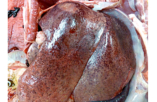
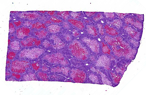
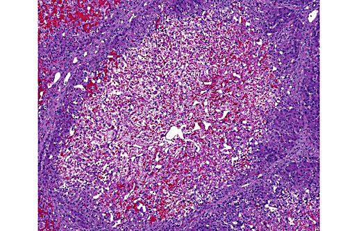
1. Liver: Necrosis, acute, centrilobular to massive, hemorrhagic, Yorkshire cross, pig.Â
2. Skeletal muscle: Degeneration and necrosis, acute, multifocal, mild to moderate, Yorkshire cross, pig.Â
Lab Results:
Condition:
Contributor Comment:
In the pig, the most obvious effect of Vitamin E/Selenium deficiency is sudden death, typically in young, fast growing weaners. Two principle presentations seen at post mortem examination are mulberry heart disease (MHD) and hepatosis dietetia (HD). Selenium is an integral part of the membrane enzyme glutathione peroxidase, reducing toxic lipid peroxidases to hydroxyl acids.(5) Vitamin E is an antioxidant and acts synergistically with selenium to protect membranes from high concentrations of lipoperoxidases.(5)
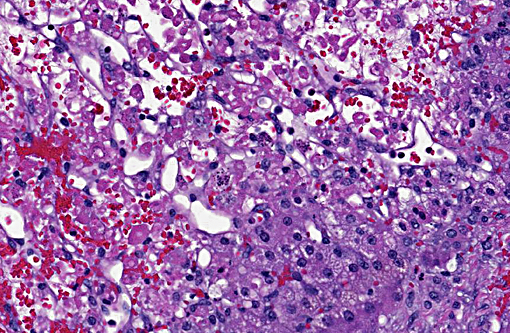
HD is characterized by hemorrhagic centrilobular to massive hepatic necrosis. Animals that survive the acute disease can develop lesions of parenchymal collapse and post necrotic scarring.(1,6) The most severe injuries are often present dorsally in the diaphragmatic liver lobes.(6) The pathogenesis of HD is not completely understood, but it is thought to be caused by a deficiency in vitamin E and/or selenium.(1) Affected animals respond to vitamin E or selenium supplementation, although, interestingly, it is difficult to experimentally induce this condition by feeding diets deficient in vitamin E and selenium.(1) It is thought that oxidative injury leads to hepatocyte necrosis since vitamin E and selenium-containing enzymes are antagonists of free radical formation and are therefore important in maintaining the stability and integrity of cellular membranes.(1,6) Experimental observations have revealed the need for concurrent deficiencies of sulfur-containing amino acids, tocopherols, and trace amounts of selenium for hepatic necrosis to develop.(6) Yellow staining of the adipose tissue is a common gross finding(6) as was seen in this case. The gallbladder may be edematous.(5) In relapsing cases, animals may be jaundice and hemorrhagic diathesis may occur with the primary manifestation of hemorrhage into or surrounding the joints.(6)
MHD (dietary microangiopathy of pigs) is characterized by fibrinoid necrosis and thrombosis of small vessels resulting in micro hemorrhages.(8) The resulting hemorrhages lead to a mulberry-like discoloration of the epicardial surface, particularly of the right atrium.(5,8) Hydropericardium is also a common finding. There is often widespread fibrinoid necrosis of small arteries and arterioles with endothelial damage and fibrin thrombi, particularly in the capillaries of the myocardium.(5,8) Typically, the following microscopic lesions are observed in a heart affected by MHD: interstitial hemorrhage associated with swollen cardiac myofibers that have lost cross striations, are hypereosinophilic, and have pyknotic nuclei.(4) In this entity, selenium concentrations within the liver and heart are often within normal limits.(5) Supplementation of selenium appears to decrease the incidence of HD, but not MHD. Therefore, vitamin E is considered to play a central role in the development of MHD.(5) An additional lesion that can be found in animals surviving greater than 24 hours is bilaterally symmetric softening of the cerebral white matter.(5)
Aside from the disease entities in pigs, vitamin E/selenium deficiency results in a broad spectrum of diseases in a variety of animal species and humans, including but not limited to: myopathy, steatitis, liver necrosis, and encephalomalacia because of increased oxidative stress on cells.(2)
JPC Diagnosis:
1. Liver, hepatocytes: Necrosis, centrilobular and midzonal, diffuse, severe, with hemorrhage and periportal hepatocellular lipidosis.Â
2. Skeletal muscle: Degeneration and necrosis, multifocal, marked.Â
Conference Comment:
When hepatic necrosis is observed in conjunction with rhabdomyocyte necrosis, vitamin E and/or selenium deficiency should be considered most likely. Selenium deficiency does not only occur in managed feed situations, but also in pasture-raised livestock. Selenium is normally present in soil and taken up by growing plants, however, in areas such as the Pacific Northwest, the soil is naturally deficient. Poor quality forages are also deficient in vitamin E, thus both must be supplemented in some situations.(7) Deficiencies of either result in the bodys reduced capacity to scavenge free radicals.Â
Free radicals are molecules with unpaired electrons rendering them highly reactive as they look to unload or oxidize that electron. They are generated as a normal product of mitochondrial respiration, absorption of radiant energy, enzymatic metabolism of drugs or chemicals, transition metals (Fenton reaction), nitric oxide production and by activated leukocytes during inflammation. Free radicals are very effective at killing cells, both those of pathogens and the normal host. Three reactions most relevant to free radical-induced cellular injury are lipid peroxidation of membranes, oxidation of proteins and DNA damage.(3) Antioxidants serve to protect the host by counteracting this oxidation. The importance of antioxidants in maintaining equilibrium is exemplified by the multitude of lesions associated with vitamin E and selenium deficiencies and nicely illustrated in this case.Â
References:
1. Cullen JM, Brown DL. Hepatobiliary system and exocrine pancreas. In: McGavin DM, Zachary JF, eds. Pathologic Basis of Veterinary Disease. 5th ed. Philadelphia, PA: Elsevier Mosby; 2012:446-448.Â
2. Kane AB, Kumar V. Environmental and nutritional pathology. In: Kumar V, Abbas AK, Fausto N, Aster JC, eds. Robbins and Cotran Pathologic Basis of Disease. 7th ed. Philadelphia, PA: Elsevier Saunders; 2005:455-456, 461.Â
3. Kumar V, Abbas AK, Fausto N, Aster JC. Cellular responses to stress and toxic insults: adaptation, injury and death. In: Kumar V, Abbas AK, Fausto N, Aster JC, eds. Robbins and Cotran Pathologic Basis of Disease. 8th ed. Philadelphia, PA: Elsevier Saunders; 2010:20-23.
4. Pallar+�-�s FJ, Yaeger MJ, Janke BH, Fern+�-�ndez G, Halbur PG. Vitamin E and selenium concentrations in livers of pigs diagnosed with mulberry heart disease. J Vet Diagn Invest. 2002;14:412414.
5. Robinson WF, Maxie MG. The cardiovascular system. In: Maxie MG, ed. Jubb, Kennedy, and Palmers Pathology of Domestic Animals. 5th ed. Vol 3. Philadelphia, PA: Elsevier Saunders; 2007:37-40.
6. Stalker MJ, Hayes MA. The liver and biliary system. In: Maxie MG, ed. Jubb, Kennedy, and Palmers Pathology of Domestic Animals. 5th ed. Vol 2. Philadelphia, PA: Elsevier Saunders; 2007:322-323, 368-373.
7. Valentine BA, McGavin MD. Skeletal muscle. In: McGavin MD, Zachary JF, eds. Pathologic Basis of Veterinary Disease. 5th ed. St. Louis, MO: Elsevier Mosby; 2012:900.
8. Van Vleet JF, Ferrans VJ. Cardiovascular system. In: McGavin MD, Zachary JF, eds. Pathologic Basis of Veterinary Disease. 5th ed. St. Louis, MO: Elsevier Mosby; 2012:581.Â