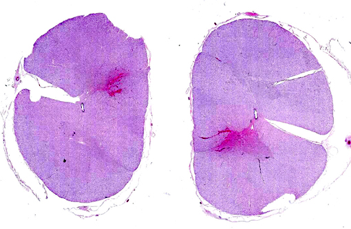
Signalment:  

7-year-old gelded male mixed breed pony, (Equus ferus caballus).The horse was a seven-year-old mixed breed gelded pony kept in a grassy pasture and fed a diet of hay and grain. Two weeks prior to euthanasia, the horse was in the proximity of strong winds and tornados which leveled several buildings on the adjacent property. For approximately 5-7 days after the storm, the pony avoided its pasture-mate (another horse) and human contact. During this time, he avoided being touched on the left and seemed lethargic. Thereafter he returned to normal behavior but two days prior to euthanasia he was unable to eat and held his head at the water trough without seeming to drink much. He progressively developed a generalized weakness and had difficulty standing. He avoided contact with his head (i.e., became head-shy) and started to have intermittent trembling of his lips and mouth. The owners tried rinsing his mouth with salt water and thoroughly examined the oral cavity (by touch). Given the progression of clinical signs and poor prognosis, owners elected euthanasia.


Gross Description:  

A seven-year-old gelded mixed breed horse with a body condition score of 2 out of 5 and mild to moderate autolysis (16 hour post-mortem interval) was received for necropsy and rabies testing. On oral exam there were moderate lingual points on the mandibular cheek teeth and buccal points on the maxillary check teeth. The spleen had an approximately 12-15 cm long laceration of the parietal surface opposite the stomach and extending transversely over the middle of the organ. The laceration site had organizing blood clots (red but with visible surrounding contraction and pallor (fibrosis)). Numerous 2-8 mm brown red nodules were scattered throughout the omentum in the area of the ruptured spleen. The cervical spinal cord had a focal area of hemorrhage and swelling at approximately the level of C1-C2.


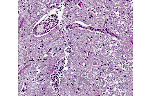
Histopathologic Description:

The section of spinal cord has multifocal perivascular cuffing by 2-4 layers of mononuclear cells dominated by lymphocytes with fewer histiocytes and occasional plasma cells. Most vessels are lined by plump endothelial cells and a focus of vessels near the tip of a dorsal funiculus is surrounded by amorphous eosinophilic material (high-protein edema fluid or an autolyzed area of hemorrhage). Axon sheaths in this area frequently contain macrophages (axonophagia) or dilated nerve processes (spheroids). Within the gray matter, neurons frequently have loss of nissl substance and nuclear material (chromatolysis) and there are multifocal small aggregates of glial cells (Babes nodules).


Morphologic Diagnosis:  

Diffuse non-suppurative and hemorrhagic vasocentric encephalomyelitis with neuronolysis with focal myelomalacia and hemorrhage.


Lab Results:  

Rabies virus fluorescent antibody was positive for rabies. Speciation of the virus was interpreted as the North Central United States and California skunk rabies.


Condition:  

Rabies


Contributor Comment:  

This case was challenging clinically because of the seemingly waxing and waning history. Nevertheless, the referring veterinarian had some concern about rabies at the time of submission. The lesion on the spleen and the history immediately after the local tornado activity are thought to represent the effects of blunt trauma from an airborne projectile. It was not until the three days preceding euthanasia that the horse was thought to be showing signs attributable to rabies.

Antemortem diagnosis of rabies remains problematic, but the disease should be considered in horses whenever there are rapidly progressing and/or diffuse neurologic signs. Differentials for rabies include hepatoencephalopathy, Eastern equine encephalitis, herpesviral encephalomyelopathy, protozoal encephalomyelitis, nigropallidal encephalomalacia, botulism, lead poisoning, cauda equine neuritis, meningitis, space-occupying masses, CNS trauma, or esophageal obstruction.(4)

Equine rabies can also be challenging to diagnose on necropsy. Numerous cases of disease confined to the spinal cord in horses have been reported making it advisable to include spinal cord in routine rabies testing in horses.(1) Horses are also unique in that their lesions often are associated with significant hemorrhage making it a consideration for focal spinal lesions associated with hemorrhage. Finally, as is demonstrated in this case, greater than half of the rabies cases in horses do not have identifiable Negri bodies.(2)


JPC Diagnosis:  

Spinal cord, gray matter: Neuronal degeneration, multifocal, moderate, with mild gliosis and lymphoplasmacytic meningitis.


Conference Comment:  

Although the contributor identified multifocal small nodules of glial cells, known as Babes nodules,(4) most conference participants did not appreciate this feature. The anatomic location as well as the subtle microscopic findings and lack of Negri bodies in this case engendered some difficulty in arriving at a diagnosis of rabies; many participants suspected an alternate viral etiology, such as Eastern equine encephalitis (alphavirus) or West Nile virus (flavivirus). 

Rabies virus is an enveloped RNA virus of the family Rhabdoviridae and the genus Lyssavirus; it causes meningoencephalomyelitis, ganglionitis and parotid adenitis, is almost invariably fatal, and is capable of affecting any mammalian species. Following infection with the virus, herbivores, unlike carnivores, are typically dead-end hosts. Reservoir hosts may vary temporally and regionally; among the most common are foxes, skunks, raccoons, feral dogs, wolves, jackals and mongoose. Fructivorous, insectivorous and vampire bats are also capable of transmitting rabies virus. Rabies viral neurotropism is due to a viral coat protein known as rabies virus glycoprotein (RVG), which binds several neural tissue receptors, including neuronal cell adhesion molecule (NCAM) and the p75 neurotrophin receptor (p75NTR). Virus inoculation typically occurs through contaminated saliva entering bite wounds inflicted by rabid animals. Initial viral replication occurs in myocytes adjacent to the bite wound, with subsequent invasion of the local neuromuscular junction and, eventually, the CNS and paravertebral ganglia via axoplasmic flow. Following viral replication in the CNS, there is centrifugal spread to salivary glands, nasal mucosa and adrenal glands.(3,4)

Three clinical manifestations of rabies are described: dumb, furious, and paralytic forms. The two most common clinical signs in affected mammals are progressive paralysis and aberrant behavioral patterns. In addition, horses in particular can have clinical signs associated with spinal cord injury, such as pelvic limb lameness, proprioceptive defects, ataxia, paralysis, and colic. Other reported species-specific features of the clinical progression of rabies include the following: cattle commonly exhibit excessive salivation and vocalization, swine are often found dead with no preceding clinical signs, sheep display passive behavior and dogs appear agitated or anxious. There are typically no gross lesions, although in horses infection may be associated with spinal cord hemorrhage. Historically, intracytoplasmic viral inclusions (i.e., Negri bodies) occur in the Purkinje cells of the cerebellum in herbivores and in neurons of the hippocampus in carnivores. In most mammalian species, Negri bodies are identified in 70% to 85% of affected animals; however, in horses this figure falls to 30% to 50%.(3,4) As an example of equine rabies with lesions limited to the spinal cord, without demonstrable Negri bodies, this case illustrates the potential variability of gross and histopathologic findings associated with rabies virus infection.


References:

1. Boone A, Susta L, Rech R, et al. Pathology in practice. J Am Vet Med Assoc. 2010;237(3):277-279.

2. Green S, Smith L, Vernau W, et al. Rabies in horses: 21 cases (1970-1990). J Am Vet Med Assoc. 1992;200:1133-1137.

3. Maxie MG, Youssef S. Nervous system. In: Maxie MG, ed. Jubb, Kennedy and Palmers Pathology of Domestic Animals. Vol 1. 5th ed. Philadelphia, PA: Elsevier; 2007:413-416.

4. Reed S, Bayly W, Sellon D, eds. Equine Internal Medicine. St. Louis, Missouri: Saunders; 2004:644-646.


Click the slide to view.


2-1. Spinal cord


2-2. Spinal cord


Back | VP Home | Contact Us |