CASE 3: S1601220 (4101207-00)
Signalment:
8-month-old, female Romney lamb, (Ovis aries).
History:
This lamb was found dead and was subsequently presented to Massey University Veterinary Pathology Department for necropsy.
Gross Pathology:
The lamb was in poor body condition and was underweight for its age. It had marked icterus of the subcutaneous connective tissues and mucous membranes. The pancreas was small, nodular and firm. Widespread crusting and ulceration were present on the ear pinnae and the skin surrounding the eyes and muzzle consistent with acute facial eczema. Four slow-release 30 g zinc boluses were present in the ruminal contents.
Laboratory results:
Liver: Zinc 161 mg/kg (Reference range 25-100 mg/kg)
Microscopic description:
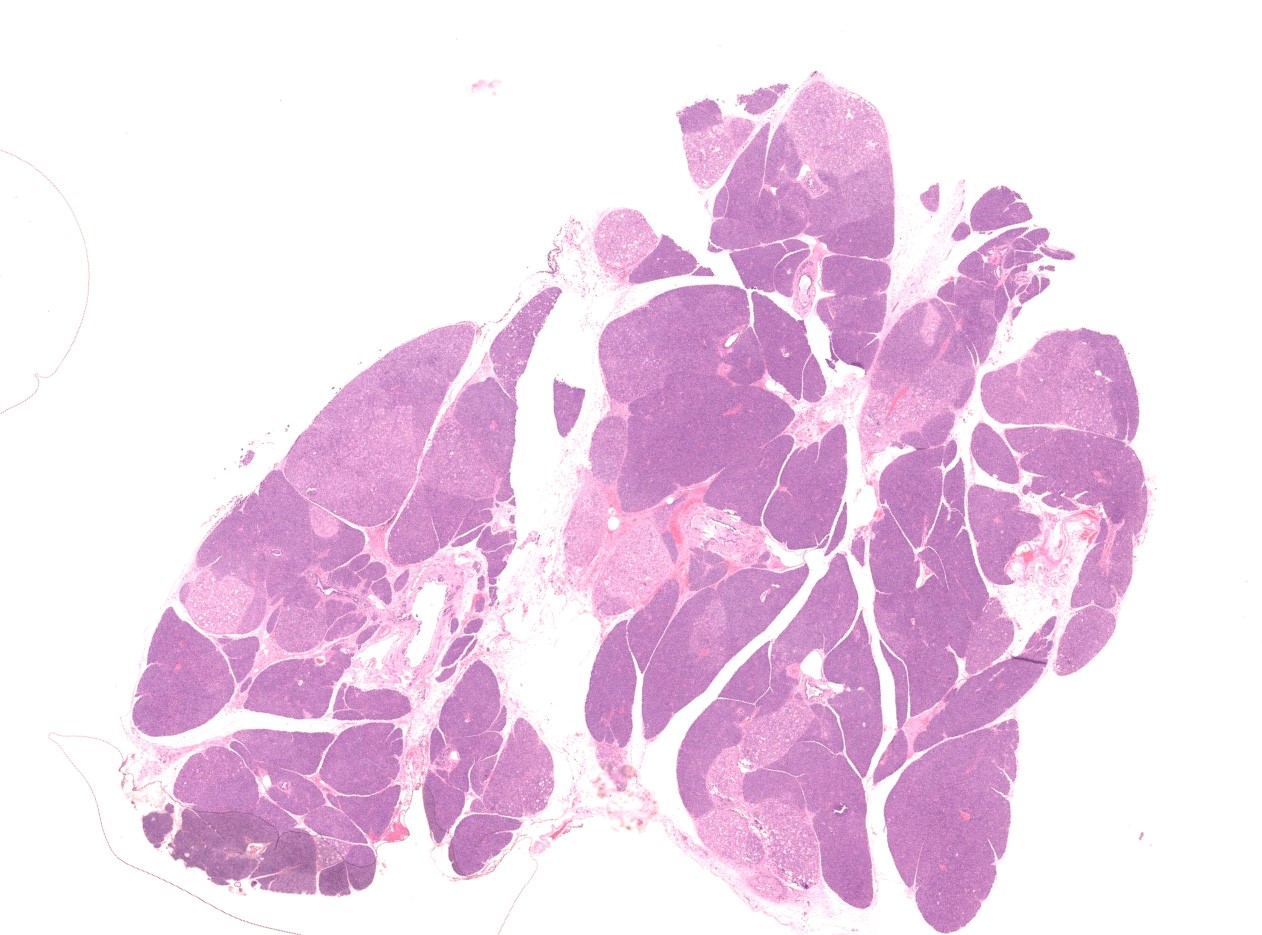
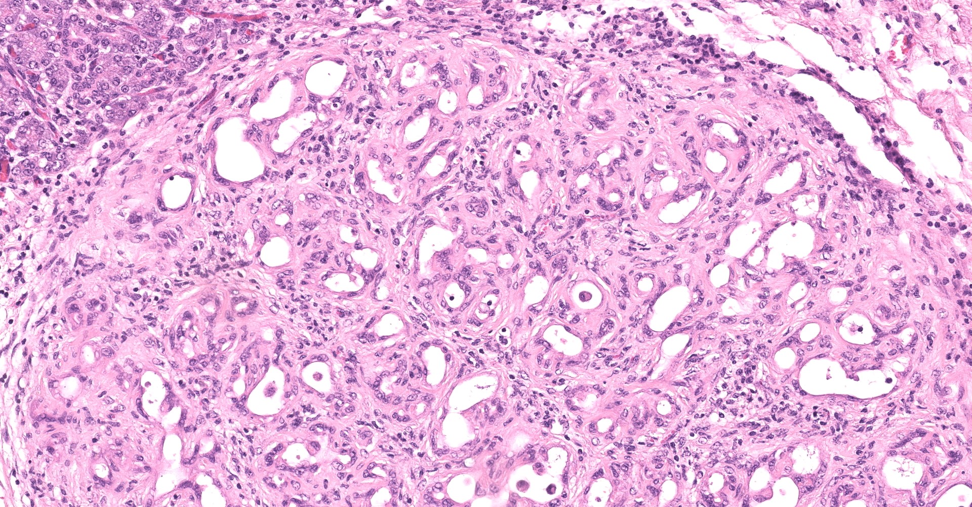
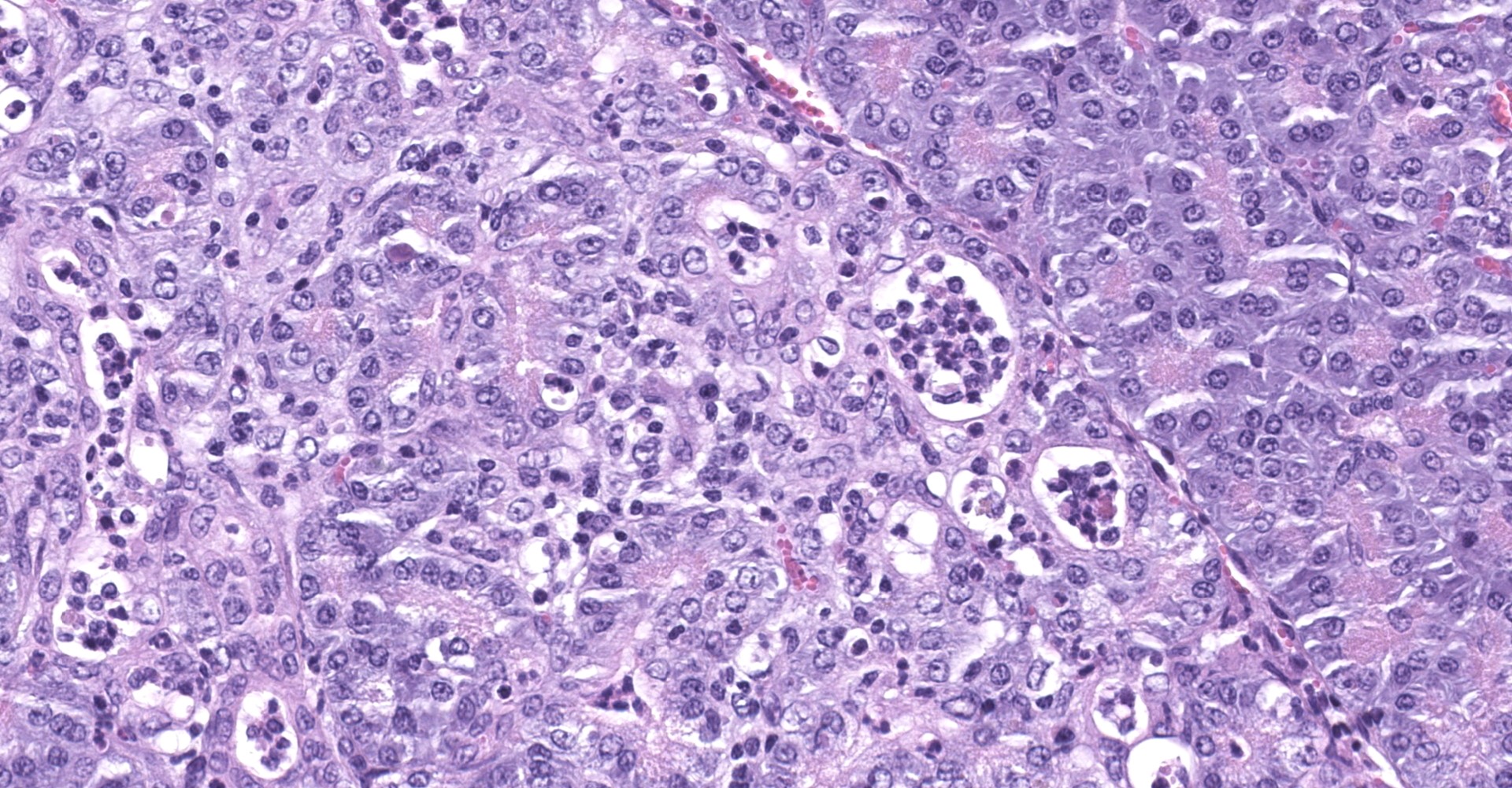
Pancreas: Within the pancreas, individual lobules show well-demarcated and varying degrees of necrosis and fibrosis. Mildly affected lobules are characterized by infiltration of the parenchyma with small numbers of neutrophils and mononuclear cells. Individual acinar cells appear shrunken with loss of zymogen granules and pyknotic nuclei. In moderately affected lobules, acinar cells show variable loss of basophilia and zymogen granules. Individual cells are swollen and vacuolated (degenerate), with other cells appearing shrunken and hypereosinophilic with pyknotic nuclei (necrosis). Acini and interlobular ducts are multifocally dilated, with enlarged luminal spaces, some containing small numbers of neutrophils. The parenchyma is infiltrated by neutrophils and mononuclear cells with corresponding disorganization and loss of associated pancreatic acini and ducts.
In severely affected lobules, the intralobular parenchyma is extensively replaced by fibrous connective tissue and fibroblasts. The few remaining acini show marked loss of basophilia and zymogen granules, and frequently contain dilated cystic luminal spaces with attenuated acinar cells. Moderate numbers of mononuclear cells and fewer neutrophils are present throughout the parenchyma.
The interlobular spaces are moderately expanded by multifocal areas of haemorrhage and oedema, and there is marked interlobular fibrosis.
Contributor's morphologic diagnosis:
Pancreas: Degeneration and necrosis, chronic-active, multifocal, severe, with intralobular and interlobular fibrosis, ovine.
Contributor's comment:
In this sheep, the diagnosis of zinc toxicity was made on the basis of similar clinical and pathological findings compared to reported experimental and natural cases of zinc toxicity, the reported increased zinc liver concentrations and the presence of four zinc bullets within the rumen.
Zinc is a trace element which has been shown to produce toxic effects in multiple species including humans, dogs, sheep, cattle and many wildlife species.1,6 In ruminants, zinc toxicity may result from administration of zinc as protection against sporidesmin toxicity.1,9,13 In other animals, zinc toxicity typically results from dietary indiscretion, and may be caused by the ingestion of zinc-coated U.S. pennies, batteries, paint, hardware, creams, and automotive parts.2,6
The largest effects are predominantly in the exocrine pancreas, where zinc excretion occurs, but other organs, including the kidney, liver, abomasum, small intestine and blood, may be affected.1,11 Clinical signs include anorexia, lethargy, vomiting and may include intravascular haemolysis, haemoglobinuria, and icterus.1,6 In large animals, poor weight gains may be the most prominent clinical sign.
In ruminants, initial effects are produced within ductular structures within the exocrine pancreas with subsequent changes in acinar cells. The endocrine pancreas is not affected. The initial reported lesions include vacuolar degeneration, necrosis and regeneration with a pronounced lobular distribution. With prolonged zinc intoxication, pancreatic necrosis exceeds the rate of regeneration, with fibrosis and atrophy becoming the predominant findings.1,14
As seen in this case, relatively normal appearing lobules may be present alongside severely affected lobules. It is hypothesized when injury of a duct occurs, drainage is impeded causing damage to the that lobule.14 This is supported by the fact that cattle, which have a dual exocrine pancreatic drainage via an accessory duct, do not show a discrete lobular pattern despite having other similar histopathological changes.14 Tissue zinc concentrations in experimental studies in sheep with similar pancreatic lesions have been reported at over 800 mg/kg, 14 and a value of 161 mg/kg, which was found in this lamb, is outside the reference range, but on low end of previously reported concentrations in experimentally induced zinc toxicity in sheep. Additionally, zinc induces a Heinz body haemolytic anaemia of which the exact mechanism is unknown. Possible mechanisms include hapten-induced immune-mediated destruction, direct cell membrane damage, or erythrocyte enzyme inhibition. Released haemoglobin may result in acute renal tubular injury, in addition to hypoxic injury caused by anaemia and reduced renal blood flow.4
Zinc salts have been shown to provide protection against sporidesmin toxicity in sheep, by forming a stable mercaptide with sporidesmin, thereby preventing the formation of free radicals from this toxin.10,12,16 Sporidesmin, a mycotoxin produced by Pithomyces chartarum, induces damage to biliary epithelium, resulting in cholestasis, and in chronic cases, hepatic fibrosis, atrophy and nodular regeneration. Phylloerythrin, a metabolite of chlorophyll, is unable to be excreted via damaged bile ducts and accumulates in the skin resulting in photodermatitis. Prior damage to the liver caused by sporidesmin toxicity has been reported to be associated with an increased likelihood of zinc toxicity.15 Metallothionein which is involved in detoxifying metal ions, is produced by the liver. In addition, serum albumin, which binds zinc in the blood, is also produced by the liver, and both albumin and metallothionein levels may be reduced in sheep with liver damage.15
Contributing Institution:
Massey University
School of Veterinary Science
Private Bag 11
222 Palmerston North 4442
New Zealand
JPC diagnosis:
Exocrine pancreas: Degeneration and necrosis, lobular, multifocal, marked, with tubular complexes and fibrosis.
JPC comment:
The contributor provides an excellent review of zinc toxicosis and its pathologic effects on the pancreas.
A previous study comparing natural and experimental cases of zinc toxicity in sheep found pathological changes may be observed in any organ involved in the absorption, excretion, or intermediary metabolism of zinc, including the abomasum, small intestine, liver, and kidney addition to the rumen and adrenal glands; however, the pancreas was the only organ consistently affected.1 This is significant in that this organ is often overlooked by pathologists, particularly when examining ruminents.1
A separate zinc related illness known as Metal fume fever (MFF) has been described in the human literature and was first described in metal workers during the mid-19th century, predominantly affecting foundry workers and welders of galvanized steel.18 The syndrome occurs due to the inhalation of freshly formed oxide fumes (predominately in the form of zinc oxide) generated from molten bronze and the welding of galvanized steel, and continues to affect an estimated minimum of 1,500-2,000 of the approximately 700,000 metal workers in the United States each year.18
Also known as "zinc shakes" and first described as "brass founders' ague", MFF results in a "flulike" illness due to the inhalation of freshly formed ZnO, resulting in numerous pro-inflammatory changes including the production of pro-inflammatory cytokines and recruitment of inflammatory cells into the lungs and is characterized by fever, cough, wheezing, chest tightness, chills, myalgia, leukocytosis with a left sift, thirst, metallic taste, and salivations.18 The diagnosis is typically based on clinical findings and history of occupational exposure and resolution is spontaneous. Treatment is symptomatic, with an emphasis on prevention of subsequent exposure. Interestingly, smoking has been found to modify the effect of welding fumes on specific markers of inflammation, with non-smokers experiencing a significant increase in circulating WBC counts in comparison to smokers.18
References:
1. Allen J, Masters H, Peet R, et al. Zinc toxicity in ruminants. Journal of Comparative Pathology. 1983; 93: 363-377.
2. Boes KM, Durham AC: Bone Marrow, Blood Cells and the Lymphoid/Lymphatic System. In: Zachary JF, ed. Pathologic Basis of Veterinary Disease. 6th ed. St. Louis, MO: Elsevier; 2017: 746-747.
3. Campbell J, Mills C. The toxicity of zinc to pregnant sheep. Environmental research. 1979;20: 1-13.
4. Cianciolo RE, Mohr FC: Urinary system. In: Maxie MG, ed. Jubb, Kennedy and Palmer's Pathology of Domestic Animals. 6th ed. St Louis, MO: Elsevier; 2016: 356-357.
5. Graham T, Holmberg C, Keen CL, Thurmond M, Clegg M. A pathologic and toxicologic evaluation of veal calves fed large amounts of zinc. Veterinary Pathology. 1988;25: 484-491.
6. Gurnee CM, Drobatz KJ. Zinc intoxication in dogs: 19 cases (1991-2003). Journal of the American Veterinary Medical Association. 2007;230: 117 4-1179.
7. Jubb KVF, Stent AW. Pancreas. In: Maxie MG, ed. Jubb, Kennedy, and Palmer's Pathology of Domestic Animals. Vol 2, 6th ed. St. Louis, MO: Elsevier; 2016:356- 357.
8. LaBonde J: Toxicity in pet avian patients. In: Seminars in Avian and Exotic Pet Medicine, pp. 23-31. Elsevier, 1995.
9. Munday R, Thompson AM, Fowke EA. et al. A zinc-containing intraruminal device for facial eczema control in lambs. New Zealand Veterinary Journal. 1997;45: 93- 98.
10. Munday R. Studies on the mechanism of toxicity of the mycotoxin sporidesmin. I. Generation of superoxide radical by sporidesmin. Chem. Biol. Interact. 41, 361- 374, 1982
11. Munday R. Studies on the mechanism of toxicity of the mycotoxin sporidesmin. 2-evidence for intracellular generation of superoxide radical from sporidesmin. J. Appl. Toxicol. 4, 176-181, 1984
12. Munday R. Studies on the mechanism of toxicity of the mycotoxin sporidesmin 3-inhibition by metals of the generation of superoxide radical by sporidesmin. J. Appl. Toxicol. 4, 182-186, 1984
13. Smith B. Toxicity of zinc in ruminants in relation to facial eczema. New Zealand Veterinary Journal. 1977;25: 310-312.
14. Smith B, Embling P. Sequential changes in the development of the pancreatic lesion of zinc toxicosis in sheep. Veterinary Pathology. 1993;30: 242-247.
15. Smith BL, Embling PP. Effect of prior sporidesmin intoxication on the pancreopathy associated with zinc oxide toxicity. New Zealand Veterinary Journal. 1999;47: 25-27
16. Smith BL, Embling PP, Towers NR, Wright DE, Payne E. The protective effect of zinc sulphate in experimental sporidesmin poisoning of sheep. New Zealand Veterinary Journal. 1977;25: 124-127.
17. Valli VEO, Kiupel M, Bienzle D: Hematopoietic system. In: Maxie MG, ed. Jubb, Kennedy, and Palmer's Pathology of Domestic Animals. Vol 3. 6th ed. St. Louis, MO: Elsevier; 2016:125-126.
18. Wardhana, Datau EA. Metal fume fever among galvanized welders. Acta Med Indones. 2014;46(3):256-262.