Signalment:
11-month-old, male castrated, dachshund, canine, (
Canis familiaris)This dog was presented to the North Carolina
State University College of Veterinary Medicine
Neurology Service for a 3-month history of stumbling and
falling which had progressed to severe vestibular ataxia,
nystagmus, and possible seizure activity. This dog had
been treated previously with antibiotics and there was no
response.
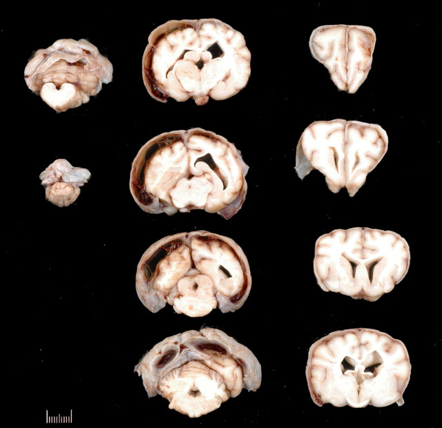
Gross Description:
The necropsy was limited to the
head only. Bilaterally, the meninges of the caudal part
of the temporal, parietal lobe and the occipital lobe
were fluctuant and markedly thickened, up to 6 mm.
Serosanginous fluid and dark red soft clots were found
within the subdural space (subdural hematoma). On the
cut surface, the arachnoid space was markedly dilated due
to marked cerebral atrophy which was most prominent
in the left occipital lobe. The lateral ventricles were
asymmetrical with the left lateral ventricle smaller than
the right lateral ventricle (
Fig. 3-1). The third ventricle
was slightly enlarged ventrodorsally and the interthalamic
adhesion was moderately thinned.
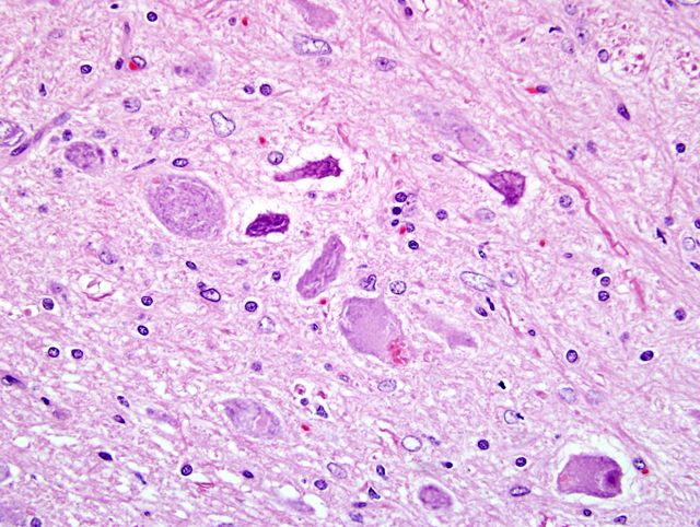
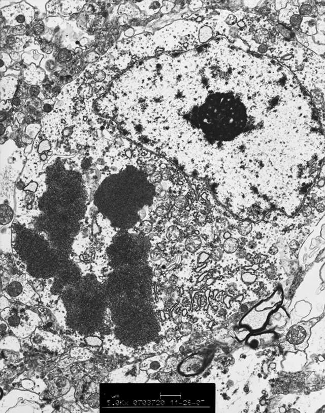
Histopathologic Description:
Cerebrum: Diffusely
in the cerebral cortical gray matter, approximately
60% of the neurons contain abundant, eosinophilic to
amphophilic, granular to globular, cytoplasmic pigment,
which occasionally displaces the nuclei peripherally
(
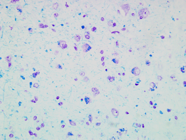
Fig. 3-4). The cytoplasmic pigment stains positively
with the PAS reaction and Sudan black and more
obviously, but less frequently, with LFB stain (
Fig. 3-5).
Moderate numbers of neurons are shrunken, rounded or
angular, and hypereosinophilic, with hyperchromatic or
pyknotic nuclei, interpreted as neuronal degeneration and
necrosis. The dura and arachnoid mater are markedly
thickened up to 5 times normal by fibroblasts, moderate
multifocal angiogenesis and additional connective tissue
matrix. Multifocally, there is a moderate amount of
subarachnoid hemorrhage. Under U.V. illumination the
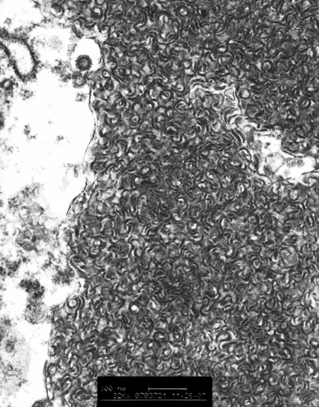
pigment is autofluorescent. Ultrastructurally, the neurons
have intracytoplasmic storage bodies which consist of
curvilinear forms (
Figs. 3-5, 3-6).
Other sections examined: Cerebellum: There is moderate
depletion of Purkinje cells and marked depletion of
granular cells accompanied by marked narrowing of the
granular layer and the molecular layer of the cerebellar
cortex. Purkinje cells also contain eosinophilic cytoplasmic
pigment which stains with PAS and LFB.
Eye: Bilaterally, there is mild depletion of the ganglion
cells in the retina. There are a few ganglion cells with
intracytoplasmic granules which stain with LFB. There is
multifocal detachment of the retina with mild hypertrophy
and hyperplasia of the pigmented epithelium (tombstone
change).
Morphologic Diagnosis:
Diffuse,
moderate, neuronal degeneration and necrosis and
abundant neuronal intracytoplasmic granular pigment
with cerebral atrophy
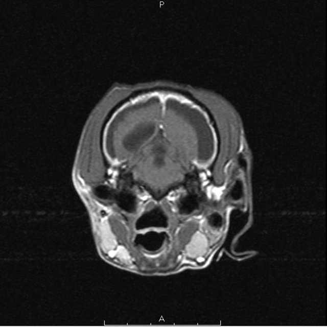
Lab Results:
MRI findings
Moderate ventricular asymmetry is present with the
right lateral ventricle being somewhat larger than the
left. The third ventricle is also enlarged. There is a large
accumulation of fluid peripheral to the cerebral cortex
(extra-axial) most apparent from the level of the optic
chiasm caudally (
Fig. 3-2). This fluid accumulation
does not suppress on the FLAIR sequence as does the
fluid within the ventricles and is slightly T1 hyperintense
relative to fluid within the ventricles. The interthalamic
adhesion is noticeably small and asymmetrical. The
cerebellum has an irregular margin and the folia within
the cerebellum have increased conspicuity. Post contrast
medium administration, there is florid enhancement of the
meninges surrounding the cerebral cortex and falx. There
is no abnormal parenchymal enhancement. There is no
evidence of increased intracranial pressure.
Condition:
Ceroid lipofuscinosis
Contributor Comment:
The neuronal ceroid
lipofuscinoses (NCLs) are inherited lysosomal storage
diseases characterized by progressive neuropathy and
accumulation of autofluorescent lipopigment in neurons
and other cells.
3 NCLs have been described in human
beings, cattle, sheep, goats, cats and in several breeds of
dogs.
1,4 Human NCLs are classified into several forms
based on the age of clinical onset, causative gene and
ultrastructure of the accumulating lysosomal storage
bodies.
3 The causative mutations in dogs have been
reported in English setters (a missense mutation in
CLN
8), border collies (a nonsense mutation in
CLN5), bulldogs
(a missense mutation in
CTSD) and juvenile dachshund
(a frame shift mutation in canine
TPP1: the ortholog of
human
CLN2). The canine TPP1 gene encodes a lysosomal
enzyme called tripeptidyl 1 peptidase
1 and is known as the
causative gene of infantile neuronal ceroid lipofuscinosis
in humans and when mutated leads to accumulation of
curvilinear-appearing cytosomes in neurons as well.
3
The major accumulating protein in this breed is unknown,
but subunit C of mitochondrial ATP is reported in English
setters, border collies and Tibetan terriers, and sphingolipid
activator proteins A and D have been identified in some
types of human NCLs.
1,3,4 The cerebral and cerebellar
cortex atrophy with cytoplasmic eosinophilic pigmen,
which stained with PAS and LFB stain in neurons, is
consistent with ceroid lipofuscinosis. The autofluorescence
and ultrastructure of the accumulating pigment in this
dog are very similar to the previous reports of juvenile
ceroid lipofuscinosis in this breed. The dilated subdural
space is considered to be secondary to the cerebral cortical
atrophy.
JPC Diagnosis:
Cerebrum: Neuronal degeneration,
necrosis and loss, extensive, with gliosis, cerebral atrophy,
meningeal fibrosis, subdural hemorrhage, and eosinophilic
neuronal cytoplasmic bodies
Conference Comment:
Neuronal ceroidlipofuscinosis,
also known as Batten disease, has been
reported in several domestic species and was recently
reported in a Vietnamese pot-bellied pig.
2 The mode
of inheritance is thought to be autosomal recessive for
this type of storage disease.
5 Although accumulation of
intracytoplasmic storage material can be found in many
organs, the most prominent pathologic manifestations
of these diseases are seen in the retina, cerebral cortex,
andcerebellum.
5
Gross lesions can vary from being nearly imperceptible to
marked cerebral atrophy. The earlier the onset of the disease
the more severe the brain atrophy.
4 Ultrastructurally,
ceroid-lipofuscinosis can take on many different structural
forms including curvilinear bodies, fingerprint bodies,
and laminated stacks of membranes.
5 Areas of cerebral
atrophy often appear to have a brown tinge.
5 The subdural
hematoma in the present case is suspected to have resulted
from trauma associated with motor disturbances.
Veterinary research into affected sheep resulted in a major
contribution to understanding the human and animal ceroid
lipofuscinoses by demonstrating that the stored material is
predominantly protein (subunit C of mitochondrial ATP
synthase) rather than lipid, as had been believed. Further
research showed that in some forms of the disease,
sphingolipid activator proteins are accumulated. Thus,
ceroid lipofuscinosis is actually a misnomer.
References:
1. Awano T, Katz LM, Brein PD, Sohar I, Lobel P, Coates
RJ, Johnson CG, Giger U and Jonson SG: A frame shift
mutation in canine TPP1 (the ortholog of human CLN2) in
a juvenile Dachshund with neuronal Ceroid lipofuscinosis.
Mol Genet Metab
89:254260, 2006
2. Cesta MF, Mozzachil K, Little PB, Olby NJ, Sills RC,
Brown TT: Neuronal ceroid lipofuscinosis in a Vietnamese
pot-bellied pig (
Sus scrofa). Vet Pathol
43:556-560, 2006
3. Haltia M: The neuronal ceroid-lipofusinoses: From
past to present, review. Biochamica et Biophysica
1762:850-856, 2006
4. Jolly DR, Palmer ND, Studdert PV, Sutton HR, Kelly
RW, Koppang N, Dahme G, Hartley JW, Patterson SJ
and Riis CR: Canine ceroid-lipofusinoses: A review and
classification. J Small Anim Prac
35:299-306, 1994
5. Maxie MG, Youssef S: Nervous system.Â
In: Jubb,
Kennedy and Palmers Pathology of Domestic Animals,
vol. 1 ed. Maxie MG, 5th ed., pp.329-330. Elsevier,
Philadelphia, PA, 2007