Signalment:
Histopathologic Description:
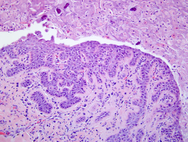
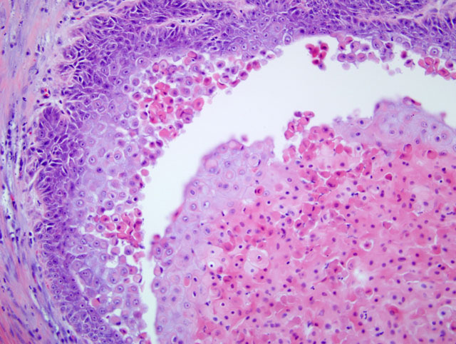
In some sections there is rupture of the cyst wall with release of acantholytic cells and keratin debris into the dermis. A mild infiltrate of neutrophils and epitheloid macrophages surround these areas.
Morphologic Diagnosis:
Condition:
Contributor Comment:
Warty dyskeratomas are rare benign tumors that are believed to arise from hair follicles. Histologically there are single to multiple cystic structures in the dermis that are lined by a stratified squamous epithelium that is hyperplastic and forms rete pegs that extend into the surrounding dermis. There is acantholysis of the stratum spinosum which can result in separation of the superficial layers of epithelium from the basal layers. There is frequent keratinocyte apoptosis or dyskeratosis. The cyst lumen is usually filled with keratin debris and acantholytic cells. Secondary inflammation from release of cyst contents into the surrounding dermis is common.(2)
JPC Diagnosis:
Conference Comment:
Warty dyskeratomas in animals can be confused with an acantholytic variant of squamous cell carcinoma originating from the hair follicle. Squamous cell carcinomas often have extensive apoptosis as a distinguishing feature. Warty dyskeratomas are benign tumors and do not infiltrate through the basement membrane.(1)
In humans, warty dykeratomas are solitary verrucous epidermal neoplasms with marked acantholysis and dyskeratosis of proliferating neoplastic cells.(3) In humans, these tumors generally are found on sun-exposed body parts, and these lesions usually involve hair follicles with some reported cases of oral involvement. Tumors appear as single, raised nodules with umbilicated centers and are usually benign tumors. Histologically, these masses are endophytic with densely packed keratin and suprabasilar clefts with marked acantholysis. Acantholytic cells are described as either corps ronds, which are suprabasilarly located, large, eosinophilic, rounded cells with perinuclear halos, or corps grains, which are small intensely eosinophilic, ovoid cells with pyknotic flattened nuclei. These two types of cells are often adjacent to an acantholytic stratum granulosum and parakeratotic stratum corneum.(3)
References:
2. Hill, JR: Warty dyskeratoma in two dogs. In: Proceedings of the 3rd AAVD/ACVD Meeting, Phoenix, (1987) p.40.Â
3. Murphy GR, Elder DE: Atlas of Tumor Pathology, Non-Melanocytic Tumors of the Skin, 3rd Series, vol 1, pp. 27-28. Armed Forces Institute of Pathology, Washington DC, 1991