WSC 22-23:
Conference 8:
CASE IV:
Signalment:
4-year-old, intact male, mix breed canine, Canis lupus familiaris.
History:
A 4-year-old, intact male, medium-sized (~15 Kg of body weight), mix breed dog was presented to the veterinary clinic with multiple (5) skin nodules in the right knee, thoracic region, dorsal aspect of the nose, and right and left thoracic limbs. In the initial clinical examination, the dog was normothermic, bright, alert and responsive, had good body condition and muscle tone, adequate hydration state, normal capillary refill time, and adequate coloration of the mucous membranes. The dog was treated empirically and unsuccessfully with enrofloxacin and prednisone. After one month of treatment, the animal was presented again to the clinic with vomiting, diarrhea, and partial loss of vision. The larger mass, located in the left thoracic limb, was surgically biopsied for histologic examination.
Gross Pathology:
A firm, raised skin nodule covered by ulcerated and alopecic epidermis.
Laboratory Results:
No laboratory results reported.
Microscopic Description:
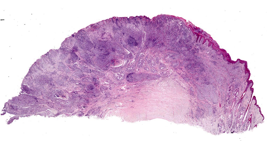
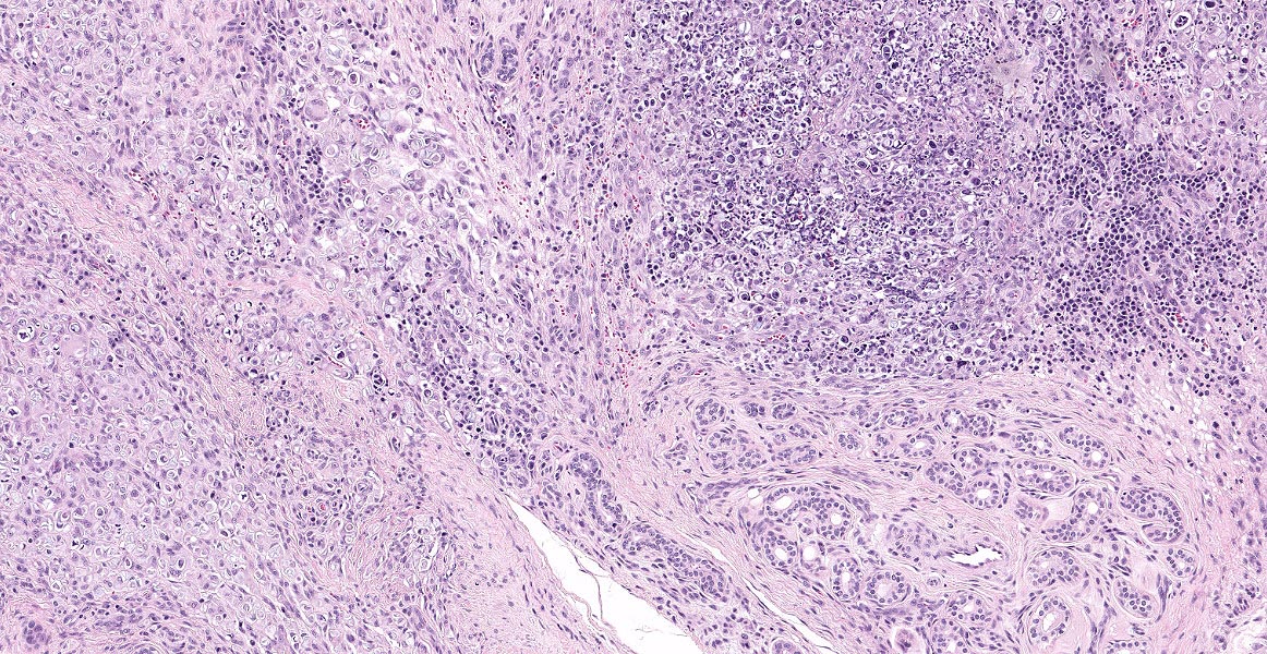
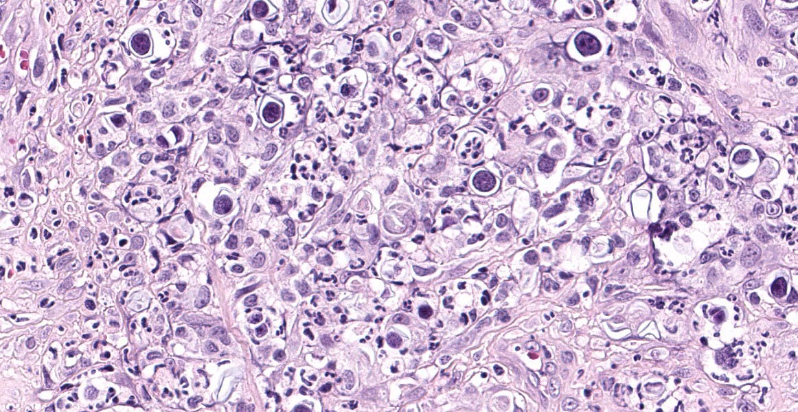
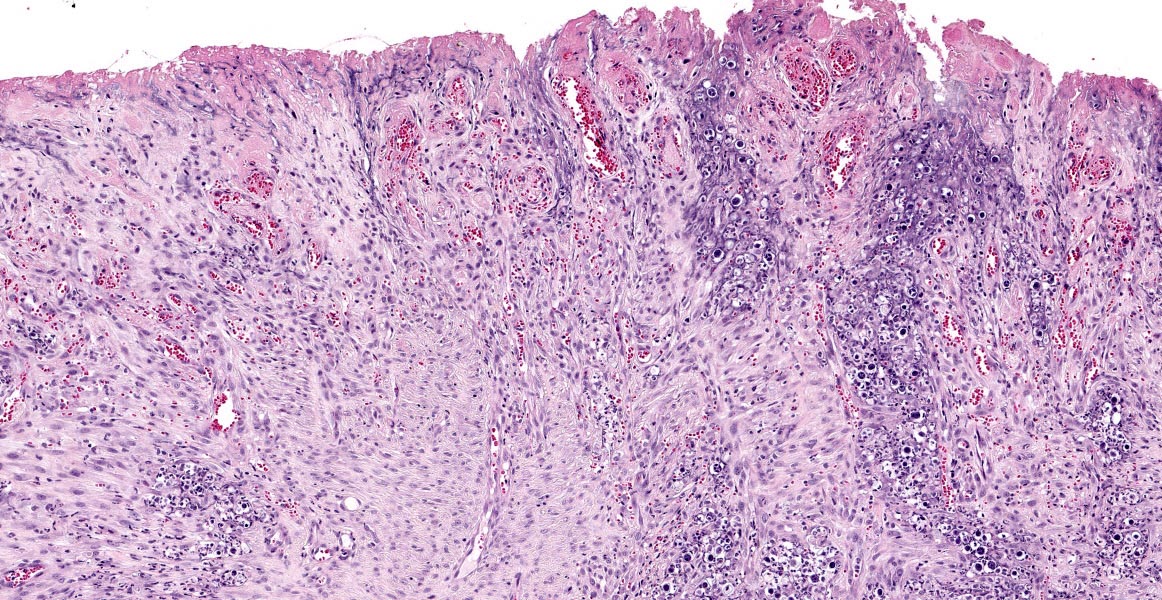
Haired skin. Diffusely expanding the dermis and subcutaneous tissue and elevating the overlying epidermis, there is a dense inflammatory cellular infiltrate composed predominantly of activated and epithelioid macrophages, occasional multinucleated giant cells, lymphocytes, plasma cells, and fewer neutrophils. Admixed with the inflammatory cells, there are numerous extracellular and intrahistiocytic microorganisms and pyknotic and karyorrhectic cellular debris (necrosis). Microorganisms (algae) are mildly pleomorphic, mostly spherical to ovoid, and range between 4 and 17 μm in diameter, with a 2 to 4 μm, often surrounded by a clear halo. Algal cells contain granular flocculent amphophilic material and a distinctly basophilic nucleus. Some algal cells present internal septation with daughter cells (endosporulation). The overlying epidermis is extensively ulcerated or lost, and the superficial dermis covered by extravasated fibrin and necrotic cellular debris, there is extensive fibrosis with perpendicularly-oriented blood vessels lined by plumped epithelium (neovascularization/granulation tissue), scattered fibrin thrombi in small-caliber superficial venules and microhemorrhage.
Algal cells are highlighted by GMS stain and PAS reaction.
Contributor’s Morphologic Diagnoses:
Haired skin: severe granulomatous and ulcerative dermatitis/panniculitis, with multinucleate giant cells and myriads of intrahistiocytic and extracellular algal microorganisms morphologically resembling Prototheca spp., Canis lupus familiars.
Contributor’s Comment:
Differential diagnoses on clinical grounds in this case included multifocal/multicentric neoplasia with cutaneous involvement, and granulomatous/pyogranulomatous dermatitis/panniculitis caused by infectious agents, mostly bacterial or fungal infections. The microscopic examination of the biopsy with HE, GMS and PAS stains revealed severe granulomatous dermatitis with intralesional algae morphologically consistent with Prototheca spp. as depicted in the submitted slides.
Additionally, as part of the diagnostic investigation, formalin-fixed paraffin-embedded skin was processed for molecular identification of Prototheca spp. at the Centers for Disease Control and Prevention (CDC) in Atlanta, Georgia. For molecular confirmation and speciation, a polymerase chain reaction-restriction fragments length polymorphism (PCR-RFLP) was performed as previously described.14 The agent was identified as P. zopfii genotype 1.
Thus, the diagnostic investigation in this case allowed for unequivocal confirmation of P. zopfii genotype 1 infection in a dog, by a combination of morphological and molecular methods.
Protothecosis is a rare disease caused by environmental algae of the genus Prototheca, saprophytes of worldwide distribution that can infect mammals, and result in either localized or systemic, life-threatening disease, particularly in immunocompromised individuals.15,21Prototheca spp. is a non-photosynthetic, achlorophyllic (colorless), aerobic alga in the family Chlorellaceae. Seven Prototheca species have been described to date,11 of these P. zopfii and P. wickerhamii have been more commonly implicated in pathogenic infections in humans and animals,21,2,16 while the novel species P. miyajii was isolated recently from a human patient with systemic protothecosis.11 Within P. zopfii species, two different genotypes (1 and 2) are currently recognized; genotype 2 is the most commonly involved in different clinical forms.2,13
Prototheca spp. have been associated with various syndromes in different species. The most commonly recognized forms of human protothecosis are disseminated, cutaneous, and olecranon bursitis.12 In cattle, mastitis is the more common manifestation,13 while rhinitis and sinusitis have been described in horses.18 In dogs, protothecosis is usually associated with colitis and/or disseminated disease, sometimes with ocular,19 or central nervous system involvement.3,10 The infection restricted only to the skin is rare in this species,5 although it appears to be the most common form in the cat.7 The cutaneous form in the dog is usually associated with P. wickerhamii infection,21,16,4 although it has recently been described in a case of P. zopfii genotype 2 infection.2 The case presented here represents the first documentation of P. zopfii genotype 1 infection in a dog.20
The pathogenesis of protothecosis in dogs is not yet fully elucidated. Ingestion has been regarded as the most likely route of transmission in most cases of disseminated protothecosis by P. zopfii;21 however, cutaneous entrance through skin trauma or penetration through mucosal surfaces have also been documented.15,21,3,10
Reports on genotypic characterization of P. zopfii strains infecting dogs in South America were limited to one description of the genotype 2 in Brazil,17 which had also been implicated in cases of bovine mastitis in this country.14 This genotype has also been detected in diseased dogs in Italy,2 and the USA.9 A large epidemiological, phenotypic and molecular analysis of 350 clinical Prototheca isolates, including 342 bovine strains from around the world, 6 canine strains (3 from Germany, 2 from Brazil and 1 from USA) and 2 human strains (1 from Austria and 1 from China), revealed that 90.6% of the bovine isolates and 100% canine and human isolates were P. zopfii genotype 2.1P. zopfii genotype 1 was only found in 2 (0.5%) of the 342 bovine isolates, both from Germany.1 Despite this overall low frequency of detection in clinical cases, P. zopfii genotype 1 has recently been linked to human protothecosis,6 which opposes to previous knowledge that considered this genotype as non-pathogenic.8
Contributing Institution:
Plataforma de Investigación en Salud Animal, Instituto Nacional de Investigación Agropecuaria (INIA), Uruguay, www.inia.uy
JPC Diagnosis:
Pawpad and haired skin: Pododermatitis, ulcerative and granulomatous, focally extensive, severe, with numerous intrahistiocytic and extracellular algae.
JPC Comment:
Since this case was submitted in 2018, Prototheca taxonomy has expanded to include 15 species, 6 of which are known to cause disease in humans or animals.12 Additionally, Prototheca zopfii genotypes 1 and 2 have been established as their own species: P. ciferrii and P. bovis, respectively.12
In addition to the species described above, P. wickerhamii can infect the nose and face of goats causing proliferative and ulcerative nodules of pyogranulomatous inflammation admixed with sporangia which may extend into the nose or underlying bone.19 Disseminated infection in goats has not been reported, and it appears sheep are either extremely rarely infected or not infected by prototheca.19
In contrast, sheep can be infected by a related chlorophyll-containing green algae, Chlorella.19 Infection may be limited to the liver and mesenteric lymph nodes or may be systemic, and the algae produces characteristic green discoloration that persists during formalin fixation but is lost during processing.19Chlorella sporangia also contain PAS-positive cytoplasmic granules (chloroplasts) that Prototheca lacks. Chlorella infection has also been documented in humans, cattle, a dromedary camel, and a dog.19
References:
- Ahrholdt J, Murugaiyan J, Straubinger RK, Jagielski T, Roesler U. Epidemiological analysis of worldwide bovine, canine and human clinical Prototheca isolates by PCR genotyping and MALDI-TOF mass spectrometry proteomic phenotyping. Med Mycol. 2012;50(3):234-243.
- Carfora V, Noris G, Caprioli A, Iurescia M, Stravino F, Franco A. Evidence of a Prototheca zopfii genotype 2 disseminated infection in a dog with cutaneous lesions. Mycopathologia. 2017; 182:603-608.
- Font C, Mascort J, Marquez M, Esteban C, Sanchez D, Durall N, Pumarola M, Lujan-Feliu-Pascual A. Paraparesis as initial manifestation of a Prototheca zopfii infection in a dog. J Small Anim Prac. 2014; 55:283-286.
- Ginel PJ, Perez J, Molleda JM, Lucena R, Mozos E. Cutaneous protothecosis in a dog. Vet Rec. 1997;140: 651-653.
- Gross TL, Ihrke PJ, Walder EJ, Affolter VK. Infectious nodular and diffuse granulomatous and pyogranulomatous diseases of the skin. In: Gross TL, Ihrke PJ, Walder EJ, Affolter VK, editors. Skin diseases of the dog and cat. Oxford, UK: Blackwell Publishing Inc; 2005:272-319.
- Hirose N, Hua Z, Kato Y, Zhang Q, Li R, Nishimura K, Masuda M. Molecular characterization of Prototheca strains isolated in China revealed the first cases of protothecosis associated with Prototheca zopfii genotype 1. Med Mycol. 2018; 56(3):279-287.
- Huth N, Wenkel RF, Roschanski N, Rösler U, Plagge L, Schöniger S. Prototheca zopfii genotype 2-induced nasal dermatitis in a cat. J Comp Pathol. 2015; 152:287-290.
- Irrgang A, Murugaiyan J, Weise C, Azab W, Roesler U. Well-known surface and extracellular antigens of pathogenic microorganisms among the immunodominant proteins of the infectious microalgae Prototheca zopfii. Front Cell Infect Microbiol. 2015; 5:67.
- Lane LV, Meinkoth JH, Brunker J, Smith SK 2nd, Snider TA, Thomas J, Bradway D, Love BC. Disseminated protothecosis diagnosed by evaluation of CSF in a dog. Vet Clin Pathol. 2012;41(1):147-152.
- Marquez M, Rodenas S, Molin J, Rabanal RM, Fondevila D, Añor S, Pumarola M. Protothecal pyogranulomatous meningoencephalitis in a dog without evidence of disseminated infection. Vet Rec. 2012; 171:100.
- Masuda M, Hirose N, Ishikawa T, Ikawa Y, Nishimura K. Prototheca miyajii nov., isolated from a patient with systemic protothecosis. Int J Syst Evol Microbiol. 2016; 66:1510-1520.
- Masuda M, Jagielski T, Danesi P, et al. Protothecosis in Dogs and Cats - New Research Directions. Mycopathologia. 2021;186:143-152.
- Mayorga J, Gómez JFB, Martínez APV, Estrada VFM, Welsh O. Protothecosis. Clin Dermatol. 2012; 30:432-436.
- Moller A, Truyen U, Roesler U. Prototheca zopfii genotype 2-the causative agent of bovine protothecal mastitis? Vet Microbiol. 2007; 120:370-374.
- Morandi S, Cremonesi P, Capra E, Silvetti T, Decimo M, Bianchini V, Alves AC, Vargas AC, Costa GM, Ribeiro MG, Brasca M. Molecular typing and differences in biofilm formation and antibiotic susceptibilities among Prototheca strains isolated in Italy and Brazil. J Dairy Sci. 2016; 99(8):6436-6445.
- Pal M, Abraha A, Rahman MT, Dave P. Protothecosis: an emerging algal disease of humans and animals. Int J Life Sci Biotechnol Pharm Res. 2014; 3:1-13.
- Papadogiannakis EI, Velonakis EN, Spanakos GK, Koutinas AF. Cutaneous disease as sole clinical manifestation of protothecosis in a boxer dog. Case Rep Vet Med. 2016;1-4.
- Ribeiro MG, Rodrigues de Farias M, Roesler U, Roth K, Rodigheri SM, Ostrowsky MA, Salerno T, Sigueira AK, Fernandes MC. Phenotypic and genotypic characterization of Prototheca zopfii in a dog with enteric signs. Res Vet Sci. 2009;87(3):479-481.
- Riet-Correa F, do Carmo PMS, Uzal FA. Protothecosis and chlorellosis in sheep and goats: a review. Jour Vet Diag Invest. 2021; 33(2): 283-287.
- Schöniger S, Roschanski N, Rösler U, et al. Protothecaspecies and Pithomyces chartarum as causative agents of rhinitis and/or sinusitis in horses. J Comp Pathol. 2016; 155:21-125.
- Shank AM, Dubielziq RD, Teixeira LB. Canine ocular protothecosis: a review of 14 cases. Vet Ophthalmol. 2015; 18:437-442.
- Silveira C, Cesar D, Keating K, DeLeon-Carnes M, Armién AG, Luhers M, Riet-Correa F, Giannitti F. A case of Prototheca zopfii genotype 1 infection in a dog (Canis lupus familiaris). Mycopathologia, 2018, in press.
- Stenner VJ, Mackay B, King T, et al. Protothecosis in 17 Australian dogs and a review of the canine literature. Med Mycolo. 2007; 45:249-266.