Signalment:
Histopathologic Description:
Morphologic Diagnosis:
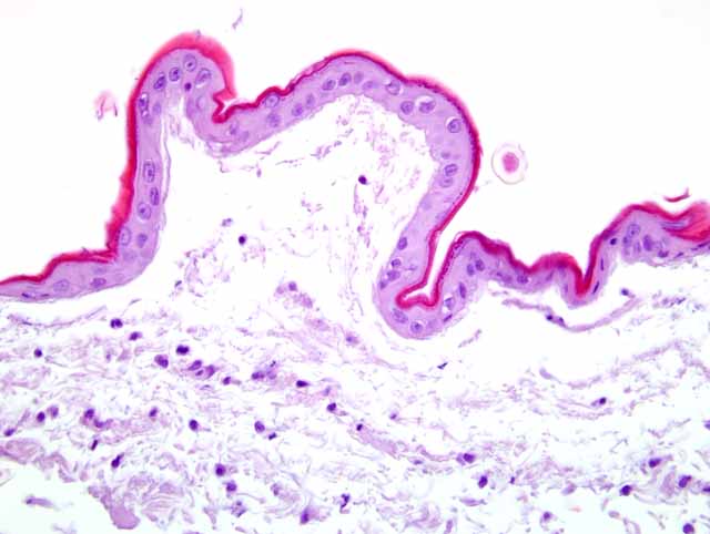
1. Skin and subcutis: Severe atrophy of epidermis and the subcutaneous panniculus with cleft formation, consistent with feline acquired skin fragility syndrome, Siamese cat, feline
2. Skin: Subacute suppurative dermatitis, superficial and perivascular, mild
Condition:
Contributor Comment:
Recently in man the usage of the term dermatoporosis has been presumed for skin lesions in aged people. In this syndrome an increased fragility of the skin is attributed to age-related alterations of the extracellular matrix metabolism.(6) A primary form due to aging or unprotected sun exposure and an iatrogenic form (after corticosteroid administration) are distinguished.
An ectodermal dysplasia with a congenital skin fragility syndrome due to a mutation in the desmosomal protein plakophilin 1 has been reported in man.(2)
Feline acquired skin fragility syndrome is a rare disease with marked skin lesions of multiple etiologies but without a genetic background. The syndrome has been reported only in cats. It has been noted in combination with several other diseases, such as diabetes mellitus, cholangiocarcinoma or hepatic lipidosis, and administration of different drugs.(4,5,8). Most commonly the syndrome is associated with iatrogenic or naturally occurring hyperglucocorticism.(5). There is one report of acquired skin fragility syndrome associated with phenytoin treatment. Since phenytoin inhibits collagen synthesis in vitro the authors assume that cats could acquire collagen disorders during treatment with phenytoin.(1)
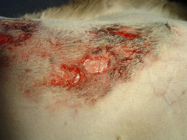
Clinical features of acquired skin fragility syndrome are striking. Affected cats show markedly thin skin which tears with minor trauma and commonly leaves great flaps of loose skin. The lesions are most commonly seen at the back. Partial alopecia occurs in most affected regions.(5)
Differential diagnosis is not problematic since the lesions are typical. The patients normally are middle-aged or older and compared to Ehlers-Danlos syndrome the skin is abnormally thin but without evidence of hyperextensibility.(5) Histologically the lesions in Ehlers-Danlos syndrome and feline acquired skin fragility syndrome are similar or indistinguishable.(4)
To confirm the histopathologic diagnosis, Massons trichrome stain was performed due to described staining abnormalities (abnormal collagen fibers, presence of segmental red staining defects, birefringence of polarized light).(3) Unfortunately fiber abnormalities are not limited to acquired skin fragility syndrome. Similar alterations can also be seen in cutaneous asthenia.(3) In our case no staining abnormalities were detectable.
JPC Diagnosis:
Conference Comment:
Cats with feline acquired skin fragility syndrome have extremely thin skin resembling tissue paper that tears very easily and is not hyperextensible. In contrast to feline acquired skin fragility syndrome, EDS is characterized clinically by the ability to stretch the skin to great lengths without tearing. The skin does not appear to be attached to the underlying subcutis. EDS is also a heritable disease, whereas feline acquired skin fragility syndrome is normally secondary to endocrine disorders, neoplasia, or improper drug administration.(5)
As the contributor stated these two entities are histologically similar. However, Dr. Goldschmidt pointed out features that allow them to be differentiated histologically in most cases. In feline acquired skin fragility syndrome, the epidermis is thin and there is also severe dermal atrophy with marked thinning of collagen fibers. Adnexal structures may also be atrophic. In contrast, the epidermis in EDS is generally unaffected, and the dermis may be of normal thickness or partially reduced in total thickness. The dermal collagen is abnormally arranged with affected fibers having red cores when stained with Massons trichrome stain.(5)
References:
2. Ersoy-Evans S, Erkin G, Fassihi H, Chan I, Paller AS, S+�-+r+�-+c+�-+ S, McGrath JA: Ectodermal dysplasia-skin fragility syndrome resulting from a new homozygous mutation, 888delC, in the desmosomal protein plakophilin 1. J Am Acad Dermatol 55:157-161, 2006
3. Fernandez CJ, Scott DW, Erb HN, Minor RR: Staining abnormalities of dermal collagen in cats with cutaneous asthenia or acquired skin fragility as demonstrated with Masson's trichrome stain. Vet Dermatol 9:49-54, 1998
4. Ginn PE, Mansell JEKL, Rakich PM: Skin and appendages. In: Jubb, Kennedy and Palmer's Pathology of Domestic Animals, ed. Maxie GM, 5th ed., vol 1, pp 389-391, Saunders, Edinburgh, 2007
5. Gross TL, Ihrke PJ, Walder EJ, Affolter VK: Skin diseases of the dog and cat, 2nd ed., pp 386-391. Blackwell, Oxford, UK, 2005
6. Kaya G, Saurat JH: Dermatoporosis: a chronic cutaneous insufficiency/fragility syndrome. Clinicopathological features, mechanisms, prevention and potential treatments. Dermatol 215:284-294, 2007
7. Parapia LA, Jackson C: Ehlers-Danlos syndrome-a historical review. Br J Haematol 141:32-35, 2008
8. Trotman TK, Mauldin E, Hoffmann V, Del Piero F, Hess RS: Skin fragility syndrome in a cat with feline infectious peritonitis and hepatic lipidosis. Vet Dermatol 18:365-369, 2007