Joint Pathology Center
Veterinary Pathology Services
Wednesday Slide Conference
2017-2018
Conference 14
January 10th, 2018
CASE III: N-1238 (JPC 4102671).
Signalment: 3-month-old, female and male, Boer, Capra hircus, caprine.
History: Progressive flaccid paralysis was observed in the hind limbs of 30 out of 40 Boer goat kids over a period of seven days. Age of onset of clinical signs ranged from five to twelve weeks of age; there was no neonatal ataxia. On examination, the kids were affected to varying degrees of severity from hind limb weakness through to an inability to stand. Four kids were found in sternal recumbency. All had normal mentation with intact reflexes and motor function. The mildly affected kids were still suckling. Six animals were euthanized. The herd had no mineral supplementation.
The farm also kept a flock of 120 mixed breed sheep; these had no mineral supplementation and showed no signs of ataxia.
Gross Pathology: Four goat kids were submitted for post-mortem examination (two males [goats A and B] and two females [goats C and D]). They were in relatively good body condition. The lungs were mildly and multifocally mottled red and there was a moderate amount of froth in the trachea (pulmonary congestion and edema). There was a moderate amount of vegetative material within the rumen and well-formed feces in the rectum. Brain and spinal cord were grossly unremarkable.
Laboratory results:
Liver mineral analysis in the four goat kids:
[Reference ranges used from ovine liver (µmol/kg DM) assuming DM 280g/kg:
Mn 130 – 286 µmol/kg DM; Fe 1919 – 19186 µmol/kg DM; Cu 1405 - 5619 µmol/kg DM; Zinc 1639 - 4096 µmol/kg DM; Se 11.3 - 67.8 µmol/kg DM; Molybdenum 11 - 45 µmol/kg DM; Cd 1 – 44 µmol/kg DM; Pb 1- 14 µmol/kg DM; Co 2 -5 µmol/kg DM]
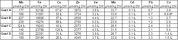
*L: Low; H: High.
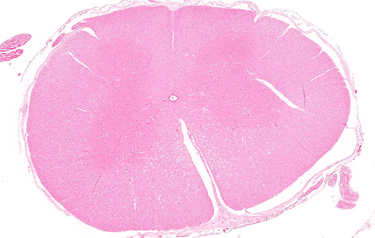
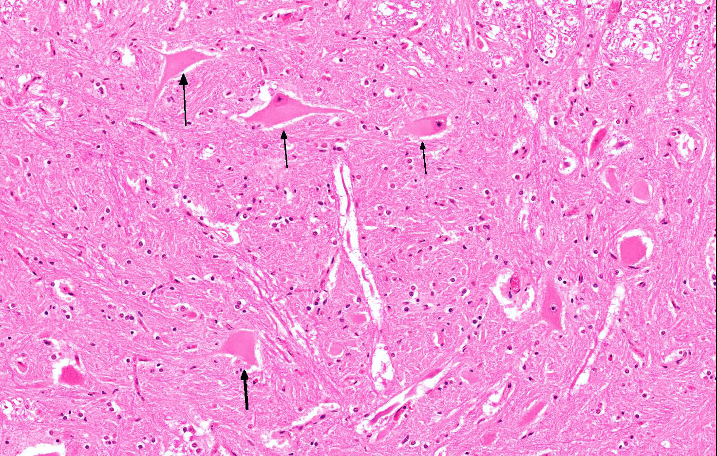
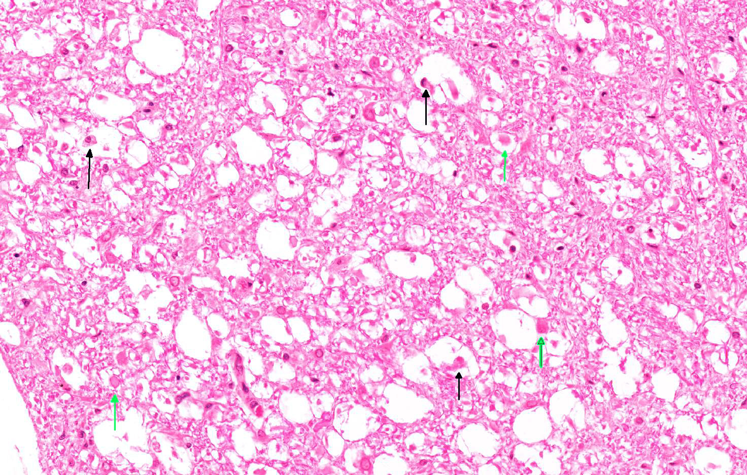
Microscopic Description: The severity of the lesions varies slightly depending on the slide. The cervical spinal cord has bilateral, symmetrical and severe degenerative lesion that affects both grey and white matter. Within the ventral funiculi (mainly the ventromedial tracts) and, less severely, the lateral funiculi, myelin sheaths are markedly distended and often contain axonal fragments and/or few macrophages within its lumen (digestion chambers, Wallerian degeneration). Rare spheroids are also observed. Moderate numbers of neurons located within both the ventral horns and the intermediate area, are swollen and rounded and show loss of Nissl granules which are only remaining at the periphery, with its nucleus displaced towards the periphery (central chromatolysis). Some neurons show karyorrhexis, karyolysis and hypereosinophilia (necrosis). Few small haemorrhages are seen within the grey matter. No inflammatory reaction is observed.
Similar lesions were observed in sections from cervical, thoracic and lumbar spinal cord from all the goat kids as well as within the brainstem from 2 of them (A and D).
Contributor’s Morphologic Diagnosis: Spinal cord (cervical): Severe, multifocal Wallerian degeneration with moderate, multifocal neuronal chromatolysis.
Contributor’s Comment: A diagnosis of enzootic ataxia was made based on the histological findings and low liver copper levels, and was supported by the signalment and clinical presentation. Caprine arthritis and encephalitis virus (CAEV) was initially considered as a differential diagnosis, but the lack of inflammatory reaction within the nervous tissue suggested that it was not involved in the development of these lesions. The pulmonary edema observed in all four goats was most likely secondary to the euthanasia.
Enzootic ataxia, also known as delayed swayback, is a copper deficiency myelopathy1. Swayback is a congenital form of the disease that affects neonates; this was not observed in this case. Ataxic kids are normal at birth, but develop clinical signs between one week and six months of age.2,4 There are differences in the metabolism of copper in sheep and goat, and between breeds within species.6 The delayed form of swayback is frequently seen in goats, while the congenital form is rare in this species.1,3 The Angora and Boer breeds of goats have been suggested as being particularly susceptible to low copper levels.1,4,5
The disease is thought to be caused by a deficiency in the intake of copper by the dam during pregnancy.1 Most commonly this is a primary deficiency due to inadequate intake from the diet, but a secondary form can occur where dietary molybdenum, zinc and cadmium impair the absorption of copper.1,6 The exact diet of the does during pregnancy in this case is unknown, but reportedly no mineral supplementation was provided. In this case, all four kids had liver molybdenum and zinc levels within the normal ranges while cadmium levels were low. In addition to the copper deficiency, all goats showed low levels of selenium and cadmium.
The exact pathogenesis of enzootic ataxia remains unclear, mainly due to the unknown mechanism of action of copper in the developing nervous system.3,6 Albeit, copper is required for the activity of several enzymes that are essential for neural function, including cytochrome oxidase and superoxide dismutase, amongst others. The effects of copper deficiency on the central nervous system occur in utero and during early neonatal life.2,3,6 Copper deficiency leads to suppression of mitochondrial respiration and reduced phospholipid synthesis. This energy failure is likely to play a role in the axonal and neuronal degeneration that is observed histologically.3 The generation of reactive oxygen species (ROS) have also been implicated in these changes. . Despite the aforementioned changes, the animal’s ability to metabolize copper is not impaired; therefore, tissue concentrations in affected animals may return to normal after dietary correction.3
Gross lesions associated with enzootic ataxia in kids are few and not consistent,3,6 correlating with the lack of relevant gross findings in this case. Microscopic lesions affect the grey and white matter of both the spinal cord and brainstem. In the spinal cord, the dorsolateral aspect of lateral funiculus and the ventromedial tracts of the ventral funiculi are most commonly affected, as evident in this case. . Goat kids show a particularly high incidence of cerebellar degeneration compared to lambs3, which was however not observed in the here presented case.
JPC Diagnosis: Spinal cord, white matter, ventral and lateral funiculi: Neuroaxonal degeneration, bilaterally symmetrical, multifocal, moderate, with ventral horn neuronal chromatolysis.
Conference Comment: Copper is an essential element for many cellular functions: antioxidant activity (superoxide dismutase), mitochondrial respiration (cytochrome oxidase), catecholamine synthesis (dopamine β-hydroxylase), melanin synthesis (tyrosinase), and iron hemostasis (ceruloplasmin). Copper deficiency causes disease in lambs, goat kids, and piglets and manifests as either absolute primary due to dietary deficiency, or conditioned secondary (most common) due to reduced intestinal absorption, reduced tissue availability, or enhanced secretion. There are several minerals that act as antagonists to copper including: molybdenum, sulfate, iron, and zinc. Ruminants are specifically affected by molybdenum and sulfate which limit copper absorption by forming complexes with copper in the rumen called thiomolybdates. Iron mechanism of antagonism is unknown.3,6,8
Clinically, there are two forms of copper deficiency in lambs and goat kids: swayback which is congenital and mainly affects lambs and enzootic ataxia which has a delayed onset and affects lambs between 1 week and several months of age. Affected animals develop progressive neurologic signs such as swaying, falling, spastic paralysis or ascending hindlimb paralysis7, ataxia, blindness, or deafness. Grossly, only lambs with congenital swayback have lesions that are apparent in the cerebral white matter, evident as bilaterally symmetrical cavitation within the occipital pole or entire corpus medullare. Lambs with delayed enzootic ataxia may develop lesions at any part of the neuraxis in the gray or white matter, but those lesions have not been clearly defined. In both forms, the microscopic changes in the spinal cord are the most characteristic and consist with Wallerian degeneration in the dorsolateral and ventromedial tracts throughout the spinal cord, a pattern suggestive of a distal axonopathy. Additionally, there are degenerative neuronal changes within the red, lateral vestibular, medullary reticular, and dorsal spinocerebellar nuclei in Clarke’s column, and in the spinal motor neurons of the intumescences.3,6,8
The pathogenesis for these syndromes is not fully understood, but most likely represents the culmination of numerous factors to include energy failure from altered function of mitochondrial cytochrome oxidase and subsequent neuronal degeneration and/or inadequate function of copper-zinc superoxide dismutase leading to oxidative damage.3,6,8
In piglets, many of the white matter changes associated with copper deficiency are similar to kids and lambs; however, there are additional neuronal changes. They also can develop skeletal and elastin abnormalities leading to fractures and arterial rupture.3,6,8 In addition to the neurological manifestations of copper deficiency, sheep also develop “steely wool” (wiry, poor hair coat), hypopigmentation of black wool, and osteoporosis. Poor hair coat and achromotrichia are particularly prominent in cattle which may develop pale colored hair around their eyes, called “spectacles”.
Contributing Institution:
School of Veterinary Medicine & Science
University of Nottingham, UK
https://www.nottingham.ac.uk/vet/servicesfortheveterinaryprofession/pathology.aspx
References:
- Allen AL, Goupil BA, Valentine BA. A retrospective study of spinal cord lesions in goats submitted to 3 veterinary diagnostic laboratories. Can Vet J. 2012;53:639–642.
- Banton MI, Lozano-Alarcon F, Nicholson SS, et al. Enzootic ataxia in Louisiana goat kids. J Vet Diagn Invest. 1990;2:70–73.
- Cantile C, Youssef S. Nervous system. In: Maxie MG, ed. Jubb, Kennedy and Palmer’s Pathology of Domestic Animals. 6th Vol. 1. St. Louis, MO:Elsevier Ltd; 2016:328–329.
- Harwood D. Diseases of dairy goats. In Pract. 2004;26:248–259.
- McKay M, Bone P, Kendall N. Establishing the plasma copper reference range in Boer goats. Vet Rec. 2010;167:499.
- Miller AD, Zachary JF. Copper deficiency. In: Zachary JF, ed. Pathologic Basis of Veterinary Disease. 6th St. Louis, MO:Elsevier Ltd; 2017:887–888.
- No authors listed. Delayed swayback diagnosed in lambs with hindlimb paresis. Vet Rec. 2015;176(5):118-121.
- Summers BA, Cummings JF, de Lahunta A. Degenerative diseases of the central nervous system. In: Duncan L, McCandless PJ, eds. Veterinary Neuropathology. Louis, MO: Mosby; 1995:273-277.