Signalment:
Gross Description:
Histopathologic Description:
The pulmonary blood vessels are congested.
Morphologic Diagnosis:
Lab Results:
The bovine virus isolation and the FA for BVD, IBR and BRSV results were negative.
Condition:
Contributor Comment:
Calf pneumonia occurs due to a complex interaction between host, environment and pathogen. Young calves with low immunity are highly susceptible to this disease. Among various bacterial agents that cause pulmonary infections, Mycoplasma and Histophilus are most common.
Mycoplasma bovis was first reported in the United States in the 1970s. The high number of new cases was reported in the summer of 2000. After that, M. bovis has been found almost in every herd. A study conducted in 2006 revealed a 46% prevalence of Mycoplasma bovis in lungs of normal cattle(3). Calves suffering from Mycoplasma pneumonia show low grade fever, tachycardia and mild depression.
H. somni is another important cause of bovine pneumonia(1,4). H. somni is also associated with a wide variety of other cattle diseases including abortion, infertility and arthritis(4). H. somni pneumonia is characterized grossly by grey to red consolidation of the cranio-ventral lung, involving from 5 to 80% of total lung volume(1). Tracheobronchial and mediastinal lymph nodes are typically edematous, with fibrinous pleuritis seen irregularly(1,3). Histologically, the suppurative necrotizing bronchopneumonia is the most common histopathologic identification of bacterial pulmonary infections.
Note: Multiple blocks were used for the slides submission; therefore not all the participants will get the same copy of the slides.
JPC Diagnosis:
1. Lung: Pleuropneumonia, fibrinosuppurative and necrotizing, diffuse, severe, with marked intralobular edema and lymphatic fibrin thrombi.
2. Lung: Bronchopneumonia, fibrinosuppurative and necrotizing, with marked suppurative bronchilitis and bronchiectasis.
Conference Comment:
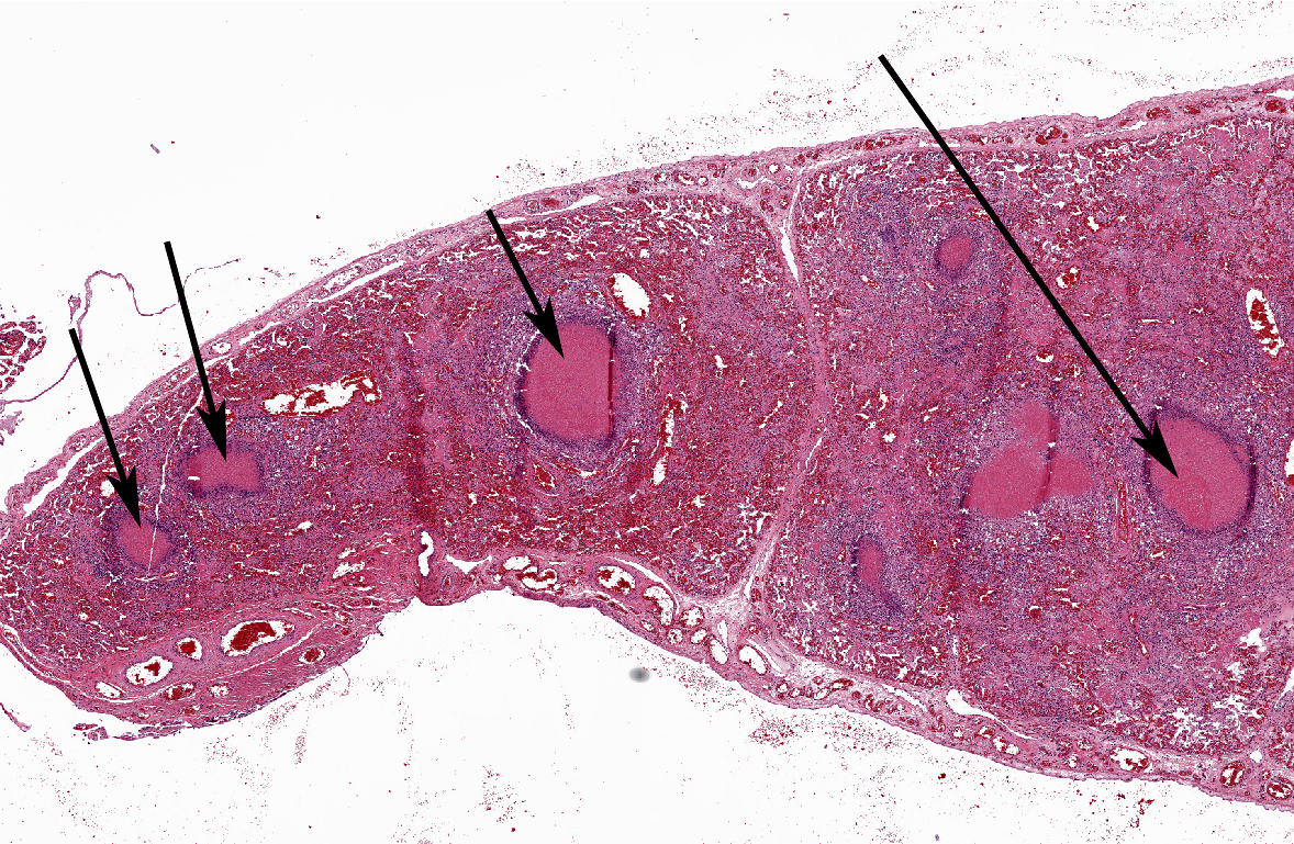
One section contains multiple ectatic bronchioles filled with caseonecrotic material within areas of profound atelectasis characteristic of mycoplasmal pneumonia.. Bronchiolar-associated lymphoid tissue (BALT) is depleted, which is characteristic of enzootic bovine pneumonia. Affected cattle often present with otitis media, as mycoplasma preferentially colonize areas of the body lined by ciliated epithelium (such as airways).
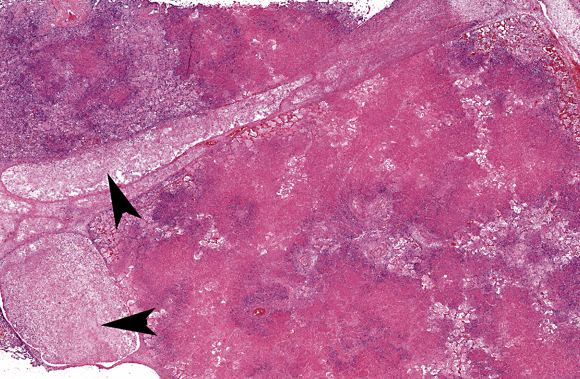
The other section of lung exhibit necrosuppurative bronchopneumonia whichis more typical of gram-negative bacteria, such as Mannheimia hemolytic or Histophilus somni. In affected lungs, alveoli are filled with fibrin and leukocytes and larger areas of coagulative necrosis may become sequestra. Grossly, affected areas of lung will be sharply demarcated from adjacent lung, and the presence of large amounts of fibrin and cellular exudate will give it a firm feel(2).
References:
2. Caswell JL, Williams KJ. Respiratory system. In: Maxie MG, ed. Jubb, Kenedy, and Palmers Pathology of Domestic Animals. 5th ed. Philadelphia, PA:Saunders Elsevier; 2007:601-5, 610-5.
3. Gagea MI, Bateman KG, van Dreumel T, McEwen J, Carman S, Archambault M, Shanahan RA, Caswell JL: Diseases and pathogens associated with mortality in Ontario beef feedlots. J Vet Diagn Invest 18:18-28, 2006.
4. Kwiecien JM, Little PB: Haemophilus somnus and reproductive disease in the cow: A review. Can Vet J 32: 595-601, 1991.