Signalment:
Histopathologic Description:
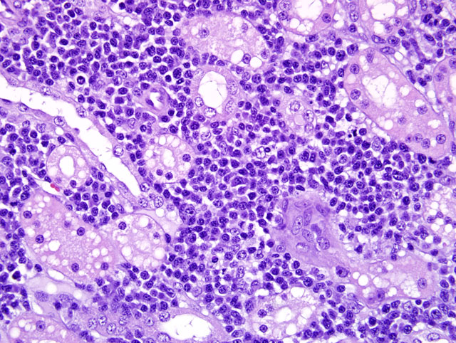
Other changes consist of occasional scattered minor proteinaceous tubular casts and diffuse moderate vacuolation of the tubular epithelium consistent with lipid accumulation that was considered excessive for the feline species and suggestive of perhaps a metabolic abnormality in addition to the obvious inflammatory disease process.
Morphologic Diagnosis:
Condition:
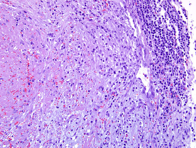
Contributor Comment:
JPC Diagnosis:
Conference Comment:
FCoV is spread via fecal-oral transmission, and the prevalence of coronavirus infection is high but the number of clinical cases of FIP is low.(1,7) The most commonly accepted theory for this peculiar finding is that FCoV undergoes a mutation and acquires virulence factors by deletions in open reading frames 3 and 7 to cause FIP.(7) Recent studies also strongly suggest that monocytes not only disseminate the virus, but also mediate the development of phlebitis associated with FIP.(7)
A strong cell mediated immune response is essential for protection against developing FIP.(1,2) Two forms of FIP, wet and dry, are generally referred to when describing FIP. If a weak cell-mediated immunity is mounted, the virus can persist in macrophages for months causing release of a variety of inflammatory mediators leading to perivascular pyogranulomatous inflammation in affected organs. This is the dry form. The wet or effusive form occurs when no cell mediated immunity is present and continued viral replication leads to the production of large quantities of non-neutralizing antibodies and immune complex deposition.(1,2)
Peritonitis is common in both the dry and wet forms. In the effusive form, copious amounts of flocculent yellow exudate may be found in the abdominal cavity, and serosal surfaces are often covered with fibrin.(1,2) Pyogranulomas are commonly found in the kidney, uvea, and peritoneum. Cervical and thoracic lymph nodes are often enlarged, and hydrocephalus is often found in cats with neurologic symptoms.(2) The classic histologic lesion of FIP is phlebitis with circumferential rings of pyogranulomatous inflammation.(2) Immunohiostochemical detection of coronavirus antigen in macrophages can be used to confirm a diagnosis of FIP.
A recent report associated FIP with papular cutaneous lesions (4) and fatal systemic coronavirus infections have recently been reported in ferrets (5) and dogs.(3)
References:
2. Brown C, Baker D, Barker I: Alimentary System. In: Jubb Kennedy and Palmers Pathology of Domestic Animals, Vol. 2, ed. Maxie M. 5th ed., pp. 1-296. Saunders Elsevier, New York, NY, 2007
3. Buonavoglia C, Decaro N, Martella V, Elia G, Campolo M, Desario C, Castagnaro M, Tempesta M: Canine coronavirus highly pathogenic for dogs. Emerg Infect Dis 12(3):492-494, 2006
4. Declercq J, De Bosschere H, Schwarzkopk I, Declercq L: Papular cutaneous lesion in a cat associated with feline infectious peritonitis. Vet Dermatol 19(5):255-8, 2008
5. Garner MM, Ramsell K, Morera N, Juan-Salles C, Jimenez J, Ardiaca M, Montesinos A, Tefke JP, Lohr CV, Evermann JF, Baszler TV, Nordhausen RW, Wise AG, Maes RK, Kiupel M: Clinicopathologic features of a systemic coronavirus-associated disease resembling feline infectious peritonitis in the domestic ferret (Mustela putorius). Vet Pathol 45:236-246, 2008
6. Maxie M and Newman S: Urinary system. In: Jubb, Kennedy, and Palmers Pathology of Domestic Animals. ed. Maxie M, vol. 2, 5th ed., pp. 425-522. Saunders Elsevier, New York, NY, 2007
7. Kipar A, May H, Menger S, Weber M, Leukert W, Reinacher M: Morphologic features and development of granulomatous vasculitis in feline infectious peritonitis. Vet Pathol 42(3)21-330, 2005