Signalment:
Gross Description:
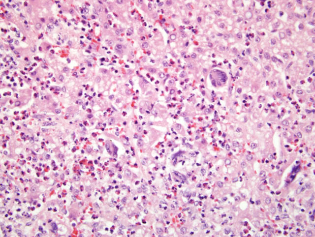
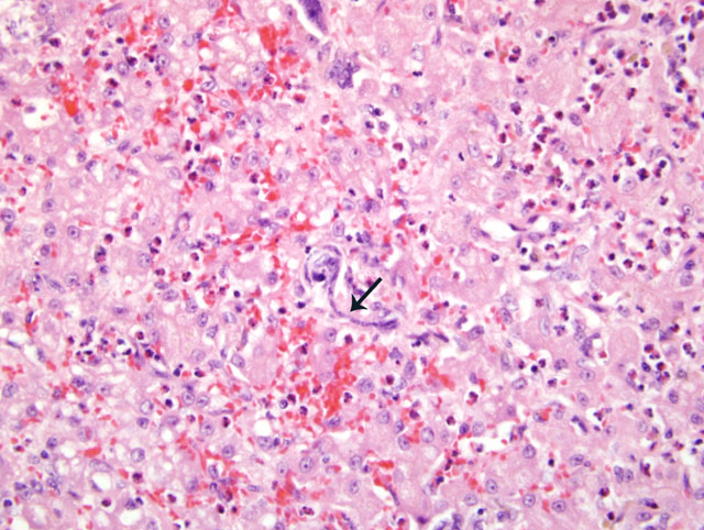
Histopathologic Description:
Morphologic Diagnosis:
Condition:
Contributor Comment:
Specific syndromes associated with heartworm disease include asymptomatic infection, glomerulonephritis, allergic pneumonitis, eosinophilic granulomatosis, pulmonary embolization, congestive heart failure, caval syndrome and aberrant migration1. Changes in this case to correlate best with eosinophilic granulomatosis, however the organ most severely affected was the liver, not the lung. Microfilaria lodged in the sinusoids causing a striking inflammatory response. While microfilaria were clearly present in the circulation, similar foci of inflammation were not observed in lung, kidney or heart, suggesting that the liver may be predilection site for this infection in otters.
JPC Diagnosis:
2. Liver, hepatocytes: Vacuolar change, glycogen-type, diffuse, mild.
Conference Comment:
There are numerous species of Dirofilaria that primarily infect particular host species. The most well known cause of microfilariasis of animals in North America is Dirofuilaria immitis, which is primarily found in the dog.4 With the exception of D. immitis, most species of Dirofilaria adults are found within the subcutaneous tissues.7 Morbidity and mortality associated with D. immitis infections have been reported in mustelids, canids, felids, otariids, and other domestic and nondomestic carnivores6, including the river otter.8 D. lutrae infections have only been reported in North American river otters, with adults occurring in subcutaneous spaces and very rarely in the cardiopulmonary vasculature.6,7 Adult forms of D. repens are found within the subcutaneous tissues of canines, other carnivores and occasionally humans.4 D. tenuis is primarily found in raccoons of the southern USA, and D. striata within bobcats.Â
Characteristic histopathologic lesions in the lungs of dogs with dirofilariasis include villous endarteritis with luminal occlusion caused by villous intimal proliferation and medial hypertrophy.2 Nitric oxide (NO) production has been implicated in the inflammatory response during filarial infections.5 NO production can be induced by a recombinant Wolbachia surface protein.5 Wolbachia is an intracellular endosymbiont bacteria that resides within some filarial organisms, including Dirofilaria immitis.4
In a recent Journal of Wildlife Diseases article, an unidentified filarial organism was isolated from wild populations of the black-footed ferret.9 Although this unidentified microfilaria elicited a positive reaction to ELISAs for D. immitis, it only shared approximately 76% molecular identity with D. immitis, while sharing approximately 97% identity with Acanthocheilonema viteae.9
Morphologic characteristics of some microfilariae of onchocercids adapted from Wisely et. al.9
| Filarial species | Length m | Sheath | Host |
| Dirofilaria immitis | 233-322 | no | Dog |
| Dirofilaria striata | 230-371 | no | Bobcat |
| Dipetalonema reconditum | 270-290 | no | Dog |
| Loaina uniformis | 285 | yes | Eastern cottontail |
| Dirofilariaeformia pulmoni | 213-288 | no | Eastern gray squirrel |
| Brugia beaveri | 300 | yes | Raccoon |
| Brugia lepori | 210-330 | yes | Swamp rabbit |
| Mansonella llewellyni | 290 ± 5 | no | Raccoon |
| Monsonella interstitium | 250 ± 5 | no | Eastern gray squirrel |
| Molinema arbuta | 280-297 | no | North American porcupine |
| Acanthocheilonema reconditum | 269-283 | no | Dog |
| Acanthocheilonema mephitis | 186-218 | no | Striped skunk |
References:
2. Kawabata A, Nakagaki K, Yoshida M, Shirota K: Histopathological comparison of pulmonary artery lesions between Raccoon Dogs (Nyctereutes procyonoides) and domestic dogs experimentally infected with Dirofilaria immitis. J Vet Med Sci 70:301-303, 2008
3. Matsuda K, Baek B, Lim C: Eurasian Otter (Lutra lutra), a definitive host for Dirofilaria immitis. J of Zoo and Wildl Med 34:200-201, 2003
4. Maxie MG, Robinson WF: Cardiovascular system. In: Jubb, Kennedy, and Palmers Pathology of Domestic Animals, ed. Maxie MG, 5th ed., vol. 3, pp. 87-89. Elsevier Limited, St. Louis, MO, 2007
5. Morch³n R, Bazzocchi C, L³pez-Belmonte J, Mart+�-�n-Pancho JR, Kramer LH, Grandi G, Sim³n F: iNOs expression is stimulated by the major surface protein (rWSP) from Wolbachia bacterial endosymbiont of Dirofilaria immitis following subcutaneous injection in mice. Parasitol Int 56:71-75, 2007
6. Neiffer DL, Klein EC, Calle PP, Linn M, Terrell SP, Walker RL, Todd D, Vice CC, Marks SK: Mortality associated with melarsomine dihydrochloride administration in two North American River Otters (Lutra canadensis) and a Red panda (Ailurus fulgens fulgens). J of Zoo and Wildl Med 33:242-248, 2002
7. Orihel TC, Eberhard ML: Zoonotic filariasis. Clin Microbiol Rev 11:366-381, 1998
8. Snyder DE, Hamir AN, Nettles VF and Rupprecht CE: Dirofilaria immitis in a River Otter (Lutra canadensis) from Louisiana. J of Wildl Dis 25:629, 1989
9. Wisely SM, Howard J, Williams SA, Bain O, Santymire RM, Bardsley KD, Williams ES: An unidentified filarial species and its impact on fitness in wild populations of the black-footed ferret (Mustela nigripes). J Wildl Dis 44:53-64, 2008