Signalment:
Gross Description:
Histopathologic Description:
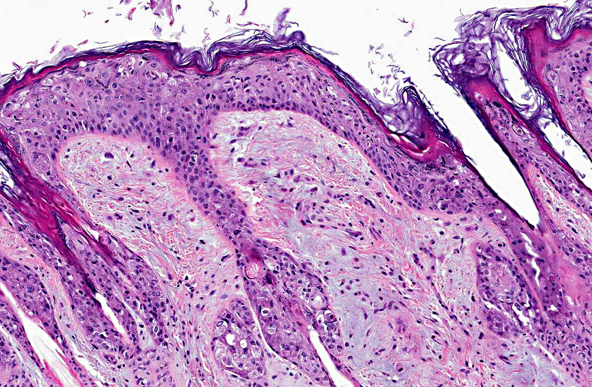
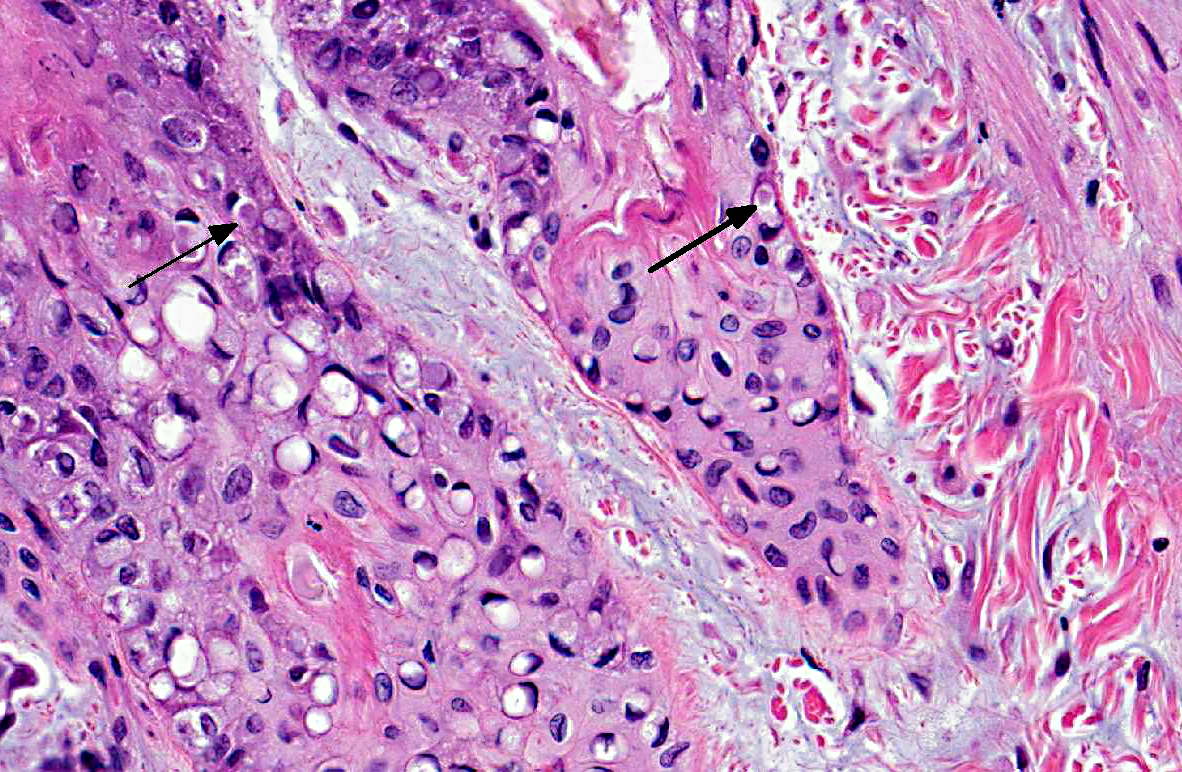
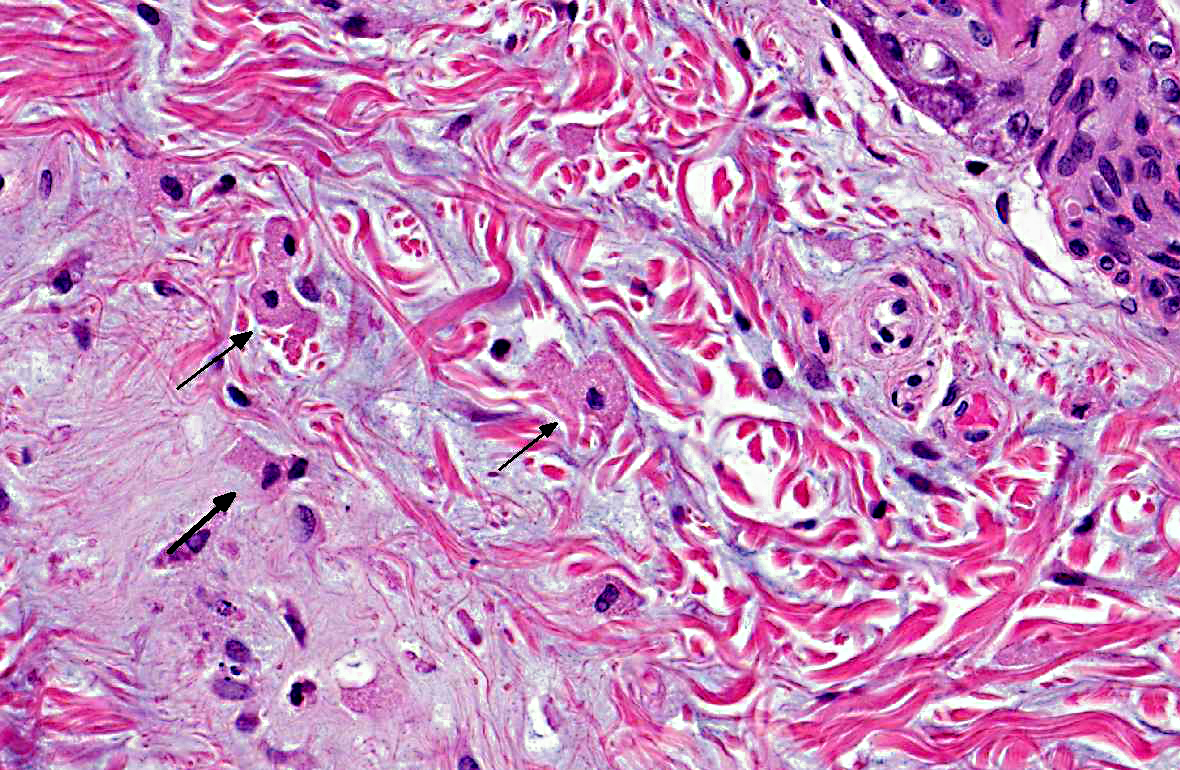
In the dermis, multiple large, stellate or polygonal cells with finely granular, basophilic cytoplasm and one or more large nuclei with finely granular to fragmented chromatin and one clear nucleolus are present. Mitoses are less than 1 per HPF. There is a slight infiltration of eosinophils, macrophages and some lymphocytes and plasma cells. The surrounding collagen fibers are loosely arranged and interspersed with a moderate amount of basophilic amorphous substance (proteoglycans).Â
Part of the conjunctival mucosa is desquamated due to autolysis.Â
Morphologic Diagnosis:
Condition:
Contributor Comment:
Three to five days after infection, affected animals first show signs of fever, depression and loss of appetite. Subsequently, edematous or firm skin nodules (mostly around the eye, mouth, nose, ear or genitalia), conjunctivitis and severe depression are seen. Death occurs within one to two weeks after the first symptoms appear.(5)
Histologically, one of the first apparent changes is an increase in number and size of the epidermal cells. After this, granular, pink cytoplasmic inclusions appear in these cells. The disease progresses until cell lysis occurs and epidermal vesicles develop. In the dermis, large stellate or polygonal cells with swollen nuclei and fragmented chromatin, polymorphonuclear leukocytes and multinucleated cells appear. Histological changes can also be seen in other organs. The lymph nodes, for example, first become activated, while, as the disease progresses, lymphocytes are replaced with the large stellate or polygonal cells previously described. The same cellular infiltration can be seen in the spleen.(5)
JPC Diagnosis:
Conference Comment:
Conference participants also discussed the pathogenesis of myxomatosis. As the contributor stated, myxoma virus is spread via arthropod vectors. The virus replicates in the host at the site of inoculation, resulting in a subcutaneous myxoid mass. In susceptible animals, the virus can then spread via cell-associated viremia (also known as leukocyte trafficking) and cause systemic disease. In less susceptible rabbit species, such as Sylvilagus brasiliensis and S. bachmani, viremia does not develop, and lesions are generally restricted to localized cutaneous fibromas.Â
A closely-related leporipoxvirus, rabbit fibroma virus (RFV, also known as Shope fibroma virus), occurs naturally in the eastern cottontail (Sylvilagus floridanus) and causes similar lesions, referred to as Shope Fibromas, in both cottontails and European rabbits.2 Shope fibromas appear grossly as flattened subcutaneous tumors on the legs, feet, and occasionally the face. Histologically, Shope fibromas are composed of a localized proliferation of reactive fibroblasts that may contain large intracytoplasmic eosinophilic inclusion bodies. The inclusion bodies can also be found in the cells of the overlying epidermis. These well-circumscribed fibrous masses are generally easily distinguishable from myxomatosis, which is characterized by proliferation of stellate mesenchymal cells on a mucinous matrix with epithelial hypertrophy.Â
Participants noted that this case features a comparatively milder atypical mesenchymal spindle cell proliferation, and more severe epithelial proliferation and loss of normal epithelial stratification than is usually observed in this disease.Â
References:
2. Kerr PJ. Myxomatosis in Australia and Europe: a model for emerging infectious diseases. Antiviral Res. 2012;93(3):387-415.
3. Percy DH, Bathrold SW. Rabbit. In: Pathology of Laboratory Rodents and Rabbits. 3rd ed. Ames, Iowa: Blackwell; 2007:256-258.
4. Sobey WR, Conolly D. Myxomatosis: the introduction of the European rabbit flea Spilopsyllus cuniculi (Dale) into wild rabbit populations in Australia. J Hyg. 1971;69:331-346.Â
5. Rivers TM. Infectious myxomatosis of rabbits. J Exp Med.1930;51:965-976.