Signalment:
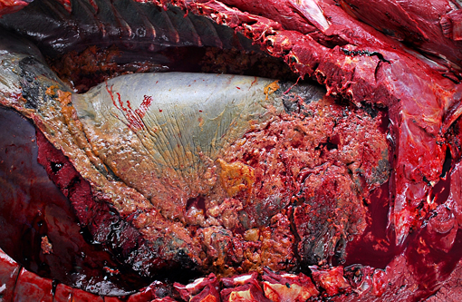
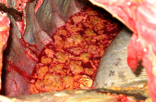
Gross Description:
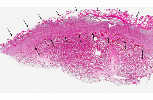
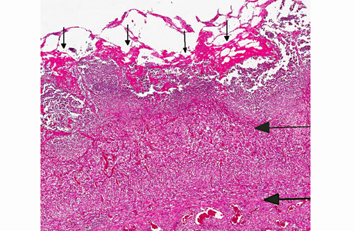
Histopathologic Description:
Morphologic Diagnosis:
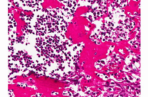
1. Lung: Pleuritis, fibrinopurulent, diffuse, severe, subacute to chronic, with intralesional cocci colonies.
2. Lung: Abscesses, multifocal, moderate, subacute, with intralesional cocci.
Lab Results:
Condition:
Contributor Comment:
Transmission is mainly via aerosol, wound contamination, oral or contagious route.(2) Aerosol transmission is the most likely route in this case. S. equi subsp. zooepidemicus is considered to be an opportunistic pathogen in horses and alpaca; it is commonly found in the nasopharynx of clinically normal horses.(1,6) There are reports of sporadic or outbreaks in many species, including humans. S. equi subsp. zooepidemicus has been associated with mastitis,(12) abscesses, meningitis, endocarditis, reproductive system disease, orchitis, arthritis, septicemia and respiratory and uterine infections.(1,3,7) Human infections caused by S. equi subsp. zooepidemicus include outbreaks of foodborne illness, meningitis, septicemia, arthritis, pneumonia, glomerulonephritis, and streptococcal toxic shock syndrome, in both immunocompromised and immunocompetent patients.(3) Severe S. equi subsp. zooepidemicus induced pleuropneumonia has been observed in horses. In Peru, infection can cause a mortality rate of 50-100% of affected alpacas (known as alpaca fever).(4,6) In China, S. equi subsp. zooepidemicus is the most commonly isolated secondary pathogen in swine since 1975. Humans may be infected via contact with sick animals, unpasteurized milk or other dairy products;(12) therefore, this bacterium poses a zoonotic health risk to animals and humans.Â
Since 2004, this pathogen has been isolated from three batches of horses imported (from the United States and New Zealand) to Taiwan. The source of infection in the present cases is presumably associated with transport stress. There have been no reports of S. equi subsp. zooepidemicus infection in livestock, zoo/wild animals or pets in Taiwan prior to this report.Â
JPC Diagnosis:
Conference Comment:
References:
1. Abbott Y, Khan S, Muldoon EG, Markey BK, Pinilla M, Leonard FC, et al. Zoonotic transmission of Streptococcus equi subsp. zooepidemicus from a dog to a handler. J Med Microbiol. 2010;59:120-123.
2. Brack M, G+�-+nther E, Gilhaus H, Salzert W, Meuthen J. An outbreak of Streptococcus equi subsp. zooepidemicus infection of probable human origin in Wanderoos (Macaca silenus)--case report. Zentralbl Bakteriol. 1997;286:441-446.
3. Chalker VJ, Brooks HW, Brownlie J. The association of Streptococcus equi subsp. zooepidemicus with canine infectious respiratory disease. Vet Microbiol. 2003;95:149-156.
4. Hewson J, Cebra CK. Peritonitis in a llama caused by Streptococcus equi subsp. zooepidemicus. Can Vet J. 2001;42:465-467.
5. Honer OP, Wachter B, Speck S, et al. Severe Streptococcus infection in spotted hyenas in the Ngorongoro Crater, Tanzania. Vet Microbiol. 2006;115:223-228.
6. Jones M, Miesner M, Grondin T. Outbreak of Streptococcus equi susp. zooepidemicus polyserositis in an alpaca herd. J Vet Intern Med. 2009;23: 220-223.
7. Lamm CG, Ferguson AC, Lehenbauer TW, Love BC. Streptococcal infection in dogs: A retrospective study of 393 cases. Vet Pathol. 2010;47:387-95.
8. Lopez A. Respiratory system, mediastinum and pleurae. In: McGavin MD, Zachary JF, eds. Pathologic Basis of Veterinary Disease. 5th ed. St. Louis, MO: Mosby Elsevier; 2012:470.Â
9. Maxie MG, Youssef S. Nervous system. In: Maxie MG, ed. Jubb, Kennedy, and Palmers Pathology of Domestic Animals. 5th ed. Vol. 1. Philadelphia, PA: Elsevier; 2007:173, 257, 358.
10. Percy DH, Barthold SW. Pathology of Laboratory Rodents and Rabbits. Ames, IA: Blackwell Publishing; 2007:229-232.Â
11. Pesavento PA, Hurley KF, Bannasch MJ, Artiushin S, Timoney JF. A clonal outbreak of acute fatal hemorrhagic pneumonia in intensively housed (shelter) dogs caused by Streptococcus equi subsp. zooepidemicus. Vet Pathol. 2008;45(1):51-53.Â
12. Pisoni G, Zadoks RN, Vimercati C, Locatelli C, Zanoni MG, Moroni P. Epidemiological investigation of Streptococcus equi subspecies zooepidemicus involved in clinical mastitis in dairy goats. J Dairy Sci. 2009;92:943-951.
13. Timoney, JF. The pathogenic equine streptococci. Vet Res. 2004;35(4):397-409.