Signalment:
Five-year-old female cynomolgus macaque (
Macaca fasicularis).The animal presented with a clinical history of lethargy and anorexia. The neurologic examination revealed mild intention tremors, anisocoria, ataxia, and left-sided facial paralysis.
Gross Description:
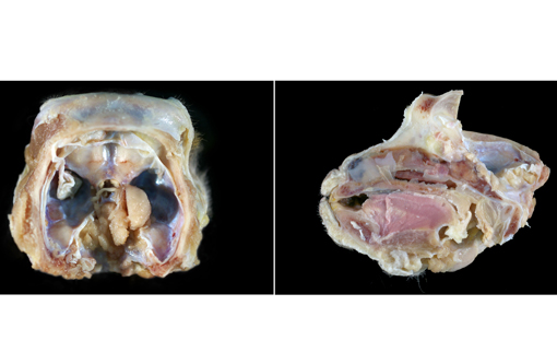
At necropsy, there was a tan to gray, granular, soft, irregularly-shaped mass that extended from the base of the skull at the level of the sella turcica, through the cribiform plate, to the upper areas of the nasal cavity and paranasal sinuses. The tumor compressed the ventral aspect of the brain from the frontal lobes to the midbrain.
Histopathologic Description:
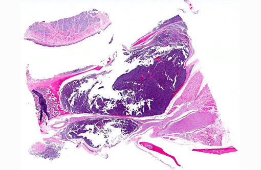
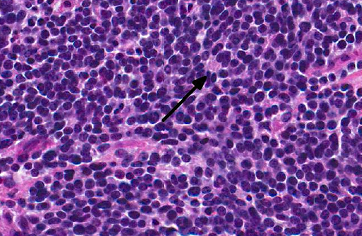
Slides from two different blocks of this tumor were submitted. This neoplasm infiltrated the nasal mucosa and propria submucosa, nasal septum (turbinate bone), flat bone (cribiform plate) and soft tissue/skeletal muscle. Sections of tongue, oral mucosa, and olfactory nerve were on some but not all slides. This infiltrative and non-encapsulated mass was composed of neoplastic cells arranged in solid clusters, sheets, and lobules that were separated by delicate fibrovascular connective tissue. Tumor cells frequently formed true rosettes or pseudorosettes and had a primitive appearance. Neoplastic cells had small, round to polygonal, hyperchromatic nuclei and scant eosinophilic cytoplasm. Mitotic figures, as well as large foamy cells (nasal clear cells), and areas of hemorrhage and necrosis were commonly observed throughout the mass.Â
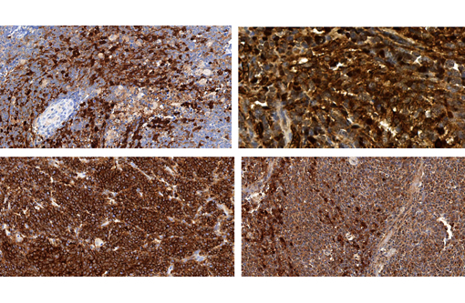
Multiple immunohistochemical (IHC) stains of this tumor were performed including ubiquitin carboxyl-terminal hydrolase L1 (PGP 9.5 neuron specific protein), CD56 (neuronal cell adhesion molecule), S-100, neuron specific enolase (NSE) and vimentin. The expression of PGP 9.5, CD56, S-100, NSE and vimentin, along with the gross and histopathologic findings, confirmed that this neoplasm was an olfactory neuroblastoma and ruled out other neoplasms such as malignant pituitary tumor, lymphosarcoma, meningioma, suprasellar germ cell tumor and intracranial schwannosarcoma.
Morphologic Diagnosis:
Olfactory neuroblastoma.Â
Condition:
Olfactory neuroblastoma
Contributor Comment:
Olfactory neuroblastomas are uncommon neuroectodermal tumors that may arise within the nasal cavity.(9) The morphology of this neoplasm is heterogenous and the histogenic origin is unclear, resulting in many different names including, but not limited to, olfactory neuroblastoma, esthesioneuroblastoma, and olfactory neuroepithelioma.(4,9) One paper describing a prospective study in humans suggests that the term "olfactory neuroblastoma" might be the most reflective of both the origin and nature of the neoplasm.(8) This neoplasm has been described in humans,(5) dogs,(9) rats,(6) cats,(4) cows,(1) a horse(11) and a cynomolgus monkey.(3) Interestingly, olfactory neuroblastomas are chemically induced in rats,(7) as opposed to other animal species and humans where this tumor is typically of spontaneous origin. Some examples of chemicals that induce olfactory neuroblastomas in rats include 1-nitrosopiperazine and nitrosomorpholine.(6) The development of olfactory neuroblastomas in rats is not related to the route of administration of the chemical but it is directly associated with chemical metabolism in nasal basal cells that leads to neoplastic transformation.Â
The differential diagnoses for this mass consisted of esthesioneuroblastoma (olfactory neuroblastoma), lymphosarcoma, suprasellar germ cell tumor, meningioma, intracranial schwannosarcoma, and malignant pituitary gland tumor. Since olfactory neuroblastomas originate from the olfactory epithelium, it is important to split the cranium along the midline into two sagittal half sections to locate the origin of the tumor in the nasal cavity with infiltration into the base of the cranial cavity.
This case was presented at the 2011 annual National Toxicology Program (NTP) Satellite Symposium, entitled Pathology Potpourri that was held in Denver, Colorado in advance of the Society of Toxicologic Pathology's 30th Annual Meeting, and published by Boorman G, et al. in the
Journal of Toxicologic Pathology.(2)
JPC Diagnosis:
Skull, nasal cavity and paranasal sinus: Olfactory neuroblastoma.
Conference Comment:
Key histological features of this case include the arrangement of neoplastic cells into parallel arrays and the presence of pseudorosettes and occasional Homer-Wright type rosettes.(5) Homer-Wright rosettes are composed of neoplastic cells surrounding a central lumen of fiber-rich neuropil. They are one of the major distinguishing characteristics of medulloblastomas, but also occur in primitive neuroectodermal tumors (PNETs) and pineoblastomas.(10) Pseudorosettes, on the other hand, are formed by neoplastic cells palisading around a centrally placed vessel.(10) Perivascular pseudorosettes are less specific than Homer-Wright rosettes in that they are also encountered in medulloblastomas, PNETs, central neurocytomas, glioblastomas and other tumors. Even with these characteristic lesions olfactory neuroblastomas can display a wide range of microscopic appearances with a large differential diagnosis. Immunohistochemically, these tumors are usually positive for some, or all, of the following: S-100 and neuroendocrine markers such as neuron-specific enolase (NSE), synaptophysin, chromagranin and CD56.(5)
References:
1. Anderson BC, Cordy DR. Olfactory neuroblastoma in a heifer. Vet Pathol. 1981;18:536-540.
2. Boorman G, Crabbs TA, Kolenda-Roberts H, et al. Proceedings of the 2011 national toxicology program satellite symposium. Toxicol Pathol. 2012;40:321-344.Â
3. Correa P, Dalgard DW, Adamson RH. Olfactory neuroepithelioma in a cynomolgus monkey (Macaca fascicularis). J Med Primatol. 1975;4:51-61.
4. Cox NR, Powers RD. Olfactory neuroblastomas in two cats. Vet Pathol. 1989;26:341-343.
5. Faragalla H, Weinreb I. Olfactory neuroblastoma: a review and update. Adv Anat Pathol. 2009;16:322-331.
6. Garcia H, Keefer L, Lijinsky W, Wenyon CE. Carcinogenicity of nitrosothiomorpholine and 1-nitrosopiperazine in rats. Z Krebsforsch. 1970;74:179-184.
7. Long PH, Herbert RA, Peckham JC, Grumbein SL, Shackelford CC, Abdo K. Morphology of nasal lesions in F344/N rats following chronic inhalation exposure to naphthalene vapors. Toxicol Pathol. 2003;31:655-664.
8. Lund VJ, Howard D, Wei W, Spittle M. Olfactory neuroblastoma: past, present, and future? The Laryngoscope. 2003;113:502-507.
9. Mattix ME, Mattix RJ, Williams BH, Ribas JL, Wilhelmsen CL. Olfactory ganglioneuroblastoma in a dog: a light, ultrastructural, and immunohistochemical study. Vet Pathol. 1994;31:262-265.Â
10. Wippold II FJ, Perry A. Neuropathology for the neuroradiologist: rosettes and pseduorosettes. Am J Neuroradiol. 2006;27:488-492.
11. Yamate J, Izawa T, Ogata K, Kobayashi O, Okajima R, Kuwamura M, et al. Olfactory neuroblastoma in a horse. J Vet Med Sci. 2006;68:495-498.