Signalment:
Tissue from a domestic duck,
Anas platyrhynchos domesticsOne duck was submitted for being lethargic with emaciation and ataxia.
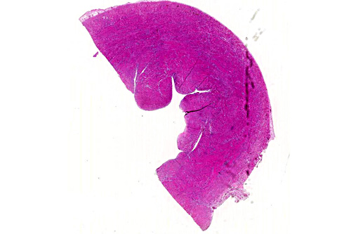
Gross Description:
No gross lesions were seen other than slight emaciation.Â
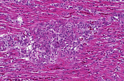
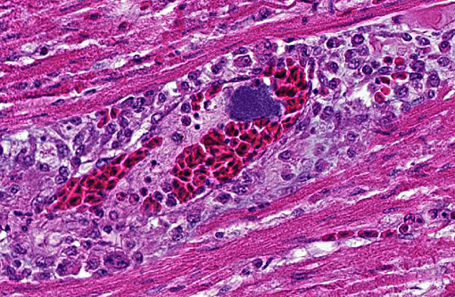
Histopathologic Description:
The heart has multifocal small areas of necrosis and moderate multifocal mixed inflammation with heterophils, lymphocytes, and some histiocytes. Bacterial colonies and emboli are throughout the myocardium. Mixed leukocytes with fibrin and a few multinucleated giant cells are on the epicardium. The lung section was congested and had a bacterial embolus but no inflammation and the liver was normal other than sinusoidal histiocytosis. No lesions were seen in the brain, intestine, and kidney sections (only heart from this case was submitted.)
Morphologic Diagnosis:
Multifocal necrotizing and subacute myocarditis
Lab Results:
(clinical pathology, microbiology, PCR, ELISA, etc.): 1+
Pasteurella multocida and 1+
E. coli were isolated from the lung and liver swabs and no bacterial growth was obtained from the heart swab.Â
Condition:
Pasteurella multocida
Contributor Comment:
This is fowl cholera septicemia (
Pasteurella multocida).Â
E. coli was not thought to be significant and
P. multocida is always considered to be significant from poultry. Chickens usually have a chronic disease with pasteurellosis, with infection in the combs and wattles. The bacteria can often also be isolated from internal organs. Turkeys and ducks are more likely to have an acute or peracute infection, often with many acute deaths.(3)
JPC Diagnosis:
Heart: Myocarditis, necrotizing and histiocytic, multifocal to coalescing, severe, with vasculitis, thrombosis, fibrinous epicarditis, and intra- and extracellular bacterial colonies
Conference Comment:
Pasteurella multocida, the causative agent of fowl cholera, remains a major problem of poultry worldwide.(4) Disease manifestation can range from a mild localized infection to peracute systemic disease with high mortality.(2) The peracute forms are characterized by septicemic lesions, with petechial and ecchymotic hemorrhages in the fat and mucus membranes and necrosis of the liver and spleen.(2) The chronic form often involves the respiratory system as fibrinonecrotic pneumonia, but arthritis, peritionitis, and salpingitis also occurs.(2) Additionally, fibrinonecrotic dermatitis is observed in turkeys and broiler chickens (see 2011 WSC 21, Case 1).Â
While all types of poultry are considered vulnerable to infection with
P. multocida, there are species and age differences in susceptibility. Turkeys and waterfowl are considered the most susceptible species while chickens are more resistant. All birds under 16 weeks of age also appear fairly resistant, though this effect is less pronounced in turkeys.(2)
Necrotizing myocarditis is not a typical finding with fowl cholera, allowing conference participants to review differentials for such a lesion in poultry. Specifically to ducks,
Riemerella anatipestifer is a frequent cause of polyserositis including pericarditis in young ducklings of intensive production systems.Â
Salmonella pullorum causes peritonitis and death in hatchling chicks or peritonitis, arthritis and pericarditis in adults. Coliform infections may lead to myocarditis, but usually with more abundant heterophilic inflammation and fibrin than present in this case.Â
West Nile Virus may perhaps be the best differential for necrotizing myocarditis of many avian species, with young chickens and geese being most likely to develop clinical disease and mortality.(1)
References:
1. Capua I. Arthropod-borne viruses. In: Pattison M, McMullin PF, Bradbury JM, Alexander DJ, eds. Poultry Diseases. 6th ed. Philadelphia, PA: Saunders Elseveier; 2008:421-422.
2. Christensen JP, Bojesen AM, Bisgaard M. Fowl Cholera. In: Pattison M, McMullin PF, Bradbury JM, Alexander DJ, eds. Poultry Diseases. 6th ed. Philadelphia, PA: Saunders Elsevier; 2008:149-154.
3. Glisson, JR, Hofacre, Cl, Christensen, FP. Fowl Cholera (Pasteurella and other related bacterial infections), Chap. 19, in Diseases of Poultry, 11th Ed., Saif, YM, editor, Iowa State Press, Ames, IA, pp. 857-676, 2003.
4. Singh R, Remington B, Blackall P, Turni C. Epidemiology of fowl cholera in free range broilers. Avian Dis. 2014;58(1):124-128.Â