Signalment:  

Male sheep (Ovis aries), dorset poll breed. Age not specified.Accreditation serology for B. ovis was inconclusive in this potential breeding animal.


Gross Description:  

The animal was euthanized, and its entire genital tract was submitted for evaluation. The tail of the right testicle was noted to be firm and increased in size.


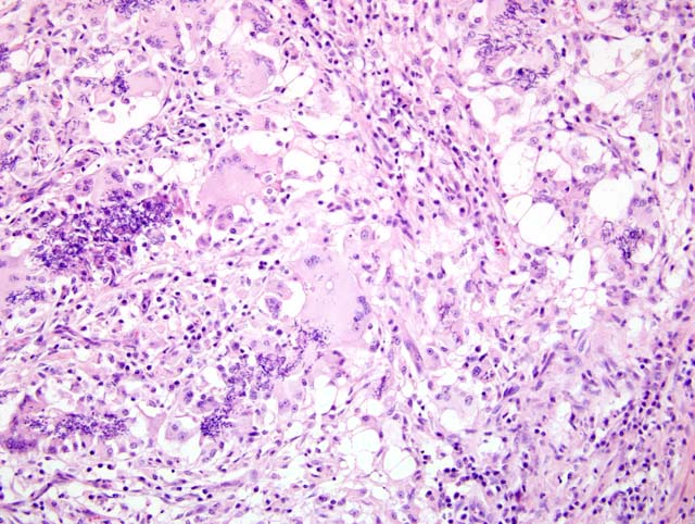
Histopathologic Description:

There is a focal (in some sections multifocal) accumulation of histiocytes and multinucleated giant cells within the epididymal interstitium, surrounded by a collar of lymphocytes and plasma cells admixed with variable neutrophils (Fig. 2-1). In numerous sections, there is distinct mineralization and Splendore-Hoeppli like reaction at the center of the focus (foci). There are myriad fragments of spermatids noted at the center of the foci, often within histiocytes. There is compression of adjacent ducts, which occasionally have hyperplastic epithelium, and there is fibrosis of the interstitial connective tissue. Large numbers of sarcocyts are noted in the cremaster muscle.


Morphologic Diagnosis:  

Multifocal or focal (depending on slide) granulomatous epididymitis with necrosis, interstitial fibrosis and epididymal epithelial hyperplasia (sperm granuloma).


Lab Results:  

Cultures for Histophilus somni, Brucella ovis were negative. Modified ZN and Gram stains of impression smears made from the gonads indicated large numbers of Gram negative rods in the seminal vesicles. Actinobacillus seminis was cultured from both seminal vesicles and ampullae.


Condition:  

Sperm granuloma


Contributor Comment:  

Spermatic granulomas are relatively common findings in rams (1) and are often associated with other conditions which induce leakage of spermatids into the interstitium. The testicle is an immunologically privileged site, and sperm are highly antigenic, containing cell walls rich in lipids and phospholipids. Epididymitis due to bacterial agents such as Brucella ovis, Histophilus somni, and Actinobacillus seminis often leads to spermatic granulomas as well as agent specific epididymitis and orchitis.

In this particular case, additional sections of epididymis, prostate gland and seminal vesicle displayed mild multifocal accumulations of neutrophils, which were presumed incited by ascending infection of Actinobacillus seminis.


JPC Diagnosis:  


1. Epididymis: Epididymitis, granulomatous, focally extensive, moderate with a sperm granuloma
2. Epididymis: Epithelial hyperplasia, multifocal, moderate


Conference Comment:  

Dr. Schlafer discussed the anatomy of the epididymis and the importance of grasping the basic anatomic features to help in identifying and understanding the pathogenesis of various entities that affect the male reproductive system. 

Sperm are produced within the testes and inside the seminiferous tubules. Sperm travel through a plexus of channels called the rete testes. From the rete testes, numerous small efferent ductules transport sperm to the head of the epididymis. Within the head and body of the epididymis sperm undergo changes that transform them into fertile cells. Sperm traverse the head and body over a period of several days, and eventually reach the tail of the epididymis for storage prior to ejaculation via the ductus deferens.(2) The contributor mentioned the highly antigenic nature of sperm; any release of sperm into the extratubular compartment leads to a foreign body type reaction. This is followed by a strong immune response resulting in an accumulation of large numbers of plasma cells, CD4 and CD8 positive lymphocytes, and an up-regulation of MHC I in epithelial cells. As in the case of a foreign body, a chronic immune response leads to fibrosis and walling off of the lesion. This often leads to spermiostasis, a spermatocele, or a sperm granuloma.(1)

The known causes of sperm granulomas were discussed; they include congenital duct anomalies, adenomyosis, trauma, and infections. Bacteria are implicated most frequently, as mentioned by the contributor, and the route of infection is via ascension from the urethra and accessory sex glands. The immunologically privileged site allows for the organism to proliferate unabated. This often leads to formation of a sperm granuloma and loss of fertility.(1)

There was extensive slide variation with this particular case. The contributor mentioned distinct mineralization and Splendore-Hoeppli like reaction at the center of the foci which was not present on all of the submitted slides.


References:

1. Foster RA, Ladds PW: Male genital system. In: Jubb, Kennedy, and Palmer's Pathology of Domestic Animals, vol 3 ed. Maxie MG, pp. 565-621. Elsevier Limited, Philadelphia, PA, 2007
2. Senger PL: Pathways to Pregnancy and Parturition, 1st ed., pp. 32-57. The Mack Printing Group-Science Press, Ephrata, PA, 1997


Click the slide to view.


2-1. Epididymis, sheep.


Back | VP Home | Contact Us |