Signalment:
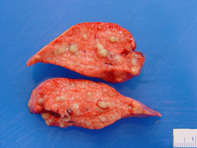
Gross Description:
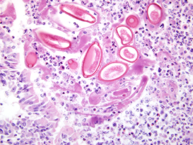
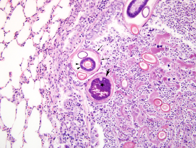
Histopathologic Description:
Morphologic Diagnosis:
Lab Results:
Condition:
Contributor Comment:
Clinical signs in infected dogs and cats are generally characterized by a low-grade chronic cough. The disease among foxes raised in earthen runs on fur farms, however, used to be much more severe, with poor growth, decreased fur quality, and substantial mortality from bronchopneumonia, particularly among young animals.7 Control of the disease was achieved by shifting the animals to raised cages with wire bottoms. In free-living wild red fox, the degree of infection may vary among animals but is probably generally higher in young-of-the-year because of their immature immune system. It is conceivable that, every year, a number of young fox die from pneumonia caused by this parasite, either because their immune system is particularly inefficient or because they are exposed to a very large number of eggs. In this case, secondary infection by Streptococcus species group G had likely contributed to the animals death. Most bacteria of this group isolated from animals are S. canis. This bacterium can be isolated from mucous membranes of asymptomatic domestic carnivores, but it can also cause opportunistic infections, including suppurative bronchopneumonia and septicemia.10
The nematode Crenosoma vulpis is another common pulmonary parasite in the red fox population of the Canadian Maritime provinces and often occurs concurrently with E. aerophilus.9 This parasite may also be a common cause of respiratory disease in domestic dogs presented with clinical signs of chronic cough.1 Crenosoma vulpis has an indirect life cycle, using snails and slugs as intermediate hosts.11 Eucoleus aerophilus is longer but more slender than C. vulpis (Table 1).8 Whereas E. aerophilus is oviparous, C. vulpis is ovoviviparous, and it also tends to inhabit deeper regions of the bronchial tree.9 Adults and first stage larvae of Crenosoma vulpis were not identified in the lungs of this fox.
Table 1: Comparison of the dimensions of adult specimens of Eucoleus aerophilus and Crenosoma vulpis.6
| length | Diameter | |||
| male | female | male | female | |
| E. aerophilus | 15-25 mm | 20-40 mm | 60-100 μ | 100-180 μ |
| C. vulpis | 3.5-8 mm | 12-16 mm | 280-320 μ | 300-480 μ |
Other potential nematode parasites of the respiratory system of red fox in this country include Oslerus (Filaroides) osleri, Dirofilaria immitis, and Angyostrongylus vasorum. Infection by O. osleri is characterized by the formation of discrete nodules typically found near the bifurcation of the main bronchi, whereas both D. immitis and A. vasorum are parasites of the pulmonary arterial tree rather than of the airways.3 Moreover, the North American distribution of A. vasorum is currently confined to the Atlantic Canadian province of Newfoundland.2
JPC Diagnosis:
Conference Comment:
The life cycle of Eucoleus (Capillaria) aerophilus can be either primarily direct or less commonly indirect involving an earthworm as an intermediate paratenic host. Eggs are deposited in the pseudostratified ciliated epithelium, work their way up the respiratory tree, are swallowed, and are then passed out with the feces.4
Physical characteristics of E. aerophilus are similar to those of other aphasmid nematodes.5 Aphasmids lack a pair of sensory papillae on their caudal end. They lack the prominent lateral cords seen in phasmid nematodes. They have a hypodermal band and one genital tract in the female.5
References:
2. Bourque A, Whitney H, Conboy G: Angiostrongylus vasorum infection in a coyote (Canis latrans) from Newfoundland and Labrador, Canada. J Wildl Dis 41:816-819, 2005
3. Bowman DD, Lynn RC, Eberhard ML: Georgis Parasitology for Veterinarians, 8th ed., pp. 190-196, 216-222, 229-233. Elsevier Science, St. Louis, MO, 2003
4. Campbell BG: Trichuris and other Trichinelloid nematodes of dogs and cats in the United States. Comp Cont Educ Pract Vet 13:769-778, 1991
5. Gardiner CH, Poynton SL: Morphological characteristics of trematodes in tissue section. In: An Atlas of Metazoan Parasites in Animal Tissues. pp. 40-42. Armed Forces Institute of Pathology, Washington, D.C., 2006
6. Hamier AN, Rupprechtt CE: A retrospective histopathologicla survey of capillariasis in raccoons from the eastern United States. J Parasitol 84:180-181, 1998
7. Hanson KB: Tests of the efficacy of single treatments with tracheal brushes in the mechanical removal of lungworms from foxes. J Am Vet Med Assoc 82:12-33, 1933
8. Levine ND: Nematode Parasites of Domestic Animals and of Man, pp. 292, 543. Burgess Publishing Company, Minneapolis, MN, 1968
9. Nev+�-�rez A, L³pez A, Conboy G, Ireland W, Sims D: Distribution of Crenosoma vulpis and Eucoleus aerophilus in the lung of free-ranging red foxes (Vulpes vulpes). J Vet Diagn Invest 17:486-489, 2005
10. Songer JG, Post KW: Veterinary Microbiology: Bacterial and Fungal Agents of Animal Disease, pp. 43-53. Elsevier Inc., St. Louis, MO, 2005
11. Soulsby EJL: Helminths, Arthropods and Protozoa of Domesticated Animals, 7th ed., pp. 282-283, 340-341. Lea & Febiger, Philadelphia, PA, 1982