Signalment:
Gross Description:
Histopathologic Description:
Morphologic Diagnosis:
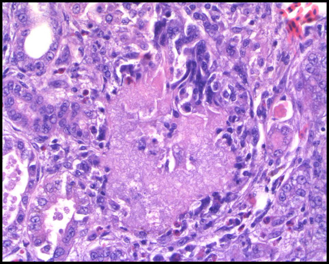
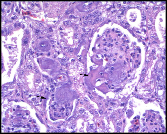
Kidney: amyloidosis, glomerular and interstitial, American flamingo (Phoenicopterus ruber)
Lab Results:
-�-� CBC revealed a leukocytosis (20.6 k/uL) with a heterophilia, lymphopenia and monocytosis. Hyperfibrinogenemia (700mg/dl), hypoglycemia (98mg/dl), hyperphosphatemia (11.2mg/dl), and hyponatremia (120mEq/L) were evident on CP. CP also showed elevations of uric acid (99.6mg/dl), CPK (3770 IU/L), and LDH (511 IU/L).
Condition:
Contributor Comment:
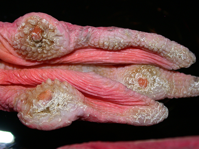
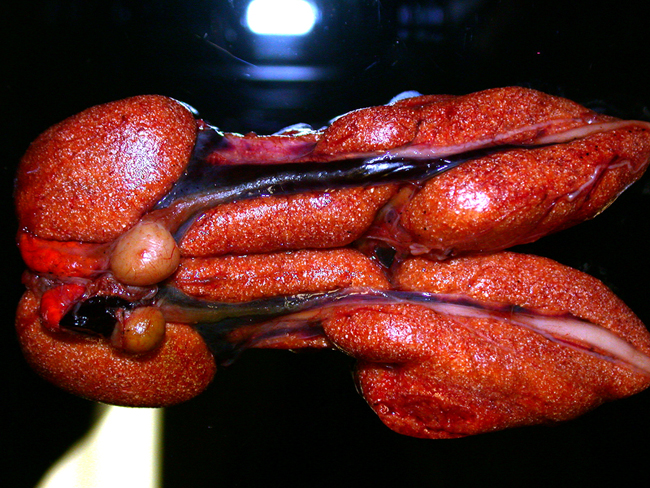
Gout occurs in two forms: articular and visceral1. Articular gout is a chronic process with an uncertain etiology that results in deposition of urates and resultant granulomatous inflammation in the joint spaces, most often of the feet. It is proposed that genetics or a high protein diet may contribute to this condition. Visceral gout occurs more acutely than articular gout and is characterized by small gouty tophi within the renal parenchyma and on the surface of the liver, heart and air sacs. In severe cases, gouty tophi may be seen within the liver and spleen parenchyma as well. These tophi incite little to no inflammatory response, as compared to deposits in articular gout, as their accumulation is more rapid. Visceral gout may occur due to dehydration or renal damage.
Renal function, in birds, is evaluated with the measurement of uric acid. This compound is the final result of nitrogen catabolism in avians and is produced in the liver1. If renal function is impaired, uric acid may build up in the bloodstream and precipitate as crystals in the tissues4. The validity of this test is not absolute as plasma uric acid may rise with normal feeding or ovulation, and may be normal in some cases of renal disease3.
We hypothesized that this aged flamingo developed articular gout over a period of time which led to severe amyloidosis in the kidney, liver and spleen. There was no other post mortem evidence of chronic disease, though this may have resolved by the time of death, leaving only amyloidosis as evidence of past pathology. The deposition of large amounts of protein in the interstitial spaces of the kidneys caused tubular degeneration and ischemia, leading to renal failure, decreased uric acid clearance, marked hyperuricemia and visceral gout.
Of the 62 American flamingos necropsied at the National Zoological Park since 1975, 21 (34%) have had amyloid deposition in at least one tissue. Amyloidosis was listed as the cause of death in 14 (23%) flamingos. This finding may indicate that this group of American flamingos, a closed flock since 1996, has a genetic predisposition to amyloid deposition.
JPC Diagnosis:
2. Kidney: Nephritis, tubulointerstitial, granulomatous and heterophilic, multifocal, moderate, with protein casts and urate tophi.
Conference Comment:
Visceral and articular gout are two separate syndromes with different etiologies, morphologies, and pathogenesis.
Extracted from Diseases of Poultry (Crespo, et al)1
| Â | Visceral Gout | Articular Gout |
| Onset | Usually acute | Usually chronic |
| Frequency | Common | Rare |
| Kidney Lesions | Almost always involved, grossly abnormal, with white chalky deposits | May become involved with dehydrations |
| Joints | May or may not be involved | Always involved, especially the feet |
| Pathogenesis | Failure of urate excretion (renal failure) | Possibly due to metabolic defect in secretion of urates by kidney tubules |
| Causes | Dehydration | Genetics |
| Â |
|
|
The diagnosis of visceral gout should not be considered a disease entity itself, but rather a sign of severe renal dysfunction leading to hyperuricemia.1
True gout must be distinguished from pseudogout in which crystals other than sodium urate, such as calcium pyrophosphate dehydrate or hydroxyapatite, are deposited in joints. Grossly, pseudogout appears as cream-colored gritty material surrounding the joint capsule. This is in contrast to urates which are found inside the joint capsule and within the synovial fluid. Tophi are not present in pseudogout. Additionally, urates are radiolucent, whereas calcium deposits are radiopaque. True gout affects the kidneys, pericardium, liver, and other internal organs, whereas pseudogout only affects the joints and does not appear to occur in other locations. Pseudogout has been reported in humans, Rhesus macaques, dogs, and turtles.5,6
References:
2. Donoghue S: Nutrition. In: Reptile Medicine and Surgery, ed. Mader DR, 2nd ed., p. 281. Saunders Elsevier, St. Louis, Missouri, 2006
3. Gregory, CR: Urinary System. In: Duncan & Proasses veterinary laboratory medicine: clinical pathology. Eds. KS Latimer, EA Mahaffey, KW Prasse. 4th ed., pp. 253-255. Iowa State University Press, Ames, IA, 2003
4. Hochleithner, M: Patient evaluation. In: Avian Medicine: eds. BW Ritchie, GJ Harrison, LR Harrison, pp. 228-241, Wingers Publishing, Inc. Lake Worth, FL. 1994
5. Jones TC, Hunt RD, King NW: Veterinary Pathology, 6th ed., pp. 60-61. Williams & Wilkins, Baltimore, Maryland, 1997
6. Mader DR: Gout. In: Reptile Medicine and Surgery, ed. Mader DR, 2nd ed., pp. 793-800. Saunders Elsevier, St. Louis, Missouri, 2006
7. Myers RK, McGavin MD: Cellular and tissue responses to injury. In: Pathologic Basis of Veterinary Disease, eds. McGavin MD, Zachary JF, 4th ed., pp. 46-47. Mosby Elsevier, St. Louis, Missouri, 2007
8. Thompson K: Bones and joints. In: Jubb, Kennedy, and Palmers Pathology of Domestic Animals, ed. Maxie MG, 5th ed., vol. 1, pp. 173-174. Elsevier Saunders, Philadelphia, Pennsylvania, 2007
9. Weisbrode SE: Bone and joints. In: Pathologic Basis of Veterinary Disease, eds. McGavin MD, Zachary JF, 4th ed., p. 1100. Mosby Elsevier, St. Louis, Missouri, 2007