Signalment:
Histopathologic Description:
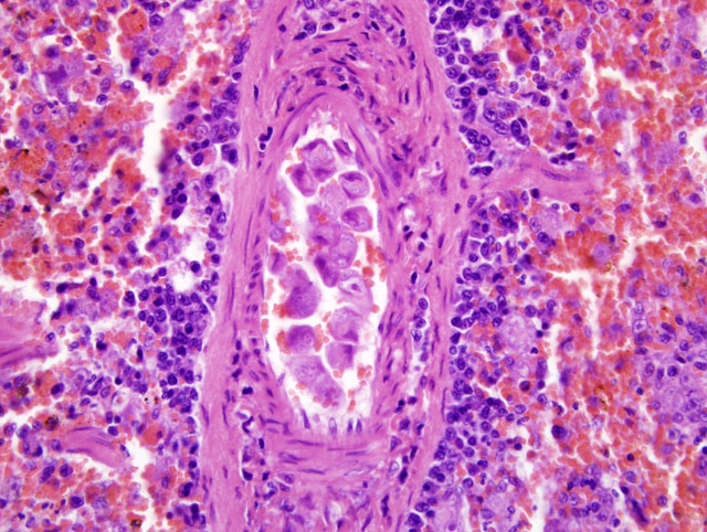
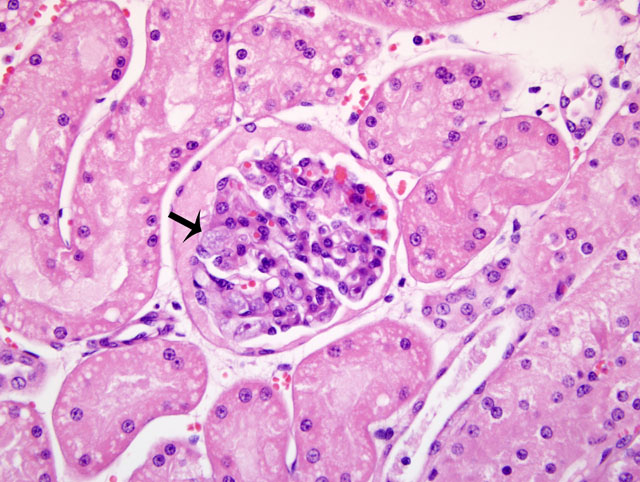
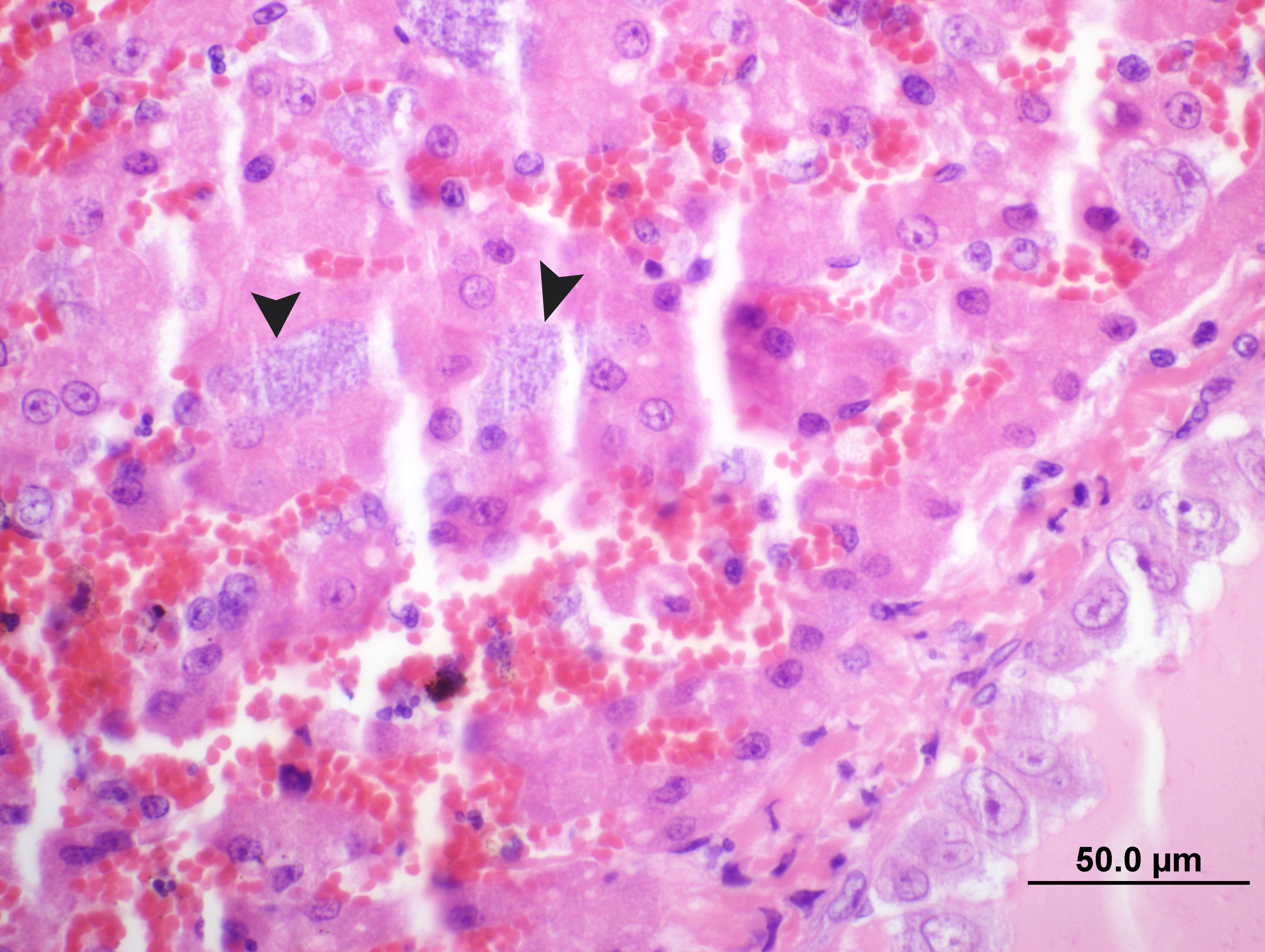
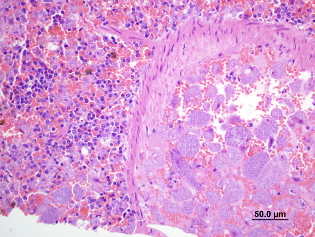
Additionally, tissues (slides not submitted) from heart, lung, stomach, small intestine, ovary and urinary bladder and gall bladder were examined. In almost all examined tissues, there were similar large schizont-laden macrophages in the blood vessels. These were numerous in the sections of liver, kidney, spleen and heart. The other findings were within normal limits.
Morphologic Diagnosis:
Condition:
Contributor Comment:
The infection is associated with both tissue (schizogonous) phases and an intraerythrocytic phase that correlates with the clinical phases of severe circulatory impairment and hemolytic anemia, respectively.(1) The clinical signs in domestic cats include depression, anorexia, pyrexia, dehydration, pallor, icterus, dark urine and occasionally dyspnea.(1,3)
The cat in the current case presented to the veterinarian, after partial abortion (aborting three of six fetuses) the previous night, in a near comatose stage with lethargy and mild gingival icterus. Initially, the cat had a reduced heart rate, which progressed to seizures, then death. A blood smear was collected immediately postmortem by cardiac puncture. Numerous schizont-laden macrophages and small round ring-shaped piroplasms in several erythrocytes were observed in the Wright-Giemsa stained smears. Generally, occasional schizont-laden macrophages may be observed on the feathered edge of blood smears in infected cats, but numbers similar to those in this case are unusual. Large numbers of schizont-laden macrophages in blood smear in the current case could be attributed to collection of blood from the heart. Because of their large size these cells are less likely to be found in peripheral circulation and are not normally found in blood smears from infected cats.(6) As in the current case, it was previously reported that high percentage of parasites are identified histologically from the spleen, liver, or lungs, suggesting sampling from these organs as the most appropriate sites for organism identification.(4) Because the tissue phase occurs prior to the erythrocytic phase, some cats can be severely ill but not have detectable parasites in their red blood cells.(1)
Three remaining fetuses were recovered at necropsy. Formalin fixed tissues from the cat and some fetal tissues (skeletal muscle, developing bone and placenta) were examined. Macrophages or piroplasms associated with the organism were not seen in fetal tissues. Whether Cytauxzoon infection in this cat would be incriminated for the partial abortion observed could not be ruled out.(6) The tissue schizonts are the phase that is responsible for clinical manifestation of cytauxzoonosis.(4) Clinical Cytauxzoon felis infection is usually fatal in domestic cats (1,4), although some infected cats survive.(1) Because of this, domestic cats (Felis domesticus) are regarded as accidental hosts.(6) Bobcats, thought to be the reservoir hosts, are persistently parasitemic, yet they rarely manifest marked clinical disease(4). Rare fatal cases of cytauxzoonosis in free-ranging bobcats has been reported.(5)
JPC Diagnosis:
Conference Comment:
| Location | Parasite | Hosts |
| Intracellular parasites (within erythrocytes) |
Hemoproteus spp.
Leukocytozoon spp. Plasmodium spp. Cytauxzoon felis Babesia cati Babesia felis Anaplasma marginale Anaplasma centrale Babesia bovis Babesia bigemina Theileria mutans Theileria annulata Theileria cervi Babesia canis Babesia gibsoni Babesia equi Babesia caballi Babesia ovis Babesia motasi |
Birds Cats Cattle Cattle Cattle Deer, Elk Dogs Horses Sheep |
| Epicellular parasites (On membrane surface of depression of erythrocytes) |
Trypanosoma johnbakeri
Hemobartonella felis (Mycoplasma haemofelis) Hemobartonella canis (Mycoplasma haemocanis) Eperythrozoon suis (Mycoplasma haemosuis) Eperythrozoon wenyoni Eperythrozoon sp. |
Birds Cats Dogs Pigs Cattle Llamas |
| Extracellular parasites (within the plasma) |
Dipetalonema reconditum
Dirofilaria immitis Setaria spp. Trypanosoma theileri Trypanosoma congolense Trypanosoma vivax Trypanosoma cruzi Trypanosoma brucei Trypanosoma evansi |
Dogs Dogs (sometimes cats) Horses Cattle Dogs |
References:
2. Brockus CW, Andreasen CB: Erythrocytes. In: Duncan & Prasses Veterinary laboratory Medicine, Clinical Pathology, eds. Latimer KS, Mahaffey EA, Prasse KW, 4th ed., pp. 19-21. Blackwell Publishing, Ames, IA, 2003
3. Maxie G: Cytauxzoonosis. In: Jubb, Kennedy and Palmers Pathology of Domestic Animals, ed. Maxie MG, 5th ed., vol 3, pp. 152-158. Elsevier Saunders, Philadelphia, PA, 2007
4. Meinkoth J, Kocan AA, Whitworth L, Murphy G, Fox JC, Woods JP: Cats surviving natural infection with Cytauxzoon felis: 18 cases (1997-1998). J Vet Intern Med (5):521-5, 2000
5. Nietfeld JC, Pollock C: Fatal cytauxzoonosis in a free-ranging bobcat (Lynx rufus). J Wildl Dis 38(3): 607-10, 2002
6. Weismann, JL, Woldemeskel, M, Smith, KD, Merill, A, Miller, D: Blood smear from a pregnant cat that died after partial abortion. Vet Clinc Pathol 36:(2), 209-211, 2007