Signalment:
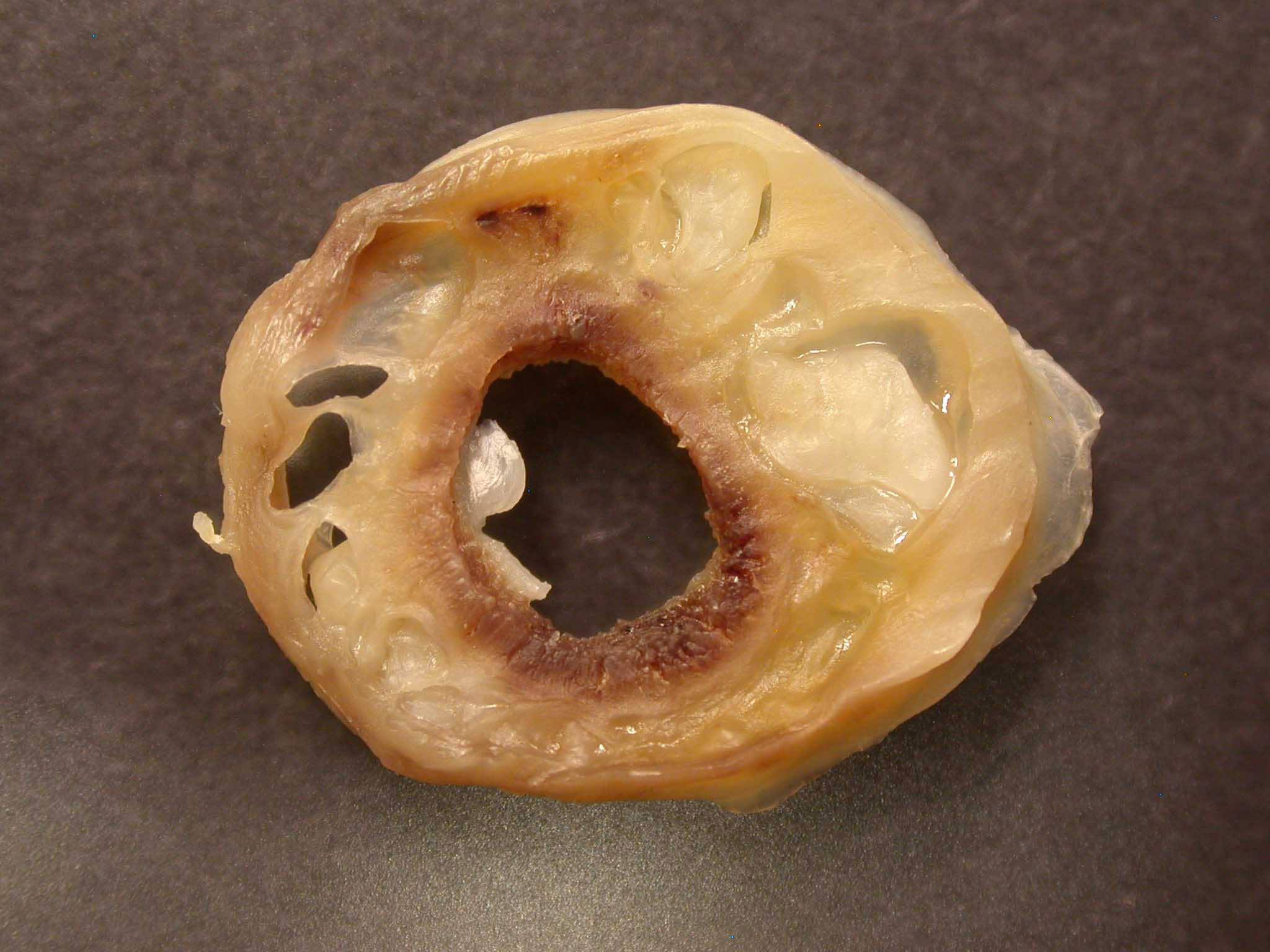
Gross Description:
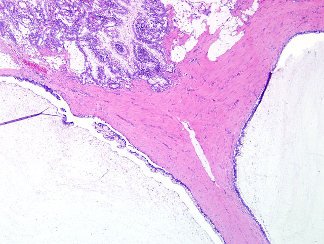
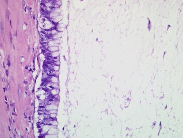
Histopathologic Description:
Morphologic Diagnosis:
1. Intestine, jejunum: Mucinous adenocarcinoma.
2. Intestine, jejunum: Moderate, sub-acute, multifocal, fibrinosuppurative, erosive enteritis with bacterial rods.
Condition:
Contributor Comment:
Transmural invasion is a differentiating feature between adenomatous hyperplasia and neoplasia.(2) Histologic patterns reported include acinar, papillary, solid, carcinoid, and mucinous (including signet-ring cell carcinoma).(6) Signet-ring cells are cells filled with copious mucin that perpheralizes the nucleus. No prognostic behavior has been attributed to the difference in histological pattern.
Intestinal adenocarcinomas metastasize widely via lymphatics, and, at times, via direct serosal implantation.(2) Serosal implantation can cause lymphatic blockage, leading to ascites.(2) Rare manifestations of metastasis include cutaneous masses and pseudomyxoma peritonei.(1,5) Cutaneous lesions in a dog with duodenal adenocarcinoma and masses in numerous organs consisted of undifferentiated, non-cohesive islands of round to polygonal cells.(5) In this case, definitive diagnosis of the undifferentiated mass was supported by identification of a primary intestinal lesion and positive staining of metastases by pancytokeratin, periodic acid Schiff, and Alcian blue. A single case of pseudomyxoma peritonei describing mucin accumulation in the peritoneum and peritoneal cavity was reported in 2003.(1) Mucin was trapped in fibrous reticulin mesh in tissue from the peritoneum and mesenteric fat and in fibrous septa in tissue from the diaphragm. To date, no pathologic basis for mucin accumulation has been identified in this patient or in humans affected by a similar condition.
JPC Diagnosis:
Conference Comment:
The following summary of the World Health Organization classification of intestinal adenocarcinoma is provided with the caveats that a given tumor often exhibits more than one growth pattern, and as mentioned by the contributor, growth pattern is not correlated with prognosis:(3)
| Classification of Intestinal Adenocarcinoma in Domestic Animals | |
|---|---|
| Type | Microscopic Features |
| Acinar (tubular) | Acini and tubules replace the intestinal mucosa |
| Papillary (polypoid, cribriform) | Multiple layers of anaplastic columnar cells line papillary projections |
| Mucinous (colloid, mucoid) | Acinopapillary growth with at least 50% of the tumor replaced by extracellular mucin |
| Signet ring cell (goblet cell, intracellular mucinous) | At least 50% of the neoplasm is composed of signet-ring cells with intracytoplasmic mucin that peripheralizes crescentic nuclei; lacks gland formation; severe desmoplasia |
| Undifferentiated | Solid sheets of anaplastic or pleomorphic cells without squamous or glandular differentiation |
| Adenosquamous (adenoacanthoma, adenocarcinoma with squamous differentiation) | Gland-forming adenocarcinoma with areas of squamous differentiation and variable keratinization |
References:
2. Brown CC, Baker DC, Barker IK: Alimentary system. In: Jubb, Kennedy, and Palmers Pathology of Domestic Animals, ed. Maxie MG, 5th ed., vol. 2, pp. 117-120. Saunders Elsevier, Philadelphia, PA, 2007
3. Head KW, Cullen JM, Dubielzig RR, Else RW, Misdorp W, Patnaik AK, Tateyama S, van der Gaag I: Histological Classification of Tumors of the Alimentary System of Domestic Animals, 2nd series, vol. X, ed. Schulman YF, pp. 89-94. Armed Forces Institute of Pathology (in cooperation with the ARP and the WHO Collaborating Center for Worldwide Reference on Comparative Oncology), Washington, DC, 2003
4. Head KW, Else RW, Dubielzig RR: Tumors of the intestines. In: Tumors in Domestic Animals, ed. Meuten DJ, 4th ed., pp. 461-468. Iowa State Press, Ames, IA, 2002
5. Juopperi TA, Cesta M, Tomlinson L, Grindem CB: Extensive cutaneous metastases in a dog with duodenal adenocarcinoma. Vet Clin Pathol 32:88-91, 2003
6. Patnaik AK, Hurvitz, AI, Johnson GF: Canine intestinal adenocarcinoma and carcinoid. Vet Pathol 17:149-163, 1980