Signalment:
2-year-old female rabbit,
Oryctolagus cuniculusThere is a ten-day history of increasing bloody discharge from the vulva. Abdominal palpation and abdominal radiographs revealed an enlarged uterus. An ovariohysterectomy was performed and the uterus was submitted.
Gross Description:
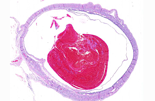
Uterus was received in formalin. Uterine horns are enlarged and filled with brown to black watery fluid and blood clots.
Histopathologic Description:
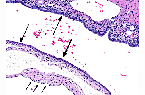
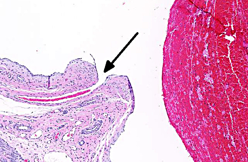
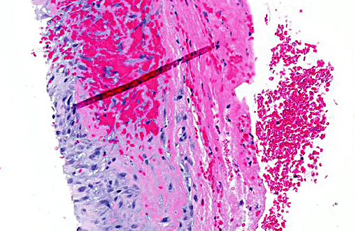
Uterus: A blood vessel in the endometrium is markedly dilated and filled with blood, laminated fibrin, and neutrophils mixed with karyorrhectic debris (thrombus). The endometrium overlying this area is thin with only 1 layer of low cuboidal cells and 2 to 3 layers of collagen separating it from the endometrium of the dilated vessel. The uterine lumen is markedly compressed by the aneurysmal vessels. In one section, the thrombus is adhered to the wall of the vessel (will vary with section). There are hemosiderin laden macrophages focally in the adjacent endometrium (in some sections).
Morphologic Diagnosis:
Uterus: Endometrial venous aneurysms.
Lab Results:
The PCV before surgery was 14%.
Condition:
Endometrial venous aneurysm
Contributor Comment:
Hematuria in rabbits has been associated with uterine adenocarcinoma, uterine polyps, renal infarction, urolithiasis, cystitis, bladder polyps, pyelonephritis and uterine endometrial venous aneurysms.(2,3,7) The most common clinical signs with venous aneurysms are hematuria or urogenital bleeding.(1,2) Occasionally, the aneurysms are associated with mild anemia and proteinuria.(1) Varices and aneurysms of uterine subserosal and myometrial venous plexi, but not of endometrial vessels, have been reported in women. Similar endometrial aneurysms have been seen in rats and mice. In rabbits, at necropsy, clotted blood may be found within the uterine lumen.(6) Non-pregnant multiparous does are most affected.(7) The defect is likely congenital.(7)
JPC Diagnosis:
Uterine horn: Endometrial venous aneurysm, with thrombosis.
Conference Comment:
In rare sections in this case, the origin of the venous aneurysm is visible within the vessel wall. By definition, an aneurysm is a localized dilatation of a vessel due to widening of the lumen which causes an abnormal attenuated vascular wall. In normal branching vessels, the elastic fibers contract but are continuous when the area of branching is cut into histologic section. In this case, when present, a small vessel empties into this large aneurysm and the elastic fibers are lost at this point of transition, enabling participants to definitively identify it as the aneurysmal origin. Aneurysms should be contrasted with false aneurysms or dissections, which are a defect in the vascular wall leading to an extravascular hematoma.(6) Both aneurysms and dissections can rupture, often with catastrophic consequences.(6)
Aneurysms occur when the structure or function of the connective tissue within the vascular wall is compromised, often during the continuous remodeling process it undergoes to maintain structural integrity.(6) These defects can occur as a congenital condition or be acquired over time with progressive weakening of the wall. Increased expression of matrix metalloproteases (MMPs) and decreased expression of tissue inhibitors of metalloproteinases (TIMPs) often contributes to this degradation.(6) MMPs, which require metal ions such as zinc for their activity, are instrumental in the process of remodeling the extracellular matrix, to include vascular remodeling.(5) Additionally, MMPs play a prominent role in tumorigenesis due to their prominent role in cell turnover and migration, and regulating signal pathways of cell growth, inflammation and angiogenesis leading to a large body of research on these proteinases.(4) Specific to angiogenesis, MMP-2 and MMP-9 are cited as both pro and anti-angiogenic while MMP-1, MMP-7, and MMP-14 are specifically pro- while MMP-12 is anti-angiogenic.(4) MMPs are produced by a variety of cell types and regulated by growth factor and cytokine secretion. Their activity is tightly controlled as they are rapidly depleted by TIMPs produced by most mesenchymal cells.(5) When this MMP/TIMP balance is altered, such as occurs with inflammation in atherosclerosis or vasculitis, the risk of aneurysm formation increases.(6)
References:
1. Allison N. Anemic virgin female rabbits. Lab Anim. 2003;32(2):23-25.
2. Bray MV, Weir EC, Brownstein DG, Delano ML. Endometrial venous aneurysms in three New Zealand white rabbits. Lab Anim Sci. 1992;42(2):360-362.
3. Garibaldi BA, Fox JG, Otto G, Murphy JC, Pecquet-Goad ME. Hematuria in rabbits. Lab Anim Sci. 1987;37(6):769.
4. Kessenbrock K, Plaks V, Werb Z. Matrix metalloproteinases: regulators of the tumor microenvironment. Cell. 2010;141:52-67.
5. Kumar V, Abbas AK, Aster JC. Inflammation and repair. In: Kumar V, Abbas AK, Aster JC, eds. Robbins and Cotran Pathologic Basis of Disease. 9th ed. Philadelphia, PA: Elsevier Saunders; 2015:105.
6. Mitchell RN. Blood vessels. In: Kumar V, Abbas AK, Aster JC, eds. Robbins and Cotran Pathologic Basis of Disease. 9th ed. Philadelphia, PA: Elsevier Saunders; 2015:501-502.
7. Percy DH, Barthold SW. Rabbit. In: Pathology of Laboratory Rodents and Rabbits. 3rd ed. Hoboken, NJ: Wiley-Blackwell; 2008:253-307.