CASE III: S2018-0004 (DVD) (JPC 4117676)
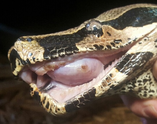
Signalment: 13 year old, female, Red-tailed boa (Boa constrictor ortonii)
History: This boa developed a mass along the inner (palatine) left maxillary dental arcade, and despite antimicrobial therapy, it continued to grow. Radiographs demonstrated widening of the bone with lysis. The mass was resected and submitted for histopathological examination.
Gross Pathology: The sample received was a 3.5 cm x 1.8 cm x 1.4
cm fleshy to firm, oblong, pink to dark red and purple mass with three teeth
embedded on one lateral side. On cut section, the mass appeared to extend to
the deep margin and was composed of multifocal, tan, soft regions and firm,
white, bony regions.
Laboratory results: NA.
Microscopic Description: Gingiva with mandibular bone:
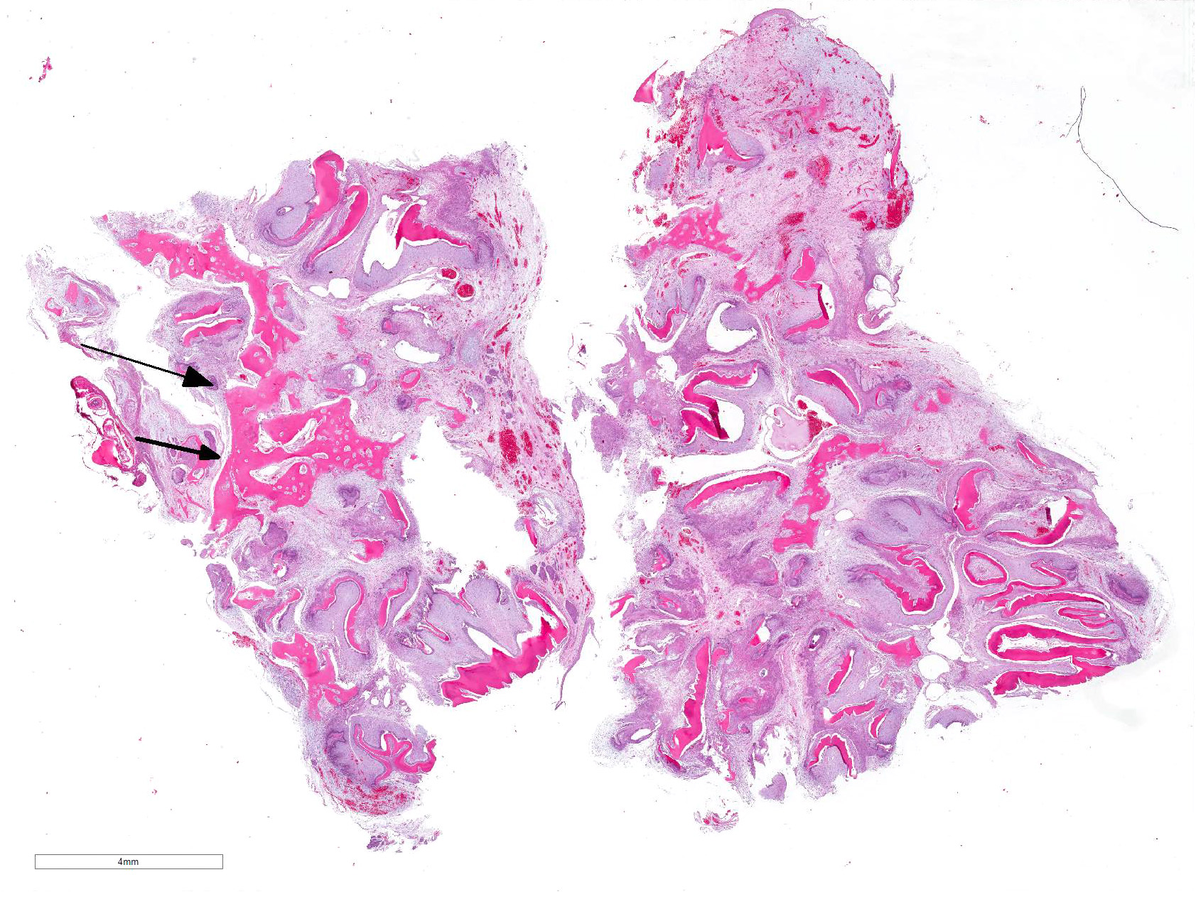
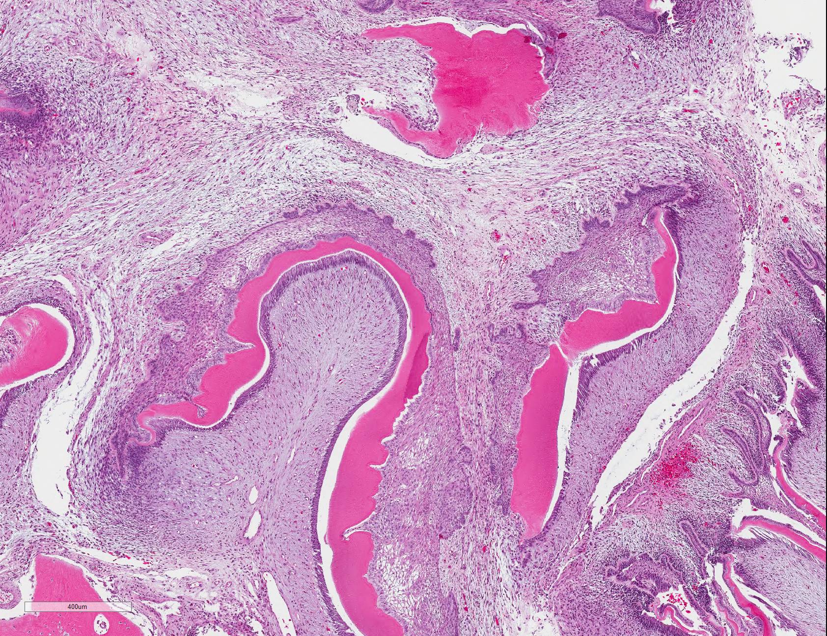
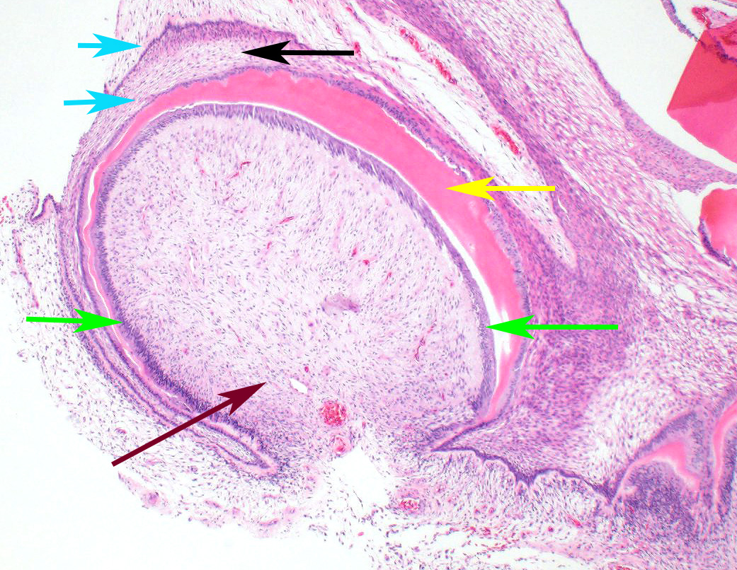
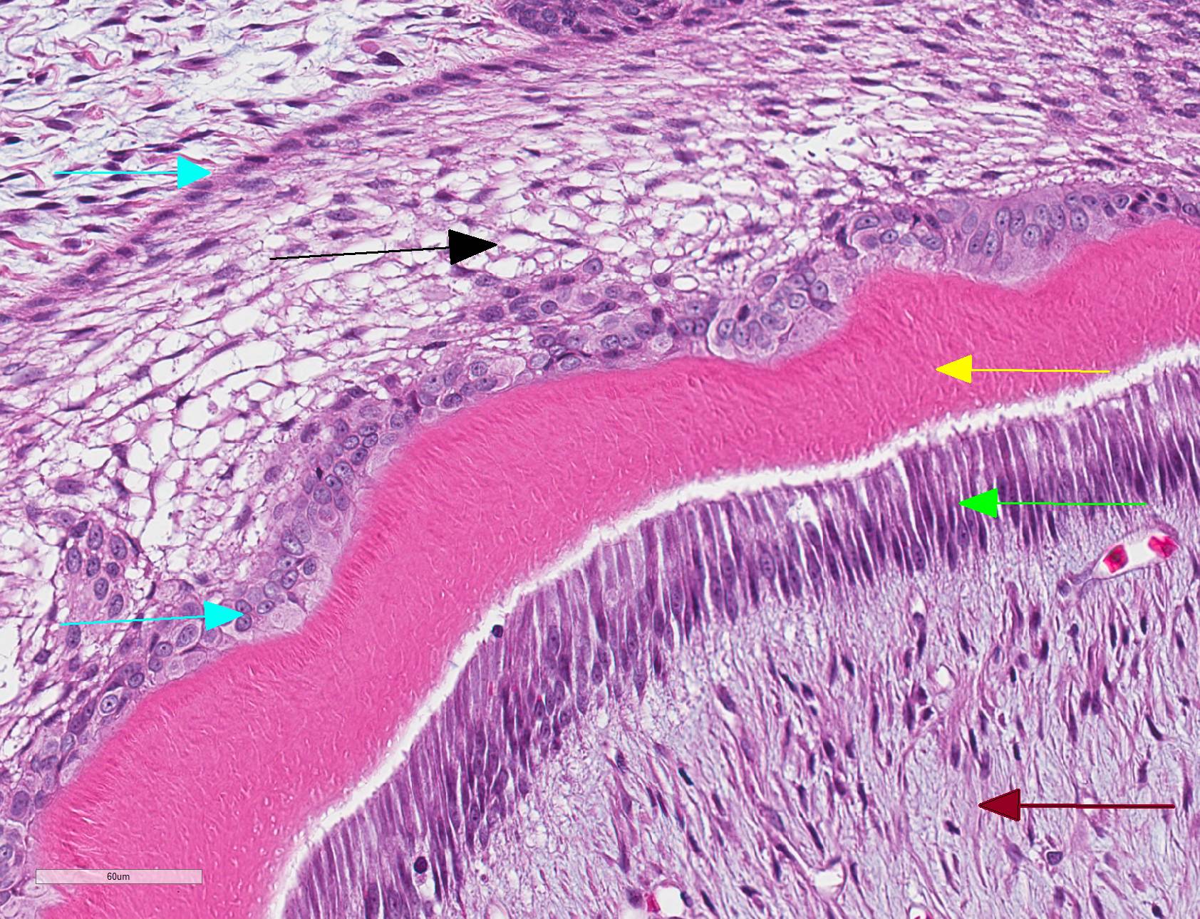
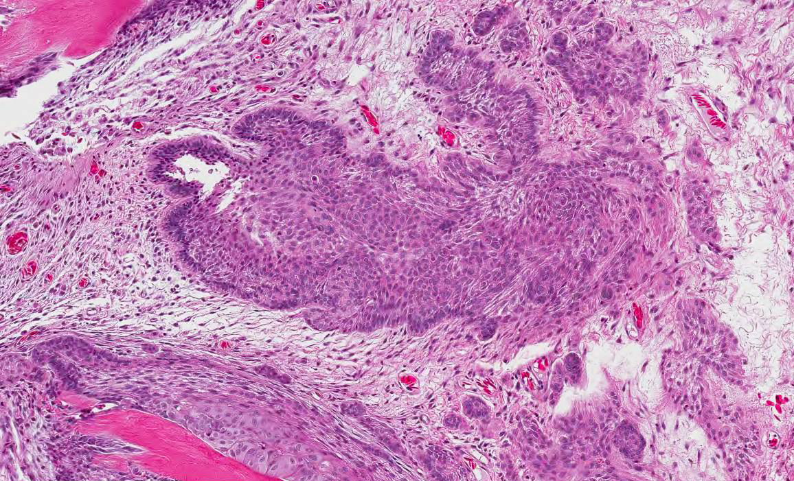
Oral mass: Expanding the submucosa and raising the overlying mucosal epithelium is a poorly demarcated, unencapsulated, neoplasm composed of epithelial and mesenchymal elements that frequently form haphazardly arranged, variably sized and shaped, tooth-like structures on a moderate fibrovascular stroma. Islands and wavy aggregates of brightly eosinophilic extracellular material with fine tubular cavities (dentin) and an occasional adjacent clear gap are surrounded on one aspect by a layer of well-differentiated, palisading odontoblasts and on the other aspect by variably thick rows and sheets of tightly packed columnar to polygonal shaped cells with apically oriented nuclei (ameloblasts). The ameloblasts are often arranged in palisading rows overlying loosely arranged stellate to fusiform cells with prominent intracellular bridging (stellate reticulum). In other regions, odontogenic epithelium is more haphazardly arranged in sheets, small follicles, or branching, anastamosing trabeculae and islands that have a peripheral palisade of cells and infiltrate into the surrounding mesenchyme. Occasionally, islands and trabeculae bud off from rows of ameloblasts bordering dentin material. Within the mass, neoplastic odontogenic epithelium occasionally form cords and nests that are not associated with dentin, with multifocal areas of prominent cellular vacuolation (degeneration) or loss of cells and accumulation of pale, basophilic wispy material admixed with sloughed cells and pyknotic nuclear debris. The neoplastic cells have distinct cell borders with small to moderate amounts of eosinophilic cytoplasm, round to oval nuclei with finely stippled chromatin and one to two, small central nucleoli. There is mild to multifocally moderate anisocytosis and anisokaryosis in this population with 6 mitotic figures in ten 400x figures. Multifocally, neoplastic cells undergo abrupt keratinization. Spindle to stellate mesenchymal cells with an accompanying vascular component (dental pulp) are often present centrally within the tooth-like structures. Infrequently, the eosinophilic dentin material is surrounded by small numbers of multinucleated odontoclasts. Multifocally, there are trabeculae of moderately cellular, woven new bone and small amounts of existing lamellar bone that are occasionally surrounded by small numbers of osteoblasts or small numbers of multinucleated osteoclasts in Howship's lacunae. The overlying mucosa is largely ulcerated and the subjacent submucosa is expanded by increased clear space and pale eosinophilic proteinaceous material (edema). In one section (slide 2), the ulcerated mucosa is covered by a dense band of eosinophilic degenerate material, with small to moderate numbers of granulocytes infiltrating into the subjacent submucosa. Scattered throughout the mass and submucosa are small to moderate numbers of histiocytes, lymphocytes and plasma cells.
Contributor Morphologic Diagnosis:
Oral mass, inner (palatine) left dental arcade: Ameloblastoma arising in an odontoma (previously odontoameloblastoma) with mucosal ulceration and proliferative new bone
Contributor Comment: Histologic evaluation of the oral mass revealed a mixed odontogenic tumor containing a neoplastic population of odontogenic epithelium (ameloblastoma) as well as a differentiated mesenchymal component and tooth-like structures (odontoma). The combination of these two findings was most consistent with an ameloblastoma arising in an odontoma (AAO) or odontoameloblastoma (OA), an uncommon odontogenic neoplasm in veterinary and human medicine. In the most recent (2017) WHO classification of head and neck tumors, the term odontoameloblastoma (or ameloblastic odontoma) was discussed.7 The current consensus concluded that, due to the lack of evidence that these tumors begin or recur as odontoameloblastomas, and that they often recur as ameloblastomas, the term ameloblastoma arising in an odontoma (AAO) was more appropriate.
Odontoameloblastomas have been reported in domestic and non-domestic mammalian species, but to our knowledge have not been recognized in reptiles.1,4,5,8,13 An ameloblastoma has been reported in a wild black rat snake, with a single mass on the mandible.2 In humans, OA or AAO are often locally aggressive with behavior and prognosis similar to ameloblastomas. In the veterinary literature, odontoameloblastomas are similarly locally aggressive and expansile with destruction of the adjacent bone of the jaw. The mandible is a common site of the primary tumor, 1,4,5,8,13 though the lesion was maxillary in this case.
Diagnosis of an odontoameloblastoma relies on the presence of three components: neoplastic odontogenic epithelium, a mesenchymal component reminiscent of dental pulp, and dental matrix (dentin and enamel). Differentiation of an odontoameloblastoma or AAO from other mixed odontogenic tumors relies not only on the presence of these components but also the relative amount. Differentials such as an ameloblastic fibro-odontoma and ameloblastic fibroma should have a predominant ectomesenchyme (pulp stroma) component, and odontomas (both complex and compound), should have less odontogenic epithelium.
In this case, the mass did extend to the margins of the examined sections, and bony lysis was suspected on radiographs. However, no gross evidence of recurrence was noted at 4 months follow up.
Contributing Institution:
Wildlife Conservation Society
2300 Southern Blvd.
Bronx, NY 10460
https://www.wcs.org/
JPC Diagnosis: Gingiva and alveolar bone: Compound odontoma.
JPC Comment: This case is an excellent example of the difficulties of subclassifying odontogenic neoplasms, especially those in exotic species. While attendees had no problem identifying odontogenic epithelium and mineralized dental matrix, the issue of differentiating ectomesenchyme of the dental papilla from the surround loose fibrous stroma of the neoplasm, especially in a reptile, proved to be a challenge.
While careful consideration was given to the contributor?s diagnosis of odontoameloblastoma, we prefer a very slightly altered diagnosis of odontoma. While we agree that this is a neoplasm composed of odontogenic epithelium, dental hard substance, and ectomesenchyme, in the sections we examined, we could not identify an overwhelming odontogenic epithelial proliferation to make the diagnosis of OA. The diagnosis of OA requires a greater proportion of odontogenic epithelium than in odontoma. In this case, the odontogenic epithelium was generally associated with prototeeth (i.e. well-formed denticles), as is seen in odontomas. While there are some areas of the tumor in which sheets of odontogenic epithelium are present without the formation of dental matrix or any apparent inductive effect on the surrounding mesenchyme, the majority of the neoplasm is composed of areas in which these three components are clearly represented.
Odontogenic neoplasms have been rarely reported in snakes, with a single previous report of ameloblastoma in a wild black rat snake (Pantherophis alleghanensis)2 and a peripheral odontogenic fibromyxoma in a red tail boa (Boa constrictor constrictor) co-infected with arenavirus (inclusions were not seen in the neoplastic cells, unfortunately.)7
Snakes, like most reptiles (with the exception of some lacertids to include bearded dragons), fish, and amphibians are polyphydont, with continuous tooth renewal as opposed to humans, which are diphyodont, or monophyodont animals with continuously growing teeth (i.e, mice.) In polyphyodon models, teeth are not regenerated following tooth loss, but are continually developed under a highly complex and coordinated process including a number of important genes and gene products to include sonic hedgehog and Wnt/b-catenin. Those readers interested in this process are referred to references 6 and 9.
References:
1. Burrough ER et al. Spontaneous odontoameloblastoma in a female Sprague Dawley rat. JVDI. 2010.22:998-1001.
2. Comolli JR et al. Ameloblastoma in a wild black rat snake (Pantherophis alleghaniensis). JVDI. 2015. 27(4): 536-539.
3. Dubielzig RR. Odontogenic tumors and cysts. In: Meuten DJ, ed. Tumors in Domestic Animals. 4th ed. Ames, IA: Iowa State University Press; 2002:402-409.
4. Dubielzig RR, Griffith JW. An odontoameloblastoma in an adult sheep. Vet Pathol. 1982. 19:318-320.
5. Elvio L, et al. Odontoameloblastoma in a calf. J Vet Dent. 2013. 30:248-250.
6. Gaete M, Tucker AS. Organized emergence of multiple-generations of teeth in snakes is dysregulated by activation of Wnt/beta-catenin signaling. PLOS One 2013; 8(9):e77784.
7. Hellebuyck T, Pasmans F, Ducatelle R, Saey V, Martel A. Detection of arenavirus in a peripheral odontogenic fibromyxoma in a red tail boa with inclusion body disease. J Vet Diagn Invest 2014; 27(2):245-248.
8. Murphy B et al. Mandibular odontoameloblastoma in a rat and a horse. JVDI. 2017. 29(4):536.
9. Murphy BG, Bell CM, Soukup JW. Tumors composed of odontogenic epithelium and fibrous stroma. In: Veterinary Oral and Maxillofacial Pathology. Hoboken, NJ: Wiley and Sons, 2020: pp. 113-118.
10. Richman JM, Handrigan GR. Reptilian tooth development. Genesis 2011; 49:247-260.
11. Uzal FA et al. Alimentary system. In: Maxie MG, ed. Jubb, Kennedy and Palmer?s Pathology of Domestic Animals. 6th ed. St. Louis, MO: Elsevier; 2016:22-24.
12. Wright JM et al. Update from the 4th Edition of the World Health Organization Classification of Head and Neck Tumours: Odontogenic and Maxillofacial Bone Tumors. Head Neck Pathol. 2017.11(1):68-77.
13. Yanai T et al. Odontoameloblastoma in a Japanese monkey (macaca fuscata). Vet Pathol. 1995. 32:57-59