Signalment:
14-month-old , female, intact, Boxer dog (
Canis familiaris).Intestine and colon biopsies were submitted from a patient with chronic diarrhea.
Histopathologic Description:
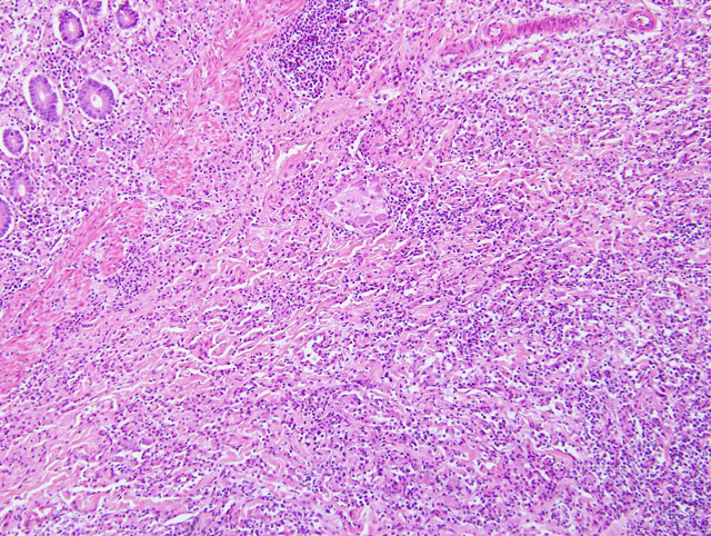
Colon: The small intestine is normal but the colonic submucosa is greatly expanded
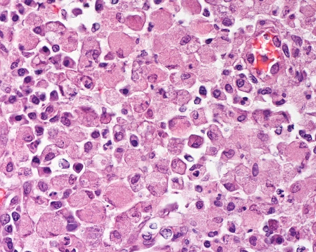
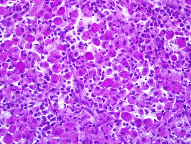
by swollen, foamy/granular histiocytes that occasionally contain a large clear vacuole (Fig 1, 2). A few of these
histiocytes are in the deep mucosal lamina propria as well, between the muscularis mucosa and the crypts. Many
scattered small lymphocytes with plasma cells and neutrophils are also in the submucosa, and the histiocytic
inflammation is also expanding into the inner muscular wall in some areas (may not be in submitted slide). The
histiocytes sometimes contain many PAS-positive granules (Fig. 3, showing PAS positive and negative histiocytes
with Goblet cells in colonic mucosa), many do not, and no fungi or acid-fast bacteria are present.
Morphologic Diagnosis:
Histiocytic ulcerative colitis of Boxer dogs.
Condition:
Histicytic ulcerative colitis
Contributor Comment:
Boxers are prone to this condition, usually before two years of age, and an altered
immunity is suspected.(1) The histiocytes sometimes contain many PAS-positive granules which are thought to be
phagocytic debris and possibly phagocytized organisms that perhaps Boxers and French bulldogs are not able to
process due to a genetic lysosomal defect.(1) In recent years, the condition has been successfully treated with
enrofloxacin(2) and a new report indicates that this treatment correlates with eradication of intramucosal
Escherichia
coli, and the few cases that dont respond have an enrofloxacin-resistant strain of
E. coli.(3)
The histiocytic influx is reportedly centered in the submucosa and into the deep mucosa and may expand through the
muscular wall to the serosa and adjacent lymph nodes.(1) Mucosal biopsies only may miss the lesions. Mucosal
ulceration progresses with chronicity from superficial erosions to patchy ulcers that stop at the submucosa to only
patchy intact islands of mucosa.
This dog was euthanized for this condition. A male littermate is normal. Interestingly, the clinician reported that he
had an unrelated (unconfirmed) case in a young Boxer about this time and it did respond well to three weeks of
enrofloxacin treatment.
JPC Diagnosis:
Colon: Colitis, histiocytic and lymphoplasmacytic, mucosal and submucosal, diffuse, severe with
intrahistiocytic granular eosinophilic material.
Conference Comment:
A number of studies over the years have noted bacteria within macrophages in histiocytic
ulcerative colitis of Boxer dogs (HUC), but recognized pathogens such as
Salmonella, Campylobacter and
Shigella
have not been detected. The very strong breed predisposition and the absence of an identified infectious cause
resulted in the conclusion that the condition is a breed specific immune-mediated disease of unknown cause.
However, some affected dogs were found to respond to treatment with chloramphenicol and, more recently, to
enrofloxacin (a fluoroquinolone antibiotic). It has been noted that HUC has features that are similar to human forms
of inflammatory bowel disease such as Crohns disease. Common features include granulomatous inflammation,
bacteria within macrophages and responsiveness to fluoroquinolone antibiotics. HUC also has similarities to
ulcerative colitis and Whipples disease. Recent studies have shown that certain adherent and invasive strains of
Escherichia coli are present in the lesional tissues of affected dogs. These strains have strong similarities to
E. coli
strains associated with some cases of Crohns disease. HUC and Crohns disease associated strains are more similar
to
E. coli associated with extraintestinal disease than to those causing diarrhea. These findings support the emerging
concept that inflammatory bowel diseases result from an overly aggressive immune response to bacterial microflora
in genetically susceptible individuals.(4)
In the sections of small intestine examined at the conference, predominantly histiocytic inflammation similar to that
present in the colon was found.
References:
1. Brown, CR, Baker, DC, Barker, IK. Alimentary System. In: Maxie MG, ed.
Jubb, Kennedy and Palmers
Pathology of Domestic Animals. 5th ed., vol. 2. Philadelphia, PA: Elsevier Ltd; 2007:112-113.
2. Davies DR, OHara, AJ, Irwin, PJ, Guilford, WG. Successful management of histiocytic ulcerative colitis with
enrofloxacin in two Boxer dogs.
Australian Vet J. 2004;82:58-61.
3. Mansfield, CS, Craven, JM, et al. Remission of histiocytic ulcerative colitis in Boxer dogs correlates with
eradication of invasive intramucosal
Escherichia coli. J Vet Intern Med. 2009;23:964-969.
4. Simpson, KW, Dogan, B, Rishniw, M et al. Adherent and invasive
Escherichia coli is associated with
granulomatous colitis in Boxer dogs.
Infection and Immunity. 2006;74:4778-4792.