Signalment:
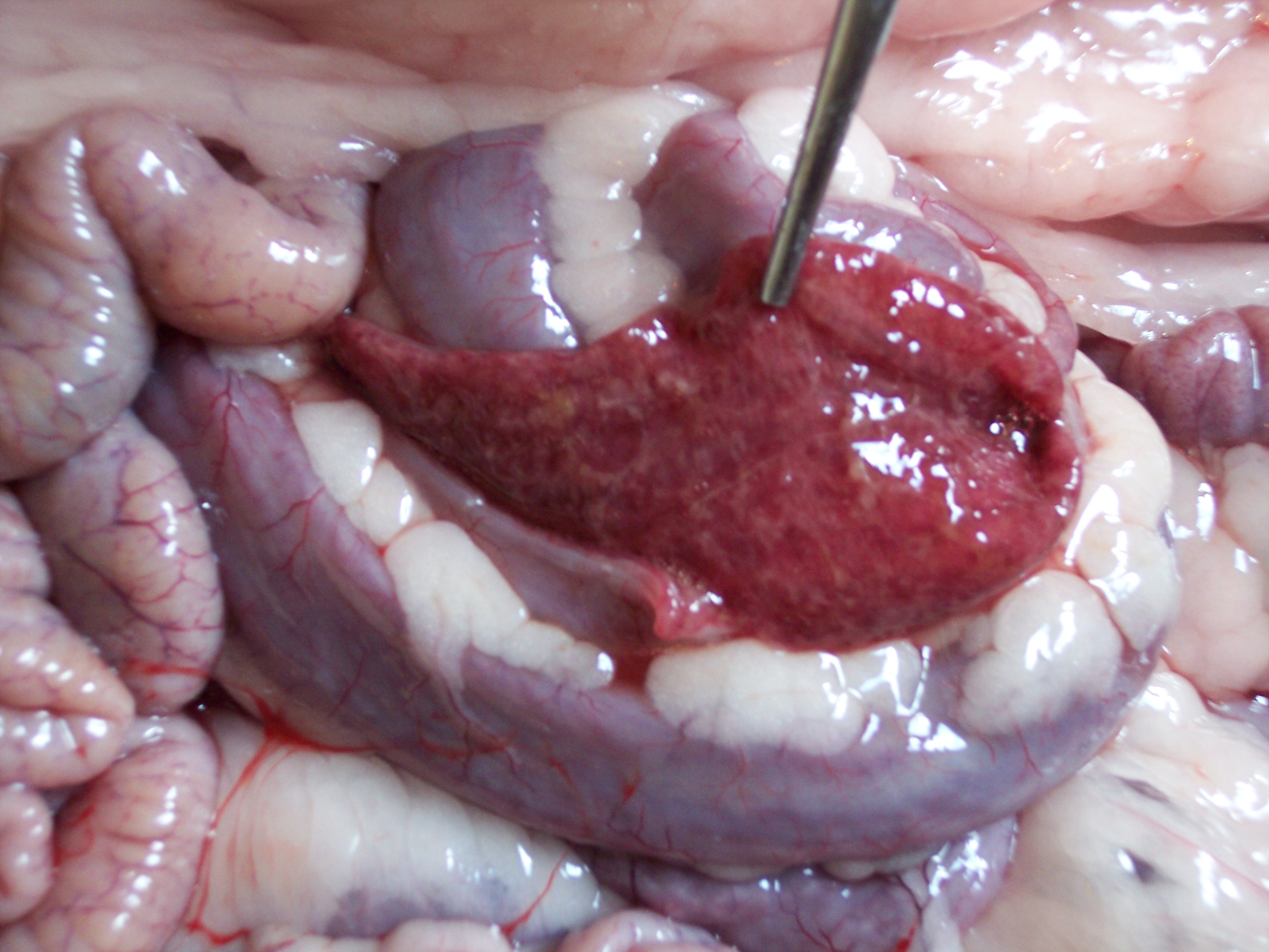
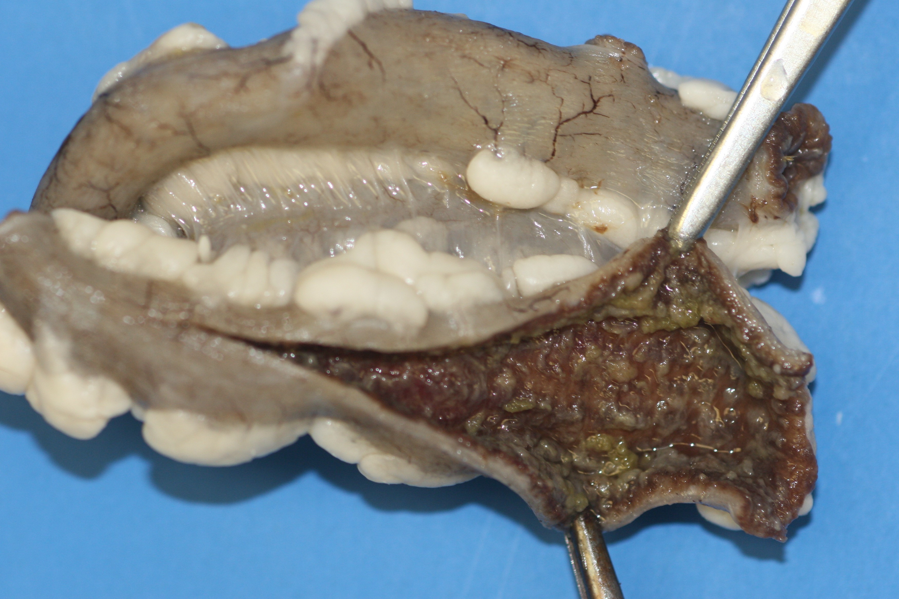
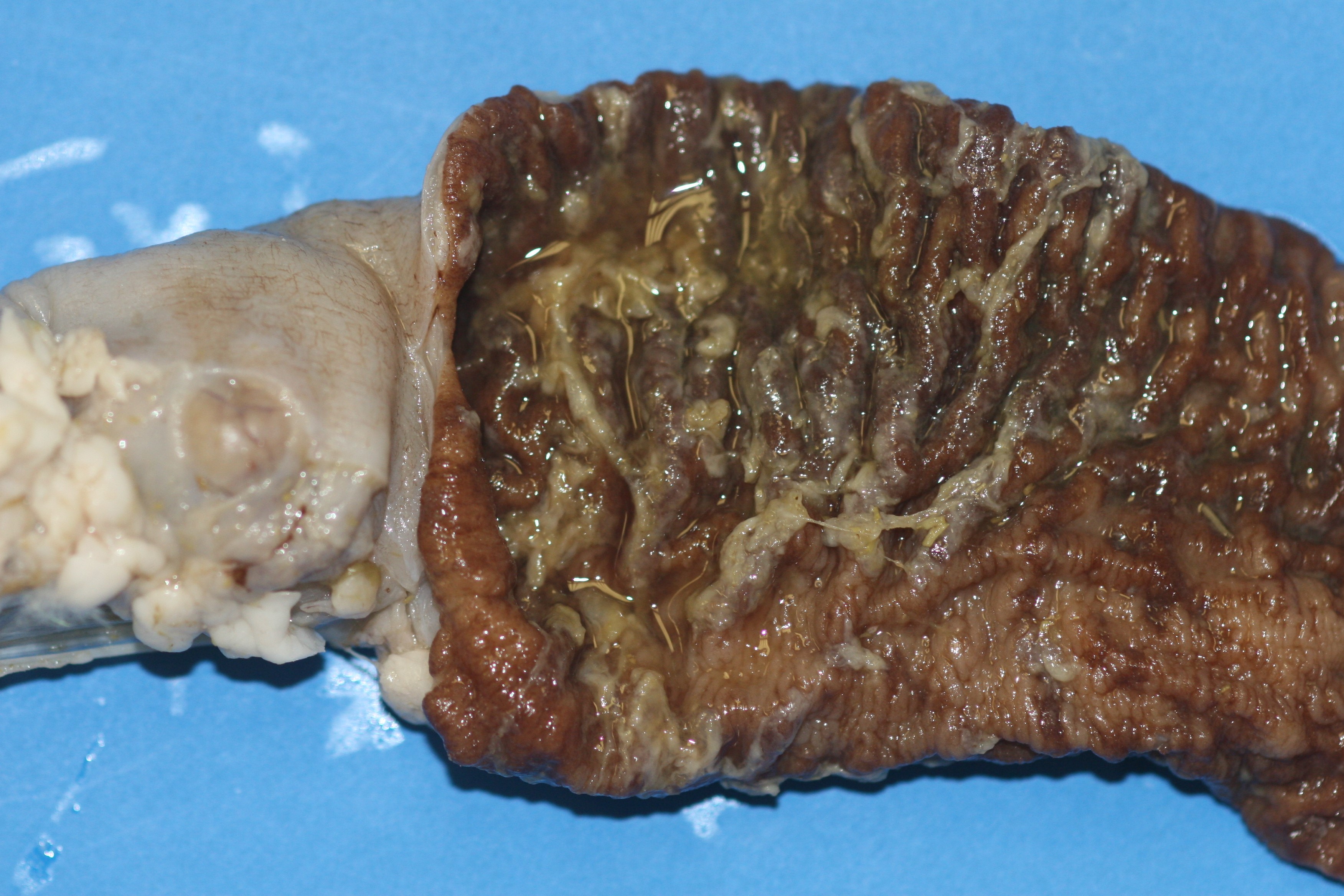
Gross Description:
Histopathologic Description:
Morphologic Diagnosis:
Condition:
Contributor Comment:
In sheep, type D enterotoxemia mostly occurs following a sudden change of diet, particularly to feeds rich in fermentable carbohydrates (resulting in large amounts of undigested carbohydrates entering the small intestine). Ovine type D enterotoxemia produces acute to chronic neurologic disease (ranging from sudden death to blindness, opisthotonus, bleating, convulsions and recumbency with paddling) frequently accompanied by respiratory signs and uncommonly accompanied by diarrhea.(3) The neurologic signs are caused by cerebral perivascular proteinaceous edema (observed in 90% of cases), and in chronic cases multifocal, bilaterally symmetrical necrosis of white matter. The respiratory signs are caused by pulmonary edema. There are usually no significant gross or microscopic changes in the intestinal tract of sheep dying from enterotoxemia.(2,3)
Less is known about predisposing factors of caprine type D enterotoxemia. Cases of type D enterotoxemia have occurred in goats on a regular hay diet.(3) In goats with type D enterotoxemia, the most consistent clinical signs are diarrhea, respiratory distress and central nervous system (CNS) signs including recumbency, paddling, bleating and convulsions. Pseudomembranous colitis, similar to that observed in this goat, is the most characteristic postmortem change in caprine enterotoxemia. Histological changes in the brain are not a consistent feature of caprine type D enterotoxemia; however, cerebral vasogenic edema can be observed in some cases of subacute type D enterotoxemia such as this. Accompanying small intestinal lesions are uncommon in goats and were not observed in this case. Cerebellar coning in this goat was considered to be a consequence of cerebral vasogenic edema.(2,3)
In summary, the major difference between type D enterotoxemia in sheep and goats is the response of the intestinal tract to the disease. The reason for the caprine-specific, selective damage to the large intestine has not been determined. It is possible the caprine small intestine is simply more resistant than the large bowel to the effects of the epsilon toxin (or possibly other C. perfringens type D toxins). Antibiotic-associated pseudomembranous colitis in humans, caused by Clostridium difficile, has a similar disease pattern; however, the reason for the lesion distribution has not been identified.(2) It is also possible that the small intestinal mucosa of sheep is more susceptible to damage by epsilon toxin through facilitating absorption of the toxin into the bloodstream with more pronounced systemic effects (CNS lesions). It has also been suggested that the selective damage of the large bowel in goats is a consequence of toxin modification by enzymes in the goat colon. However C. perfringens epsilon toxin, produced as an inactive prototoxin, is activated following cleavage by trypsin. Because trypsin is secreted in the small intestine, epsilon toxin should be activated in the small intestine of both sheep and goats.(2) Transit speed of the intestinal content should not be a factor with regard to the different disease pattern of type D enterotoxemia in sheep and goats because of the similar transit time in both ruminants (normally approximately three hours for small intestine, and 18 hours for large bowel).(2)
JPC Diagnosis:
Conference Comment:
There are four clinical forms of Clostridium perfringens type D enterotoxemia in goats: peracute, acute, chronic and subclinical. The peracute form manifests as sudden death with little to no warning. The acute form is characterized by diarrhea and colic of a few days duration, and this turns into the chronic form if the animal does not either fully recover or die within a few days of clinical onset. Mentioned by the contributor, the distal small intestine, cecum, and large intestine are most severely affected in goats. Histologically, affected areas are hyperemic and may be covered by a layer of fibrin with a moderate number of inflammatory cells present in the lamina propria.(1)
References:
2. Uzal FA, Kelly WR: Experimental Clostridium perfringens type D enterotoxemia in goats. Vet Pathol. 35:132-140, 1998
3. Uzal FA, Songer JG: Diagnosis of Clostridium perfringens intestinal infections in sheep and goats. J Vet Diagn Invest 20:253-265, 2008