Signalment:
2-year-old, intact male ferret (
Mustela putorius furo).This animal was presented at consultation for a slow growing nodular mass on the right side of the trunk.
The animal was doing well, with no other clinical problems.
Gross Description:
The formalin-fixed specimen was a 15 x 15 x 10 mm mass with a smooth surface. Epidermal
ulceration was present. On cut section, a white irregular firm mass was present in the dermis, extending into the
subjacent adipose tissue.
Histopathologic Description:
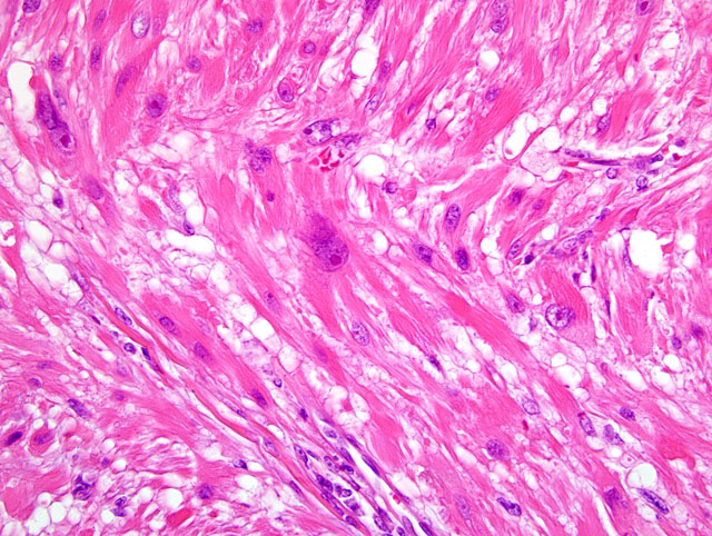
The mass is ovoid, well demarcated, unencapsulated, lying between an ulcerated
epidermis (not present on all sections) and the superficial muscular layer of the skin. Two or three hair follicles are
embedded within the proliferation. These are spindled to strap-cells arranged in interwoven bundles. Some of them
are contiguous with the arrector pili muscle cells. They have an abundant, fibrillar or vacuolated, eosinophilic
cytoplasm with indistinct borders. The nucleus is large, generally ovoid or fusiform with blunt ends and a large
basophilic nucleolus. Anisocaryosis is moderate to marked and anisocytosis is moderate. Bi- or multinucleated
cells are few. The mitotic index (1 to 2 mitoses per 10 high power fields) is low. Multiple lymphoplasmacytic
inflammatory foci, with some hemosiderophages are present in and at the periphery of the mass. On some sections,
a giant cell granuloma around a fragmented hair shaft is present.
Morphologic Diagnosis:
Piloleiomyosarcoma, well differentiated, dermal, ferret (
Mustela putorius
furo).
Condition:
Piloleiomyosarcoma
Contributor Comment:
Originating from the arrector pili muscle, this rare tumor in all animals is now well
recognized in the ferret.(1,2) The histological and cytological patterns are typical. In spite of a low mitotic index
(tumor with mitotic index ⥠2 per 10 HPF is considered to be malignant) and a moderate anisocytosis, this tumor
was classified as malignant because of a frank nuclear pleomorphism. However, the complete excision of this well
demarcated nodule was curative and no local recurrence occured three months after surgery. These tumors are
strongly vimentin, desmin and smooth muscle actin positive (not available in our unit), and negative for cytokeratin.
(2)
JPC Diagnosis:
Haired skin and subcutis: Leiomyosarcoma, low-grade (piloleiomyosarcoma).
Conference Comment:
Conference participants concurred with the contributors diagnosis. Classification schemes
in humans use mitotic rate to distinguish piloleiomyosarcoma from piloleiomyoma, with a mitotic rate of ⥠2 per 10
HPF indicative of malignancy; the degree of nuclear pleomorphism does not correlate with malignancy in human
classification schemes for this neoplasm.(2) In this case, the mitotic rate averages 1 per HPF, and there is marked
anisokaryosis, as noted by the contributor. Additional microscopic findings noted by participants were follicular
atrophy overlying the neoplasm with many follicles in telogen, mild superficial dermal edema, mild superficial
lymphoplasmacytic dermatitis, and minimal parakeratosis.
Basal cell tumors and mast cell tumors are the most commonly reported cutaneous neoplasms in ferrets. Reports of
piloleiomyosarcoma in ferrets suggest that it behaves similarly to its human equivalent, i.e., it carries a good
prognosis with no reports of distant metastasis and infrequent local recurrence.(1,2) This case illustrates the
potential confusion that may result from classifying a neoplasm as malignant versus benign based on histologic
assessment alone, particularly when histomorphology may not be predictive of a significant difference in biological
behavior.
In humans, superficial leiomyosarcoma is classified by its location and origin; the dermal variety (i.e.
piloleiomyosarcoma) originates from arrector pili muscles, while its subcutaneous counterpart (i.e.
angioleiomyosarcoma) originates from vascular smooth muscle. The distinction between the two variants, unlike
the distinction between piloleiomyoma and piloleiomyosarcoma, is clinically significant because, while
piloleiomyosarcomas have only a 30% incidence of local recurrence and no reported metastasis,
angioleiomyosarcomas have a 54% incidence of local recurrence and a 39% incidence of distant metastasis.(2)
References:
1. Mikaelian I, Garner MM: Solitary dermal leiomyosarcomas in 12 ferrets. J Vet Diagn Invest
14:262-265, 2002
2. Rickman BH, Craig LE, Goldschmidt: Piloleiomyosarcoma in seven ferrets. Vet Pathol
38:710-711, 2001