Signalment:
7-year-old warmblood mare (
Equus caballus).The mare presented with a 6-month history of coughing. The veterinarian suspected respiratory infection and allergic pneumonitis; however, the horse did not improve with treatment. Thoracic radiography and pulmonary endoscopic examination were performed (see below).
Gross Description:
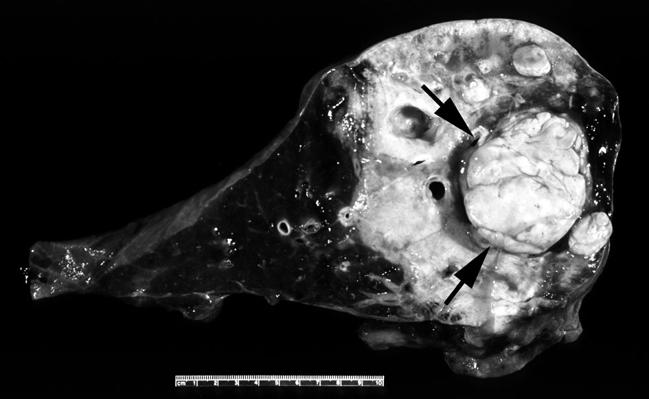
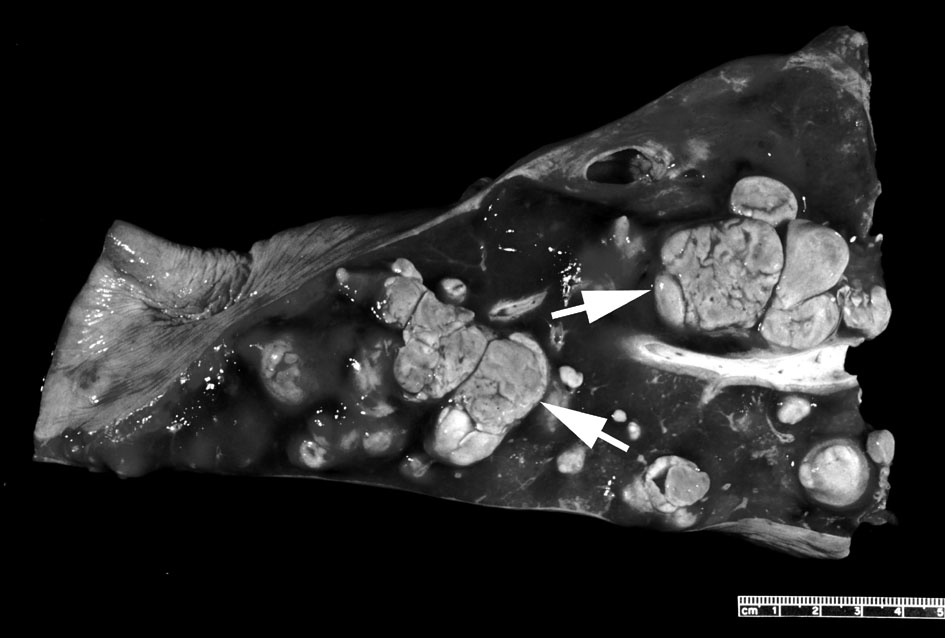
The right lung was surgically excised. The resected lung had a large intraluminal pale yellow-to-tan firm mass expanding and distorting the right caudal lobar bronchus
(Fig. 3-1) and had many variably-sized masses protruded into the airways and within the pulmonary parenchyma
(Fig. 3-2).
Histopathologic Description:
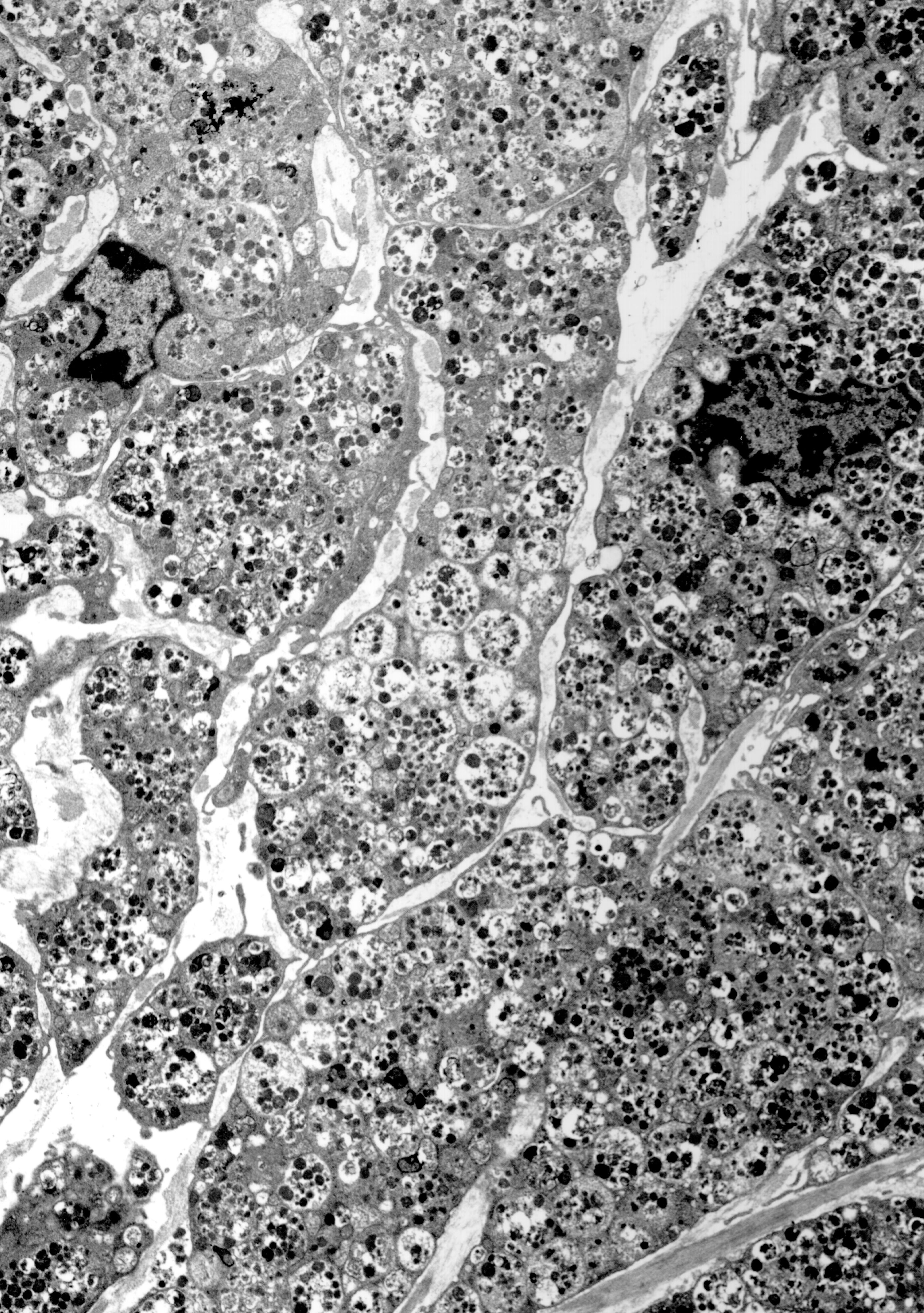
[Submitted tissue: Tissue from one of the lung masses fixed in glutaraldehyde]. Transmission electron micrograph. The tumor
(Fig. 3-3) consisted of a homogenous population of neoplastic mononuclear cells. The neoplastic cells were elongate to polygonal with moderate to abundant amounts of cytoplasm, thin elongate cytoplasmic extensions, irregularly shaped nuclei and small nucleoli. The cytoplasm was filled with single membrane-bound secondary lysosomes filled with slightly electron dense amorphous granular material, membranous debris, and spherical moderately electron dense material. Some of the lysosomal contents were consistent with degenerate organelles. Cell-cell junctions, basement membranes, and angulate bodies were not identified. Cytoplasmic organelles were difficult to evaluate because of the abundant number of secondary lysosomes. The neoplastic cells were separated by electron lucent spaces and extracellular bundles of fine fibrils.
Morphologic Diagnosis:
Lung mass: Granular cell tumor.
Condition:
Granular cell tumor
Contributor Comment:
Granular cell tumor (GCT) has been reported to occur in multiple species, including horses.
2,6,8 In the horse GCT occurs primarily in the lungs.
8 Similar to the horse of the current case, affected horses may be dyspneic and may be misdiagnosed with allergic airway disease. GCT in horses typically presents as single to multiple masses that may obstruct airways. Masses protruding into the airways can be visualized and sampled via endoscopic examination of the bronchial tree. The clinical course is typically benign, and in the current case surgical excision of the affected lung was curative.Â
The exact cell of origin of GCTs is not known.
1,3,6,8 Morphologic and immunohistochemical analyses of a variety of GCTs in multiple species suggest that at least some GCTs may be of neural origin (Schwann cells, neuroectoderm).
6,7 The cell of origin may depend on where in the body the tumor originated. Immunohistochemistry was not performed in the current case.
The ultrastructural features of GCT are similar across species.
1-3,6-8 The main morphologic finding is that of neoplastic cells with abundant amounts of secondary lysosomes containing granular to membranous to amorphous material. The contents likely are remnants of degraded organelles and cell membranes (autophagosomes). In the current case a few lysosomes were found to contain degraded mitochondria. Cell-cell junctions, basal lamina, extracellular collagen, and angulate bodies are variably present in GCTs of animals and humans; however, in the current case cell-cell junctions, basal lamina, and angulate bodies were not identified. The amounts of collagen was minimal(not present in the submitted micrograph). The identity of the extracellular finely fibrillar material present in the current case is not known but may be a product of the neoplastic cells.
JPC Diagnosis:
Lung (per contributor): Granular cell tumor, Warmblood (
Equus caballus), equine.
Conference Comment:
In the horse, granular cell tumors (GCTs) are found primarily within the lower trachea and bronchi as airway associated peri-and endo-bronchial tumors.
4,8 They are often slow growing, benign neoplasms that over time may result in airway obstruction.
4 GCTs can arise in any tissue, and some are thought to be of neuroectodermal origin. They are characterized by neoplastic cells containing abundant cytoplasm with numerous small, eosinophilic, PAS positive, diastase resistant, non-argyrophilic granules identified as secondary lysosomes or phagosomes (myelin figures) on transmission electron microscopy.
4,8 Small secondary lysosome granules have been associated with active Golgi apparatus, while larger granules are characteristic of multivesicular autophagocytic vacuoles.
8
Although granular cell tumors have been reported to occur in many locations, in dogs they generally occur in the oral cavity, particularly the tongue, while in rats they occur within the meninges and brain.
6 They have also been reported in the reproductive tract of rodents and a rabbit.
6,8
References:
1. Barnhart KF, Edwards JF, Storts RW: Symptomatic granular cell tumor involving the pituitary gland in a dog: A case report and review of the literature. Vet Pathol 38:332-336, 2001
2. Facemire PR, Chilcoat CD, Sojka JE, Adams SB, Irizarry AR, Weirich WE, Morrisset SS, Dutweiler VA: Treatment of granular cell tumor via complete right lung resection in a horse. J Am Vet Med Assoc 217:1522-1525, 2000
3. Higgins RJ, LeCouteur RA, Vernau KM, Sturges BK, Obradovich JE, Bollen AW: Granular cell tumor of the canine central nervous system: Two cases. Vet Pathol 38:620-627, 2001
4. Irizarry-Rovira AR, Lennox AM, Ramos-Vara JA: Granular cell tumor in the testis of a rabbit: Cytologic, histologic, immunohistochemical, and electron microscopic characterization. Vet Pathol 45:73-77, 2008
5. Kagawa Y, Hirayama K, Tagami M, Tsunoda N, Yoshino T, Matsui T, Furuoka H, Taniyama H: Immunohistochemical analysis of equine pulmonary granular cell tumours. J Comp Pathol 124:122-127, 2001
6. Markovits JE, Sahota PS: Granular cell lesions in the female reproductive tract of aged Sprague-Dewley rats. Vet Pathol 37:439-448, 2000
7. Pusterla N, Norris AJ, Stacy BA, Smith P, Fielding CL, Moore PF, Watson JL: Granular cell tumors in the lungs of three horses. Vet Rec 153:530-532, 2003
8. Wilson DW, Dungworth DL: Tumors of the respiratory tract. In: Tumors in Domestic Animals, ed. Meuten DJ, pp. 365-399. Iowa State Press, Ames, IA, 2002