Signalment:
1-year-old intact male Argentinian
Mastiff dog, (Canis familaris).
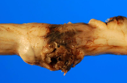
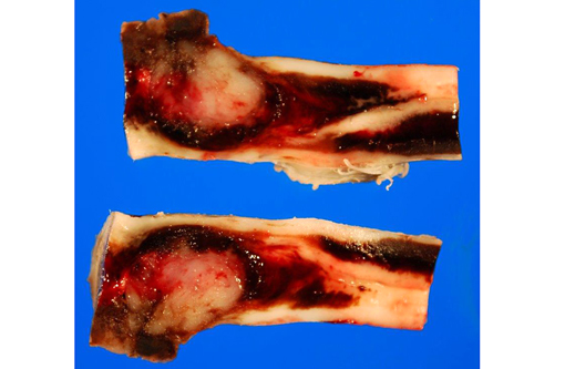
Gross Description:
Over the dorsal vertebral
processes, centered on the thoracolumbar
junction, is a 12 cm long incision that is closed
with surgical staples. Subcutaneous tissue deep to
the incision is dark red and contains large
aggregates of friable, dark red material (clot,
fibrin). The right dorsal pedicles and lamina of
the lumbar vertebrae 1 and 2 are absent (surgical
artifact), and the surrounding tissue is dark red.
Dura overlying the mass is mostly intact with a
single small defect (surgical artifact). At the level
of L1-L2, the spinal cord is moderately enlarged
and dark brown to red. Protruding from subdural
space into the spinal cord is a 1 cm diameter,
well-demarcated, soft, white mass. The tissue
immediately surrounding the mass is markedly
expanded by abundant dark red material
(hemorrhage).
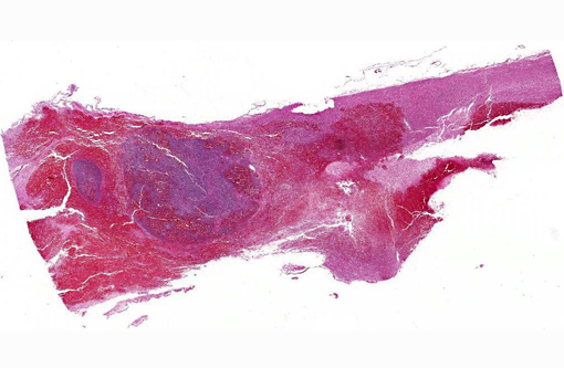
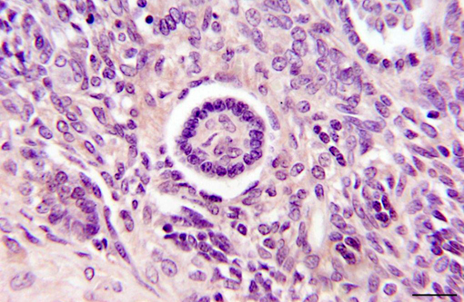
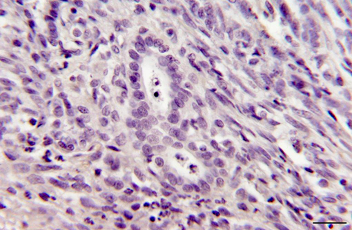
Histopathologic Description:
Spinal cord, L1-
L2: Expanding from within and beneath the dura
and infiltrating the underlying spinal cord is a
densely cellular, well-demarcated, partially
encapsulated neoplasm. Neoplastic cells are of
two distinct cell populations. One population
forms tubules lined by cuboidal cells that have a
small amount of eosinophilic cytoplasm and a
single round nucleus with finely stippled
chromatin. Surrounding the neoplastic tubules are
sheets of polygonal blastemal cells that have
minimal cytoplasm and a round, deeply basophilic
nucleus with slight vesicular chromatin. The
mitotic rate is high with 25 mitoses in 10 highpowered fields. Surrounding the neoplasm is
abundant hemorrhage that extends cranially and
caudally, obliterating large portions of the spinal
cord and surrounding few remaining neurons.
Scattered throughout the areas of hemorrhage are
few small aggregate of neutrophils. Cranial and
caudal to the mass, white matter tracts are
markedly vacuolated with many spheroids.
Immunostaining of the mass demonstrates that
neoplastic epithelial cells are immunoreactive on
staining with cytokeratin and mesenchymal cells
are immunoreactive on staining with vimentin.
Neoplastic cells are not immunoreactive on
staining with neuron specific enolase or glial
fibrillary acidic protein.
Morphologic Diagnosis:
Spinal
cord, L1-L2: Nephroblastoma.
Condition:
Nephroblastoma
Contributor Comment:
Spinal nephroblastomas occur in
young dogs, and German Shepherds may be overrepresented.2
These neoplasms typically arise at
the thoracolumbar junction and are thought to
arise from ectopic rests of embryonal renal tissue
entrapped in the subdural space.2
Metastasis is
not typical4
though possible intraspinal metastasis
has been reported.5
Affected animals typically
present with hindlimb ataxia, paresis, and
proprioceptive deficits.4,5
The classic histomorphology of spinal
nephroblastoma is similar to that of renal
nephroblastoma. Neoplastic cells form
embryonic glomeruli, primitive tubules, and
primitive acini, surrounded by a mesenchymal
stroma.2 Cytologic evaluation shows a
characteristic triphasic pattern with mesenchymal
cells, epithelial cells, and undifferentiated
hyperchromatic cells.1
The submitted neoplasm
demonstrates characteristic primitive tubules, acini, and mesenchymal stroma, though it lacks
classic embryonic glomeruloid structures.
Differential diagnoses include ependymoma,
primitive neuroectodermal tumor, or poorly
d i f f e r e n t i a t e d a s t r o c y t o m a . 5
Immunohistochemistry can aid in establishing a
definitive diagnosis. The epithelial cells within
spinal nephroblastomas are immunopositive for
cytokeratin, and the blastemal cells and stroma
are immunopostive for vimentin.2,5 The
neoplastic cells are immunonegative for NSE,
GFAP, neurofilament, or chromogranin.2,5 In
a d d i t i o n , t h e s e n e o p l a s m s m a y b e
i m m u n o p o s i t i v e o n s t a i n i n g w i t h a
nephroblastoma-specific marker, Wilmâs tumor
gene protein product (WT1).3 Immunostaining of
this neoplasm was consistent with spinal
nephroblastoma; immunostaining for Wilmâs
tumor gene protein product was not performed.
JPC Diagnosis:
Spinal cord: Nephroblastoma.
Conference Comment:
Nephroblastoma, also
known as Wilmsâ tumor, is an important human
pediatric neoplasm. The term ââblastomaââ defines
the neoplastic population as embryonic, rather
than mature terminally differentiated cells;
histologically, nephroblastoma recapitulates the
embryologic development of the kidney.1 The
protein product of the Wilmsâ tumor suppressor
gene-1 (WT-1) is a DNA binding protein
important in normal renal development;
inactivation of this gene likely prevents
differentiation of primitive metanephric cells and
i s d o c u m e n t e d i n s o m e p e d i a t r i c
nephroblastomas.3
A similar intradural
extramedullary spinal cord neoplasm occurs
between the tenth thoracic (T10) and second
lumbar (L2) spinal cord segments in large breed
dogs, typically less than three years old, and is
thus referred to as thoracolumbar spinal tumor of
young dogs.5 Although the histogenesis of this
tumor has not been firmly established, it is
thought to originate from ectopic metanephric
blastema trapped between the dura and the
developing spinal cord.5 The Wilmsâ tumor gene
protein product, WT1, has been identified
immunohistochemically in some of these âcanine
spinal cord nephroblastomas.â3
As noted by the contributor, the microscopic features (a poorly
differentiated blastemal component, mesenchymal
stroma, and an epithelial component forming
tubules and vague glomeruloid structures) and
immunohistochemical staining characteristics
(cytokeratin positive epithelial cells and vimentin
positive blastemal and mesenchymal cells) in this
case are consistent canine spinal cord
nephroblastoma (or perhaps more accurately,
thoracolumbar spinal tumor of young dogs);
however, there is significant slide variation and
tissue identification for some conference
participants was difficult, as some sections did not
contain any identifiable spinal cord.
References:
1. De Lorenzi D, Baroni M, Mandara MT. A true
"triphasic" pattern: thoracolumbar spinal tumor in
a young dog. Vet Clin Pathol. 2007;36:200-203.
2. Meuten DJ. Tumours of the urinary system. In:
Meuten DJ, ed. Tumors in Domestic Animals.
Ames, IA: Iowa State Press; 2002:519-520.
3. Pearson GR, Gregory SP, Charles AK.
Immunohistochemical demonstration of Wilms
tumour gene product WT1 in a canine
"neuroepithelioma" providing evidence for its
classification as an extrarenal nephroblastoma. J
Comp Pathol. 1997;116:321-327.
4. Summers BA, deLahunta A, McEntee M,
Kuhajda FP. A novel intradural extramedullary
spinal cord tumor in young dogs. Acta
Neuropathol. 1988;75:402-410.
5. Terrell SP, Platt SR, Chrisman CL, Homer BL,
de Lahunta A, Summers BA. Possible intraspinal
m e t a s t a s i s o f a c a n i n e s p i n a l c o r d
nephroblastoma. Vet Pathol. 2000;37:94-97.