Results
AFIP Wednesday Slide Conference - No. 27
26 April 2000
- Conference Moderator:
MAJ Jo Lynne Raymond
Department of Veterinary Pathology
Armed Forces Institute of Pathology
Washington, DC 20306-6000
- Return to WSC Case Menu
-
- Case I - 99/1240 (AFIP 2694720)
-
- Signalment: Three to four-year-old ox.
-
- History: Six out of 65 intensively housed cattle,
1-4 years old, kept in different pens, developed severe nervous
symptoms characterised by staggering, circling, blindness, head-pressing
and collapse within 2-3 days of receiving a home-mixed salt lick
consisting of 50% coarse feed-grade sodium chloride and 50% calcium
diphosphate.
-
- The outbreak occurred during mid-winter, when the feeding
of salt lick was resumed after an interval of about 2 months
during which time lick constituents were unavailable on the farm.
A concentrated production ration containing 1% salt was fed during
the same period. As far as could be established, there was also
restricted access to drinking water in some of the pens. Three
of the most severely affected animals were euthanized by intravenous
injections of barbiturate for necropsy, histopathological and
toxicological examinations.
-
- The remaining 3 animals were successfully treated by parenteral
administration of diuretics, corticosteroids, B-complex vitamins,
as well as by dosing regulated quantities of water and supportive
oral remedies via stomach tube. In the latter animals the nervous
symptoms and blindness persisted for about 2 weeks after which
complete recovery occurred.
-
- Gross Pathology: There was marked congestion and edema
of the brain and meninges, as well as petechiae and ecchymoses
within the gray- and white matter of the cerebrum, cerebellum
and brain stem, and a moderate to severe congestion of the abomasal
mucosa.
-
- Laboratory Results: Sodium levels were determined
in the brain of 3 cases by atomic absorption photospectrometry.
In 2 cases, the levels were exceedingly high, viz. 3060 ppm and
3885 ppm, and in the third slightly high, 1839 ppm. According
to Osweiler, levels of >2000 ppm is supportive of sodium toxicosis.
-
- Determinations for organophosphate and organochlorine pesticides
and lead, as well as bacteriological examinations on the brain
of one of the animals were negative.
Contributor's Diagnosis and Comments: HE stained transverse
sections (3 different sections, marked A, B and C) of the dorsolateral
cerebrum, approximately at the level of the midbrain are presented.
-
- Morphologic Diagnosis: Poliomalacia, multifocal,
acute, moderate, with vasculitis, fibrinoid, multifocal, moderate
and leucocytosis, (neutrophilic and monocytic); microhaemorrhages,
scattered, multifocal, mild.
-
- Histopathological changes are present within the meninges
and gray and white matter and comprised varying degrees of vasculitis
and edema with accompanying neuronal and glial cell changes.
The vascular changes included: endothelial cell swelling, medial
fibrinoid change and accumulation of a homogenous eosinophilic
material within lumens of capillaries, venules and arterioles,
mononuclear leucocyte (neutrophil and occasionally eosinophil)
infiltrations into the walls of some vessels (more pronounced
in sections marked 99/1240 C), or extravasations into the extravascular
spaces.
-
- Marked edema with dilatation of perivascular spaces, vacuolation
of gray and white matter, as well as eosinophilic degeneration
and necrosis of neurons, and glial cell cytoplasmic swelling
are evident within the gray matter of the cerebral and cerebellar
cortices. Degenerative and necrotic changes of neurons comprise
swelling, chromatolysis, increased eosinophilia, fading of nuclear
membranes and nuclear pyknosis. Perivascular and neuropil hemorrhages
are occasionally present in both the gray and white matter.
-
- The clinical signs and histopathological changes of the poliomalacia
described here, appear to be similar to those of cerebrocortical
necrosis (CCN) associated with thiamine deficiency in cattle
and sheep, as well as lead poisoning in cattle. However, the
extremely high levels of sodium detected in the brains of two
of three of the animals that were euthanized together with a
history of salt engorgement as well as possible water deprivation,
favors a diagnosis of water deprivation/sodium ion toxicosis.
-
- In contrast to pigs where eosinophilic meningoencephalitis
is common and together with laminar neuronal changes is regarded
to be pathognomonic for salt poisoning in this species, eosinophils
are only rarely encountered in cases of salt poisoning in cattle
and sheep.
-
- The pathogenesis of the edema underlying the poliomalacia
is attributed to passive diffusion of sodium into the brain substance
from the blood and cerebrospinal fluid when the sodium levels
in these compartments rise. This is counteracted by an energy
dependent process (anaerobic glycolysis), which is inhibited
by high levels of sodium entering the brain, resulting in trapping
of sodium ions within the brain. When water intake and renal
excretion of sodium reduces the blood sodium levels to normal,
an osmotic gradient results which leads to cerebral edema.
-
- Sodium chloride poisoning has been classified as acute/direct
salt poisoning where there has been ingestion of excessive salt
in feed or drinking water or as delayed/indirect when there has
been a restriction in water intake with or without ingestion
of excessive salt. These cases probably resemble indirect salt
poisoning due to the delay in onset of clinical symptoms (a few
days) and gastrointestinal symptoms, although there was no definite
evidence of water restriction in all the cases affected.
-
- Hypernatremia with resultant brain edema and neurological
disease has also been induced in calves by injudicious use of
sodium bicarbonate and oral electrolyte solutions during treatment
of diarrhoea with dehydration and acidosis.
-
- AFIP Diagnosis: Cerebral cortex: Neuronal necrosis,
laminar and segmental, with fibrinoid vascular necrosis and edema,
breed not specified, bovine.
-
- Conference Note: The contributor has provided an excellent
discussion of this case.
-
- Contributor: Pathology Section PO Box 12502 Onderstepoort
0110 South Africa.
-
- References:
- 1. Angelos JM, Smith BP, George LW: Treatment of hypernatremia
in an acidotic neonatal calf. J Amer Vet Med Assoc 214:1364-1367,
1999
- 2. Butler R: Salt poisoning/water deprivation in sheep?
Control & Therapy series: Post Graduate Foundation in Veterinary
Science of the University of Sydney, Australia: Mailing 207:1078,
1999
- 3. Jubb KVF, Huxtable CR: The Nervous System. In: Pathology
of Domestic Animals, eds Jubb KVF, Kennedy PC, Palmer N, vol
1, 4th ed, pp. 340-347. Academic Press, San Diego, 1993
- 4. Osweiler GD, Carr TF, Sanderson TL: Water deprivation-sodium
ion toxicosis in cattle. J Vet Diag Invest 7:583-585, 1995
- 5. Osweiler GD: Toxicoses resulting from sodium-water imbalance.
In: Toxicology, pp. 355-357. Williams & Wilkins, Philadelphia,
USA, 1996
- 6. Penrith M-L: A case of salt poisoning in grower pigs.
Newsletter of the Veterinary Laboratory Diagnosticians Group
S Afri Vet Assoc 3:2-3, 1995
- 7. Scarratt WK, Collins TJ, Sponenberg DP: Water deprivation-sodium
chloride intoxication in a group of feeder lambs. J Amer Vet
Med Assoc 186:977-978, 1985
- 8. Summers BA, Cummings JF and De Lahunta A: Salt poisoning.
In: Veterinary Neuropathology, pp. 254-255. Mosby, St Louis,
MO, 1995
- 9. Trueman KF, Clague DC: Sodium chloride poisoning in cattle.
Aust Vet J 5: 89-93, 1978
- 10. Van Leeuwen JA: Salt poisoning in beef cattle on coastal
pasture on Prince Edward Island. Can Vet J 40: 347-348, 1999
-
-
- Case II - TAMU 99-1 (AFIP 2694696)
-
- Signalment: Five-year-old, Aberdeen Angus bull
-
- History: Lesions of 3 months duration on all four
distal limbs
-
- Gross Pathology: Gray, proliferative, symmetric, moist
lesions were observed in circumferential bands extending 15-cm
proximally from the coronary band on both front feet. Nodular,
dry alopecic lesions were seen on the fetlocks of both rear limbs
and the distal scrotal skin. Serpentiginous, dry alopecic lesions
were on the sternal skin.
-
- Contributor's Diagnosis and Comments: Chronic proliferative
dermatitis/pododermatitis with epithelial and mesenchymal proliferation,
epidermal hydropic degeneration, focal vasculitis and intraepithelial
inclusion bodies, hyperkeratosis, bacterial colonies
Etiology: uncharacterized bovine parapoxvirus
-
- This case represents a good example of an unpublished condition!
There are numerous parapox virions in the lesions (parapox are
different in appearance from other poxviruses on EM), and the
lesions are typical of poxvirus diseases. Of course, cutaneous
lesions are not usually described in cattle with parapoxvirus.
The recognized parapoxviruses include: contagious ecthyma of
sheep, bovine papular stomatitis, parapoxvirus of red deer and
pseudocowpoxvirus.
-
- At necropsy, the serpentiginous lesions of the sternal skin
made us suggest parapoxvirus in the gross report (honest!).
Some of the gross lesions looked like papillomas, and the moist
lesions of the front limb were reminiscent of the lesions in
cattle attributed variably to spirochetes (none seen) or papillomavirus.
Vasculitis can be seen with lumpy skin disease and bovine papular
stomatitis and is seen in this case. In order to find out if
this is a new parapox disease, samples have to be typed, but
the lab that does this is in New Zealand, and we could not legally
send the material into that country.
-
- A couple of interesting features of ovine parapoxvirus infection
are that the virus is resistant to both alpha and gamma interferon,
and is believed capable of modulating the Th-1 immune response
of the infected host through an IL-10-like gene product of the
virus. The virus also makes an endothelial growth factor. Parapoxviruses
are suggested to occur in musk ox, gray seals, and the Japanese
serow (these have not been confirmed in THE lab). This group
of viruses is responsible for a nuisance category (due to the
mild signs) of zoonoses called "farmyard pox". This
type of human pox is described to occur also in handlers of reindeer
and other cervids.
-
- AFIP Diagnosis: Haired skin: Dermatitis, proliferative,
chronic-active, focally extensive, severe, with hyperkeratosis,
epithelial ballooning degeneration, and eosinophilic intracytoplasmic
inclusion bodies, Aberdeen Angus bull, bovine.
-
- Conference Note: Parapoxvirus is a genus of the family
Poxviridae. Parapoxviruses are large, enveloped, highly epitheliotropic,
DNA viruses that are cocoon shaped and measures 260 x 160 nm.
There are over 100 polypeptides in the virion. The core proteins
include a transcriptase and several other enzymes. There is
extensive cross-neutralization and cross-protection between viruses
belonging to the same genus, but not between those of different
genera. Even though parapoxviruses exhibit a highly restricted
host range, most can infect humans, producing generally benign
cutaneous lesions limited to the site of inoculation.
-
- By electron microscopy parapoxviruses are ovoid with a regular
spiral arrangement of "tubules" (surface rodlets) on
the outer membrane. There is no nucleocapsid conforming to either
icosahedral or helical symmetry; hence it is called a "complex"
virion. An outer membrane encloses a dumbbell-shaped (biconcave)
central core and two "lateral bodies" of unknown nature.
-
- Some sections contain an organizing thrombus in the subcutis.
-
- Contributor: Texas A&M University, Department
of Veterinary Pathobiology, College of Veterinary Medicine, Texas
A&M University, College Station, TX 77843-4467.
-
- References:
- 1. Haig DM: Poxvirus interference with the host cytokine
response. Vet Immunol & Immunopath 63(1-2):149-56, 1998
- 2. Haig DM, Mercer AA: Ovine diseases. Orf. Vet Res. 29(3-4):311-26,
1998
- 3. Murphy FA, Gibbs EBJ, Horzinek MC, Studdert MJ: Veterinary
Virology, 3rd ed., pp. 278-291. Academic Press, San Diego, CA,
1999
- 4. Robinson AJ, Mercer AA: Parapoxvirus of red deer: Evidence
for its inclusion as a new member in the Genus Parapoxvirus.
Virolo 208(2):812-815, 1995
- 5. Smith KJ, Skelton HG, James WD, Lupton GP: Parapoxvirus
infections acquired after exposure to wildlife. Arch of Dermatol
127(1):79-82, 1991
-
-
- Case III - 989-4037 (AFIP 2694690)
-
- Signalment: Tissue is from a 5-year-old female mule
deer (Odocoileus hemionus).
-
- History: This mule deer was showing signs of weakness,
excessive salivation and emaciation.
-
- Gross Pathology: Gross lesions included extensive
serous atrophy of adipose tissues, mild muscular atrophy, bronchopneumonia,
mild gastric ulceration and enlarged adrenal glands.
-
- Laboratory Results: Pasteurella sp. was isolated
from the pneumonic portion of the lung. No significant growth
was cultured from the gastric ulcer.
-
 Immunohistochem
Mab89/160.1.5
Immunohistochem
Mab89/160.1.5
- Case 27-3. This photomicrograph is of the dorsal motor
nucleus of the vagus nerve of this deer stained with a monoclonal
antibody (Mab 89/160.1.5). The positive red staining is interpreted
to be scrapie-associated prion protein or an antigenically similar
protein that has been found with chronic wasting disease of deer
and elk. (Legend and image from Colorado State
University Diagnostic Lab, Ft. Collins, CO, USA and reproduced
with permission.)
-
- Contributor's Diagnosis and Comments: Spongiform encephalopathy,
severe, brain, compatible with chronic wasting disease (etiology-thought
to be a prion protein).
-
- This slide is from the obex region of the medulla oblongata
of the brain stem of a 5-year-old female mule deer. The primary
histological lesions at this level of the medulla oblongata are
found within the dorsal motor nucleus of the vagus nerve with
milder lesions of the hypoglossal nucleus, nucleus of the spinal
tract of the trigeminal nerve, nucleus ambiguus and the olivary
nuclei. The histological lesions are characterized by intracytoplasmic
neuronal vacuolation, microcavitation of the neuropil and mild
astrocytosis. Many of the sections contain variably-sized areas
of mineralization at the vagal nucleus. This is a common incidental
finding with no known association with the observed vacuolar
changes. This condition is confined to North Central Colorado
and Southeast Wyoming.
-
 20x
obj
20x
obj
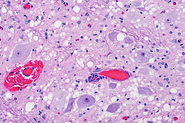
- Case 27-3. Brain stem. Multifocally, neurons and neuropil
contains round clear vacuoles (spongiform degeneration). There
is focal perivascular hemorrhage (probably an artifact) and mild
gliosis.
-
- AFIP Diagnosis: Brain stem, medulla oblongata: Neuronal
degeneration, vacuolation, and loss, with spongiform change of
gray matter neuropil, and astrocytosis, consistent with spongiform
encephalopathy, mule deer (Odocoileus hemionus), cervid.
Conference Note: Chronic Wasting Disease (CWD) is classified
as a transmissible spongiform encephalopathy (TSE). As early
as 1967 chronic wasting disease was diagnosed in captive mule
deer and mule deer / white-tailed deer hybrids. It was later
discovered in captive elk and white-tailed deer and within the
last few years it has been diagnosed in significant numbers of
free-ranging deer and elk. CWD shares many epizootiologic, clinical
and pathologic features of other TSE's (scrapie, bovine spongiform
encephalopathy (BSE), etc.).
-
- Although the cause of these diseases is still speculative
and controversial, prions (proteinaceous infectious particles)
are generally accepted as the etiologic agents. Prions are isoforms
of a normal cell surface sialoglycoprotein, prion protein (PrP),
that are protease-resistant and differ in conformation from the
normal PrP. The normal prion protein (PrP) consists of alpha
helices, which are regions in which the molecular backbone twists
into a specific kind of spiral. The abnormal form contains beta
strands, which are regions in which the backbone is fully extended
and form beta sheets when aggregated. The abnormal conformation
may result from a mutation, which predisposes the aberrant PrP
to "flip", or from the physical interaction between
the abnormal and normal PrP. For these reasons, spongiform encephalopathies
are considered inherited as well as communicable diseases.
-
- The exact mechanism of infection is still controversial and
speculative. It is generally accepted that BSE is caused by
the accumulation of abnormal protease-resistant PrP within neurons.
This occurs either via a mutational alteration of the PrP gene
and subsequent translation of abnormal PrP, or by infection of
abnormal PrP which then causes a cascading conformational alteration
of normal PrP. In the infectious scenario, the abnormal PrP
replicates in lymphoid tissues and lower intestinal tract. The
agent continues to replicate in the lymphoid tissues and intestine
for months to years. It then spreads via hematogenous and/or
axonal routes to the central nervous system (CNS) where it localizes
in the medulla oblongata and diencephalon. The reason for this
tropism is unclear, but retrograde migration via visceral and
peripheral nerves has been proposed. It later spreads to other
parts of the CNS, replicates and causes clinical disease usually
at three to four years of age. Recent studies suggest that lysosomes
are the site of abnormal PrP production, and it is the accumulation
of this abnormal PrP, which eventually leads to rupture of the
lysosomal membrane. The liberation of hydrolytic lysosomal enzymes
is proposed to cause the characteristic spongiform change. Like
scrapie, transmission is thought to occur most commonly by ingestion.
CNS and placental tissue are especially rich in abnormal PrP.
-
- Even though the TSE's are a heterogenous group of neurodegenerative
disorders, the common feature of deposition of a shared abnormal
isoform of a native sialoglycoprotein (prion protein), has resulted
in the development of reliable immunohistochemical markers.
These markers yield positive results with both formalin fixed
and autolytic tissues.
-
- Contributor: Colorado State University, Department
of Pathology, Ft. Collins, CO 80523 USA.
-
- References:
- 1. Clark WW, Hourrigan JL, Hadlow WJ: Encephalopathy in
cattle experimentally infected with the scrapie agent. Am J
Vet Res 56(5): 606-612, 1995
- 2. Kuezius T, Haist I, Groschup MH: Molecular analysis of
bovine spongiform encephalopathy and scrapie strain variation,
J Infect Dis 178(3): 693-699, 1998
- 3. O'Rourke KI, Baszler TV, Miller JM, Spraker TR, Sadler-Riggleman
I, Knowles DP: Monoclonal antibody F89/160.1.5 defines a conserve
epitope on the ruminant prior protein. J Clin Microbiol 36:1750-1755,
1998
- 4. Spraker TR, Miller MW, Williams ES, Getzy DM, Adrian
WJ, Schoonvelt GG, Spowart RA, O'Rourke KI, and Merz PA: Spongiform
encephalopathy in free-ranging mule deer, white-tailed deer and
Rocky Mountain elk. J Wildl Dis 33:1-6, 1997
- 5. Williams ES, Young S: Chronic Wasting Disease of captive
mule deer: a spongiform encephalopathy. J Wildl Dis 16:89-98,
1980
-
-
- Case IV- 17077-99 (AFIP 2689003)
-
- Signalment: Perinate, crossbred, domestic pig
-
- History: An entire litter of pigs was born with generalized
skin lesions. No other litters had been affected previously and
none have been affected since this litter had been born.
-

- Case 27-4. This newborn piglet (note umbilical cord)
has multiple, diffusely distributed, slightly raised, reddish-brown
epidermal papules.
-
- Gross Pathology: One dead neonatal pig was submitted.
The pig submitted was slightly gaunt. There were multiple ulcers
on the skin, which measured approximately 3 to 10 millimeters
in diameter and were randomly distributed over the entire body
as well as on the palatine surface of the tongue. Several of
the hooves were partially sloughed. Small amounts of fibrinous
exudate were present in the peritoneal cavity. Individual lung
lobules had a dark-red appearance.
-
- Laboratory Results: Poxvirus was identified in a homogenate
of skin by means of electron microscopy. Streptococcus suis
was isolated from an abdominal swab and from the lung.
-
- Contributor's Diagnosis and Comments: Severe diffuse
suppurative necrotic epidermitis with ballooning degeneration
and intracytoplasmic eosinophilic inclusion bodies.
-
- There is full thickness necrosis of epithelium, which extends
into hair follicles. At the margins of the necrotic epithelium,
there is ballooning degeneration of epithelial cells. Numerous
eosinophilic intracytoplasmic inclusion bodies can be seen in
degenerated epithelial cells. The ulcerated surface is covered
by a crust of serum proteins, degenerate inflammatory cells,
and mixed bacterial colonies. The dermis is diffusely infiltrated
by large numbers of histiocytes, neutrophils, and fewer lymphocytes.
-
- This represents an unusual case of swinepox infection, since
there had not been a history of prior episodes of swinepox on
the farm. There have been no new cases of swinepox in six months
since the initial occurrence. Typically, swinepox virus is thought
to be transmitted mainly by the sucking louse, Haematopinus suis
via mechanical transmission. Pigs on this farm were devoid of
louse infestation. Congenital swinepox infection is thought
to occur as a result of transplacental infection, although placentas
were not available for examination in this case.
-
- AFIP Diagnosis: Haired skin: Dermatitis and folliculitis,
necrotizing, subacute, focally extensive, severe, with vasculitis,
acanthosis, epithelial ballooning degeneration and eosinophilic
intracytoplasmic inclusion bodies, crossbred domestic pig, porcine,
etiology consistent with a poxvirus.
-
- Conference Note: Postnatal infection through contact
with Haematopinus suis resulting in mild or subclinical disease
is the most common form of swinepox. Congenital infections occur
much less frequently. Although all of the piglets in this litter
were affected, frequently only one pig in a litter is affected,
demonstrating that the compartmentalization of the placenta can
restrict spread of infection as it does in many other intrauterine
infections.
-
- Congenital swinepox cannot be conclusively differentiated
from congenital infection with vaccinia virus infection by histopathology,
but the presence of intranuclear vacuoles strongly suggests infection
with swinepox virus.
-
- Contributor: Veterinary Diagnostic Center, Fair Street
and East Campus Loop, Lincoln, NE 68583-0907.
-
- References:
- 1. Borst GHA, Kimman TG, Gielkens AU, van der Kamp JS: Four
sporadic cases of congenital swinepox. Vet Rec 127:61-63, 1990
- 2. House JA, House CA: Swine Pox. In: Disease of Swine,
ed. Straw BE, 8th ed., pp. 29 1-294. Iowa State University Press,
Ames Iowa, 1999
- 3. Murphy FA, Gibbs EBJ, Horzinek MC, Studdert MJ: Veterinary
Virology,
3rd ed., pp. 278-291. Academic Press, San Diego, CA, 1999
- 4. Neufeld JL: Spontaneous pustular dermatitis in a newborn
piglet associated with a poxvirus. Can Vet J 22:156-1 58, 1981
- 5. Paton DJ, Brown IH, Fitton J, Wrathall AE: Congenital
pig pox: A case report. Vet Rec 127:204, 1990
-
-
- J Scot Estep, DVM
Captain, United States Army
Registry of Veterinary Pathology*
Department of Veterinary Pathology
Armed Forces Institute of Pathology
(202) 782-2615; DSN: 662-2615
Internet: estep@afip.osd.mil
-
- * The American Veterinary Medical Association and the American
College of Veterinary Pathologists are co-sponsors of the Registry
of Veterinary Pathology. The C.L. Davis Foundation also provides
substantial support for the Registry.
- Return to WSC Case Menu
 Immunohistochem
Mab89/160.1.5
Immunohistochem
Mab89/160.1.5
 20x
obj
20x
obj
