Results
AFIP Wednesday Slide Conference - No. 25
April 5, 2000

Conference Moderator:
Dr. Michael Goldschmidt, Diplomate, ACVP
Laboratory of Pathology, School of Veterinary Medicine
University of Pennsylvania
Philadelphia, PA 19104
Return to WSC Case Menu
Case I - 3413/98 (AFIP2694788)
 
Signalment: Canine, Labrador retriever, 7-year-old, male.
 
History: The dog was presented to the hospital with a history of generalized skin necrosis. Mucosa of the oral cavity was also involved. Three days later, the dog became anorexic, showed increasing resistance to being touched, and was euthanized.
 
Gross Pathology: The dog had large areas of alopecia, with multifocal ulcerations, distributed over the whole body surface with the exception of distal extremities and tail. Multiple ulcerations were present on the ventral portion and margins of the tongue. In the esophagus there were multiple linear ulcerations.
 
Laboratory Results: CBC revealed mild leukocytosis with a marked left shift and lymphopenia. Blood chemistry was within normal limits.
 
Contributor's Diagnosis and Comments: Skin: epidermal necrosis (surface and follicular keratinocytes), multifocal, acute, severe, with serocellular crusts, leukocytic exocytosis, many apoptotic keratinocytes and hydropic degeneration of basal cells.
 
This case was originally diagnosed with a surgical biopsy as erythema multiforme, but because of the extent and severity of this lesion, it was then interpreted to represent a case of toxic epidermal necrolysis (TEN). TEN, often caused by drugs, is a generalized disease with systemic illness. Death is a common sequela. The role of concurrent infections and neoplasia is under discussion.
 
AFIP Diagnosis: Haired skin: Necrosis, epidermal and follicular, diffuse, with apoptotic keratinocytes, parakeratosis, mild superficial and periadnexal lymphoplasmacytic dermatitis and congestion, Labrador retriever, canine.
 
Conference Note: The differential diagnosis discussed included toxic epidermal necrolysis (TEN), erythema multiforme (EM) and chemical/thermal burns. While TEN and EM may be different extremes of the same process, this lesion was considered more consistent with TEN because of the full thickness epidermal necrosis and severity of the clinical disease. EM is usually characterized by apoptosis of individual keratinocytes. Both TEN and EM are often associated with drug administration. Chemical/thermal burns can also cause full thickness epidermal necrosis, but are almost always associated with dermal necrosis, which is not a feature of TEN.
 
The following chart highlights the most significant features that differentiate these three conditions.

 Condition

 Location

Depth of Necrosis

 Inflammation

 TEN
 mucosal and cutaneous, extensive  full epidermal with +/- subepidermal bullae, does not effect dermis  none or minimal except when ulcerated

 EM
 mucocutaneous, ventral neck, proximal limbs, nose, footpad  full epidermal with numerous apoptotic keratinocytes and +/- subepidermal bullae  lymphohistiocytic, perivascular and interface

 burns
 variable  coagulative necrosis extends into dermis  peripheral neutrophils and macrophages

Contributor: Institut für Tierpathologie, Universität Bern, Länggasse 122, CH-3012, Bern, Switzerland.
 
References:
1. Gross TL, Ihrke PJ, Walder EJ: Veterinary Dermatopathology, pp. 44-46. Mosby-Year Book Inc, St Louis, MO, 1992
2. Yeager AJ, Scott DW, Wilcock BP: The Skin and Appendages. In: Pathology of Domestic Animals, eds. Jubb KVF, Kennedy PC, Palmer N, vol. 1, 4th ed, pp. 624-625. Academic Press, San Diego, CA, 1993
3. Yager JA, Wilcox BP: Color Atlas and Text of Surgical Pathology of the Dog and Cat, vol. 1, p. 175. Wolfe, London, 1994
 
 
Case II - PA-3579(AFIP2694670)
 
Signalment: 7-year-old neutered male Collie mix dog
 
History: This dog developed multiple, firm, non-painful nodules on the planum nasale and muzzle.
 
Gross Lesions: not provided.

Contributor's Diagnosis and Comments: Dermatitis and panniculitis (some sections), granulomatous and pyogranulomatous, multifocal and coalescing (primarily peri-adnexal), marked

Etiology: Consistent with sterile granuloma and pyogranuloma syndrome (idiopathic peri-adnexal multinodular granulomatous dermatitis).

Sterile granuloma and pyogranuloma syndrome is a disorder seen not uncommonly in the dog associated with multiple, firm, well-demarcated alopecic nodules generally involving the muzzle region. Some breed predilection is noted (great Dane, boxer, English bulldog, dachshund, golden retriever) but any breed may be affected. Age and sex predilections have not been observed. Some lesions regress spontaneously, while others may wax and wane. Associated clinical signs are generally not apparent. Several histopathological subtypes have been described. Differential considerations include infectious processes (bacterial and fungal), that can be ruled out via culture and special stains, foreign body reaction and variations of cutaneous histiocytosis. The condition is idiopathic, but the histiocytic inflammatory response in conjunction with the often marked clinical response to glucocorticoid therapy suggests the likelihood of an immune dysfunction, possibly associated with persistent antigenic drive.
 
AFIP Diagnosis: Haired skin, subcutis and panniculus carnosus: Dermatitis and panniculitis, histiocytic, lymphoplasmacytic, and neutrophilic, periadnexal and perivascular, multifocal and coalescing, severe, collie mix, canine.

Conference Note: Sterile granuloma and pyogranuloma syndrome (SGPS), idiopathic peri-adnexal multinodular granulomatous dermatitis (IPMGD) and cutaneous histiocytosis share many clinical and histopathologic features. They may represent different manifestations of the same disease. Based on the deep infiltration, frequent mitoses, angiocentricity and absence of distinct granulomas, we favor cutaneous histiocytosis over the other conditions in this case. SGPS and IPMGD are usually dramatically responsive to glucocorticoids. While lesions of cutaneous histiocytosis may regress spontaneously and some respond well to immunosuppressive therapy, most are slowly progressive and warrant a guarded prognosis.

Special stains for bacteria and fungi (Brown and Brenn, Brown and Hopps, GMS) performed at the Armed Forces Institute of Pathology did not reveal infectious agents.
 
Contributor: Division of Laboratory Animal Resources, S-1040 BioMedical Science Tower, University of Pittsburgh, Pittsburgh, PA, 15261
 
References:
1. Carpenter JL, Thornton GW, Moore FM, King NW: Idiopathic periadnexal multinodular granulomatous dermatitis in dogs. Vet Pathol 24:5-10, 1987
2. Gross, TL, Irhke, PJ and Walder, EJ: Veterinary Dermatopathology, pp. 194-198, Mosby Yearbook, St Louis, MO, 1992
3. Scott DW, Miller WH, Griffin CE: Small Animal Dermatology, 5th ed., pp. 552-555. WB Saunders, Philadelphia, PA, 1998
 
 
Case III - 194705(AFIP AFIP# 2677563)
 
Signalment: 4-year-old, 20 kg, male, mixed breed dog.
 
History: The dog was presented to the Koret School of Veterinary Medicine of the Hebrew University. On physical examination, the dog had lymphadenopathy, anorexia and fever (40.0 C°). The dog had ulcerative skin lesions on the nose, lips, and ears and on large parts of the back. The initial laboratory work included a complete blood count (CBC), cytological examination of the skin lesions and lymph node, and screening for antibodies against Leishmania spp.

Gross Pathology: Submitting clinician described ulcerative skin lesions.
 
Laboratory Results: The CBC revealed a moderate non-regenerative anemia. The cytology for the presence of protozoal parasites was negative and the serological test was positive for Leishmania spp.
 
Contributor's Diagnosis and Comments: Skin, dermatitis, ulcerative, granulomatous mainly histiocytic, chronic diffuse severe with intrahistiocytic and intralesional protozoal parasite: Leishmania spp.

Visceral leishmaniasis is a disease of humans and members of the Canidae family. In the Middle East, leishmaniasis is caused by L. infantum; the vector is the sandfly Phlebotomus sp. and the reservoir is mainly dogs. Visceral leishmaniasis is a fatal zoonotic disease if the patient does not receive the proper treatment. Classical visceral leishmaniasis in dogs appears as a chronic wasting disease with anemia, generalized lymphadenopathy and skin lesions.

The most common findings in cutaneous lesions are hyperkeratosis with massive scaling and ulcerative dermatitis with massive infiltration of mainly histiocytes in which the amastigote form of Leishmania sp. can be found.
 
AFIP Diagnosis: Skin: Dermatitis and panniculitis, ulcerative, histiocytic, lymphoplasmacytic, and neutrophilic, chronic, diffuse, severe, with intrahistiocytic amastigotes, mixed breed, canine, etiology consistent with Leishmania spp.
 
Conference Note: Leishmaniasis is a zoonotic disease caused by many pathogenic species of the genus Leishmania (order Kinetoplastida, family Trypanosomatidae). Differentiation among species cannot be made based only on their morphological features in tissue sections. Differentiation is based on clinical signs and pathological changes, morphology of the parasite, and on specific laboratory tests (eg., serology, immunohistochemistry, indirect immunofluorescence). Leishmania spp. occur in two parasite forms: the intracellular amastigotes (found in parasitophorous vacuoles within macrophages of the vertebrate host) and the promastigote (found predominantly in the sandfly vector). The amastigotes are spherical, 2.5 to 5 mm in diameter, have a prominent, eccentric nucleus, and a transversely oriented kinetoplast. Leishmania amastigotes are the only known protozoa that survive and replicate in the macrophage phagolysosome.
 
Visceral leishmaniasis occurs naturally in man, dogs, cats, cattle, horses, sheep, and a variety of wildlife species. The geographic distribution is wide, with most cases originating from countries bordering the Mediterranean, large areas of Africa, India, China, Mexico, Argentina, Columbia, Brazil and Venezuela. Most cases reported in the United States originated elsewhere, although a few foci of endemic infection have been recognized. Phlebotomid flies exists in the United States. Recent media reports have described leishmaniasis in a number of foxhounds in New York State. The causative agent was identified as Leshmania of the donovani species complex. It was suggested that the foxhounds might have been infected while hunting in other states. Testing has not revealed any associated human cases, but seropositive dogs have been discovered in Virginia, Michigan, and Maryland.
 
Histoplasmosis, toxoplasmosis, blastomycosis, and trypanosomiasis should be considered in the differential diagnosis. A kinetoplast and a small nucleus can be identified in Leishmania organisms in tissue sections. Kinetoplasts are absent in the causative organisms of the first three diseases. The tissue phase of Trypanosoma cruzi has a kinetoplast, but the tissue distribution and lesions of trypanosomiasis (Chagas disease) differ from those of leishmaniasis.
 
Although conference participants diagnosed leishmaniasis, the high mitotic rate and multifocal atypia of the macrophages lead most to at least consider a malignant round cell tumor.
 
Contributor: Kimron Veterinary Institute, Department of Pathology, Bet Dagan 50250, PO Box 12, Israel.
 
References:
1. Bettini S, Gradoni L: Canine leishmaniasis in the Mediterranean area and its implication for Human Leishmaniasis. Insect Sci Applic 7:241-245, 1986.
2. Ciaramella P, Oliva G, De Luna R, Gradoni L, Ambrosio R, Cortese L, Scalone A, Persechino A: A retrospective clinical study of canine leishmaniasis in 150 dogs naturally infected by Leishmania infantum. Vet Rec 22: 539-542 1997
3. Font A, Roura X, Fondevila D, Closa JM, Mascort J, Ferrer L: Canine Mucosal Leishmaniasis. J Amer Anim Hosp Assoc 32:131-136, 1996
4. Gardiner CH, Fayer R, Dubey JP: An Atlas of Protozoan Parasites in Animal Tissues, 2nd ed., pp. 3-10, 53-60. Armed Forces Institute of Pathology, Washington DC, 1998
5. Hervas J, Mendez A, Carrosco L, Gomez-Villamandos JC: Pathological study of visceral leishmaniasis in jackal (Canis aureus). Vet Rec 21:293-295, 1996
6. Jones TC, Hunt RD, King NW: Diseases caused by protozoa. In: Veterinary Pathology, 6th ed., pp. 549-600, Williams and Wilkins, Baltimore, 1997
7. Rab M A, Frame IA, Evans DA: The role of dogs in the epidemiology of human visceral Leishmaniasis in northern Pakistan. Transact Roy Soc Trop Med Hyg 89:912-615 ,1995
8. Ramos-Vara JA, Ortiz-Santiago B, Segales J, Dunstan RW: Cutaneous leishmaniasis in two horses. Vet Pathol 33(6):731-4, 1996
 
 
Case IV - 98-3637 (AFIP 2678063)
 
Signalment: One-year-old female pet rat
 
History: The animal was purchased as a juvenile from a pet store in Las Cruces, New Mexico, US in June 1996. The owners described the hair coat as becoming gradually thinner over the six months from June to December 1996. In 1997, the owner described the coat as dispersing, with bumpy skin. The animal was presented to a veterinary practitioner in March 1997 for evaluation of hair loss; differential diagnosis was ringworm and endocrinopathy. Treatment was ineffective, and euthanasia was requested in May 1997.
Case 25-4. Hair loss is nearly complete over most of the body. The skin over the trunk and thorax forms large thick, rings composed of hundreds of pearlescent nodules, mostly 2-4mm in diameter.

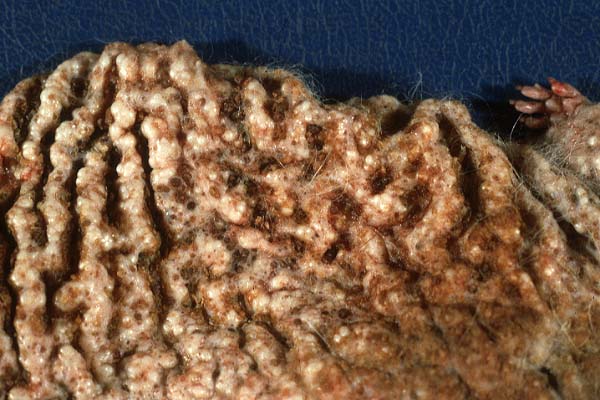
Gross Pathology: The animal has an armadillo appearance, with thick concentric folds forming large rings over the trunk and thorax, starting at the tail and smoothing out somewhat toward the neck. Large folds are also found in the axillary and perineal areas. Hair loss appears complete over most of the body, but there is still some dark grey fuzz on the dorsocranial neck, face, proximal tail and proximal forelimbs. Yellow brown, abundant crust fills the folds between the thick rings of skin, especially on the back. Hundreds of pearlescent nodules, most 2-4 mm in diameter, are scattered in the skin of the abdomen and thorax. The face, ears and lips also have many nodules. Fewer nodules are found in the skin of the limbs, and the tail and pads appear unaffected. The animal has little body fat. The adrenal glands appear grossly normal. The thyroid glands appear slightly small.
 
Laboratory Results:
Fungal culture (skin) - No fungi isolated.
Bacterial culture (skin) -
1 - moderate Staph coagulase negative, 2 colony types.
2 - few Escherichia coli.
3 - rare diphtheroid.

Contributor's Diagnoses and Comments: Cystic keratin dilatation of follicular infundibula, and possibly of sebaceous ducts as well; epidermal hyperplasia with orthokeratotic hyperkeratosis; mild chronic dermatitis.
In the thyroid glands, approximately half of the follicles were collapsed, with no colloid. Mild neutrophilic infiltrates were noted in the endometrial epithelium and glands. Minimal lymphocytic infiltrates were noted in the renal medullary interstitium, with focal tubular mineralization.
 
This rat is thought to be similar to the "rhino mouse", in which there is an inability of follicles to form hair and abnormal follicular cornification with formation of cystic hair follicles. The rhino mouse was first described in 1940. New genotypes (mutations in the hairless gene) have been described. They are models of the human condition papular atrichia, in which hair loss is accompanied by the formation of comedones. The rhino mouse has also been used as a model of human acne, and for the study of skin physiology, pharmacology and pathology.
 
AFIP Diagnosis: Haired skin: Hyperkeratosis, orthokeratotic, infundibular, diffuse, severe, with alopecia, comedones, adnexal atrophy, and mild lymphoplasmacytic dermatitis.

Conference Note: Spontaneous mutations resulting in hairless mice have been recognized since the mid 1800's, and new mutations with similar gross and histologic features continue to arise. The hairless locus is on chromosome 14. Two hairless alleles are recognized, the hairless (hr) and the hairless rhino (hr-rh). Other mutations related to hair loss include naked (N), ragged (Ra), nude (nu), asebia (ab), and ichthyosis (IC).
 
Contributor: New Mexico Veterinary Diagnostic Services, PO Box 4700, Albuquerque, NM 87196-4700.
 
References:
1. Benirschke K, Garner FM, Jones TC: Pathology of Laboratory Animals. Chapter 22, Hereditary Disease, pp. 1997-1998. Springer Verlag, New York, NY, 1978
2. Sundberg JP, Boggess D: Rhino-9J (hr rh9J). A New Allele at the Hairless Locus. Vet Pathol 35:4:297-299, 1998
3. Panteleyev AA, Paus R, Ahmad W, Sundberg JP, Christiano AM: Molecular and functional aspects of the hairless (hr) gene in laboratory rodents and humans. Exp Dermatol 7(5):249-267, 1998
 
 
J Scot Estep, DVM
Captain, United States Army
Registry of Veterinary Pathology*
Department of Veterinary Pathology
Armed Forces Institute of Pathology
(202)782-2615; DSN: 662-2615
Internet: estep@afip.osd.mil
 
* The American Veterinary Medical Association and the American College of Veterinary Pathologists are co-sponsors of the Registry of Veterinary Pathology. The C.L. Davis Foundation also provides substantial support for the Registry.
 
Return to WSC Case Menu