Results
AFIP Wednesday Slide Conference - No. 20
16 February 2000
- Conference Moderator:
Dr. F. Yvonne Schulman
Diplomate, ACVP
Department of Veterinary Pathology
Armed Forces Institute of Pathology, Washington, DC 20306-6000
-
- NOTE: Click on images for larger views. Use
browser's "Back" button to return to this page.
Return to WSC Case Menu
-
- Case I 405/98 (AFIP 2677385)
-
- Signalment: Equine: Six-year-old, half-breed, bay
gelding
-
- History: Treated 6 months ago for conjunctivitis.
More recently eye became cloudy, blood-shot and increased in
size. Eyelids were also swollen. Ocular examination, including
ultrasound, revealed the presence of an intraocular mass.
-

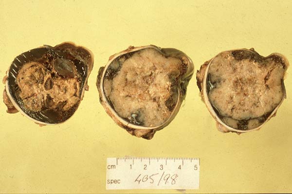
- Case 20-1. Gross Eye. The globe is filled by a variegated
tan mass.
-
- Gross Pathology: The globe of the left eye was submitted
for examination. Grossly it was enlarged and contained a nodular,
soft tissue mass. This mass was white/pale gray in color, friable
and contained small cystic foci. It expanded both the anterior
and posterior chambers with minimal displacement of the lens.
The mass appeared to be contained within the globe.
-
- Laboratory Results: Routine hematology, including
differentials, failed to reveal any significant abnormalities.
Urea, creatinine and GGT were within normal concentrations.
-
- Contributor's Diagnosis and Comments: Diagnosis: Medulloepithelioma
-
- Microscopic examination reveals a poorly demarcated densely
cellular mass partially bordered by ocular sclera and ocular
muscle bundles. Remnants of choroid and retinal pigmented epithelium
are also observed. The cells forming the mass are mononuclear
containing oval or polygonal medium-sized nuclei (occasionally
a large nucleus is observed). These nuclei are pale staining
with a speckled chromatin pattern. Both single cell necrosis
and cell mitosis are seen. Cell borders and cytoplasm are not
discernible. Cells are arranged in loose sheets or aggregated
into pseudostratified formations to resemble duct-like structures
(neuroepithelial rosettes). Cells within these latter formations
rest on thickened hyalinized membranes. Separating many of these
neuroepithelial rosettes are dense bands of connective tissue.
In addition, large expanses of necrotic tissue and regions of
hemorrhage are also noted.
-
- Follow-up: After surgery this animal returned home. Over
a couple of months there was weight loss and a general deterioration
in the animal's condition. No additional investigations were
performed and the animal was euthanized. At death, the local
veterinary surgeon sampled the submandibular lymph node, which
appeared enlarged. This tissue was submitted for microscopic
examination and revealed metastatic spread of the tumor.
-
- In the horse intraocular primary or secondary neoplasms are
very rare. Primary melanomas, astrocytomas, microgliomas, neuroepithelial
tumors of the optic nerve, medulloepitheliomas and secondary
lymphosarcomas have been reported (Davidson, 1991; Ueda et al.,
1993). A medulloepithelioma is a ciliary body tumor arising from
undifferentiated embryonal medullary epithelium of the forebrain
and optic vesicles (Eagle et al., 1978; Davidson et al., 1981).
They may be classified as teratoid medulloepitheliomas if they
also contain undifferentiated mesenchyme, cartilage, striated
muscle or tissue resembling brain (Collins & Moore, 1991).
-
- Medulloepitheliomas are also observed in man where they usually
manifest early in life (up to 5 years of age (cited in Ueda et
al., 1993)). However, in the horse they have been reported in
young and middle-aged animals (Bistner, 1974; Eagle et al., 1978;
Ueda et al, 1993).
-
- AFIP Diagnosis: Eye: Medulloepithelioma, breed not
specified, equine.
Conference Note: Embryologic invagination of the optic
cup results in a bilayered medullary epithelium with the inner
layer forming the iridial pigmented epithelium, ciliary body
non-pigmented epithelium and the neurosensory retina, while the
outer layer forms the outer iridial pigmented epithelium, ocular
ciliary pigmented epithelium, iridal dilator muscle, ciliary
pigmented epithelium, and the retinal pigmented epithelium. Tumors
arising from the undifferentiated embryonal medullary epithelium
that form primitive neuroectodermal structures (retina, ciliary
epithelium, vitreous, and neuroglia), as well as differentiated
mesenchymal tissues (cartilage, skeletal muscle, brain, etc.)
in some cases, are classified as medulloepitheliomas. Ciliary
tumors developing from more differentiated cell types form adenomas
or adenocarcinomas.
Contributor: Department of Veterinary Pathology, University
College Dublin, Ballsbridge, Dublin 4, Ireland.
-
- References:
- 1. Bistner SI: Medulloepithelioma of the iris and ciliary
body in a horse. Cornell Vet 64:588-595, 1974
- 2. Collins BK, Moore CP: Canine anterior uvea. In: Veterinary
Ophthalmology, ed. Gelatt KN, 2nd ed., pp. 385-387. Lea and Febiger,
Philadelphia, PA, 1991
- 3. Davidson MG: Equine ophthalmology. In: Veterinary Ophthalmology,
ed. Gelatt KN, 2nd ed., p. 598. Lea and Febiger, Philadelphia,
PA, 1991
- 4. Eagle RC, Font RL, Swerczek TW: Malignant medulloepithelioma
of the optic nerve in a horse. Vet Pathol 15:488-494, 1978
- 5. Ueda Y, Senba H, Nishimura T, Usui T, Tanaka K, Inagaki
S: Ocular medulloepithelioma in a thoroughbred. Eq Vet J 25:558-561,
1993
-
-
- Case II - 990663.5 (AFIP 2694700)
-
- Signalment: 8-year-old, male, Bernese mountain dog.
-

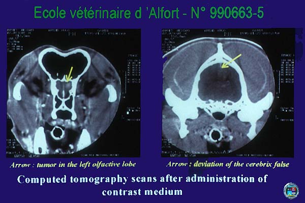
- Case 20-2. CAT scans.
-
- History: According to the owner, the dog exhibited
three generalized tonic/clonic seizures and behavioral changes
for ten days. The seizures occurred at various times of the day.
Neurologic examination revealed that vision was normal in right
eye and absent in the left eye. Pupillary light reflexes were
lower for this eye. Computed tomography images obtained after
administration of contrast media showed two lesions, one (7 mm
diameter) in the left olfactory lobe and the second (25 mm diameter)
in the right temporal area. Stereotactic biopsy was performed
three days later. Histopathological features revealed an idiopathic
granulomatous encephalitis. The dog was treated by immunosuppressive
therapy. Generalized seizures occurred a few weeks later. Continued
decline resulted in euthanasia.
-
- Gross Pathology: Necropsy examination was normal except
for the encephalon. Leptomeninges were thickened. The encephalon
was fixed two days in buffered formaldehyde and cut in 0.5 mm
thick sections. Two regular and uniform nodules, one of 6 mm
diameter and the second of 8 mm diameter were respectively located
in the left olfactory lobe and in the right parahippocampal gyrus.
-
- Laboratory Results: Laboratory evaluations (glucose,
alkaline phosphatase, serum urea nitrogen, alanine aminotransferase,
calcium, and analysis of blood and urine) did not reveal any
abnormalities.
-
- Contributor's Diagnosis and Comments: Brain: Poorly
differentiated large round cell tumor, Bernese mountain dog.
-
- The meninges and subpial parenchyma are focally infiltrated
by neoplastic cells. The neoplasm is composed of pleomorphic
round cells. They also infiltrate Virchow--Robin spaces. They
are characterized by distinct-cell borders. Their cytoplasm,
abundant and eosinophilic, is occasionally vacuolated. Mitotic
activity is high (8 per HPF). Large binucleated and multinucleated
cells are seen in great number. Variations in nuclear size and
shape are marked.
-
- The silver stain for reticulin doesn't demonstrate characteristic
pattern seen in primary lymphomas of the CNS. It is not associated
with the angiocentric growth pattern.
-
- Immunohistochemically, neoplastic cells in the tumor of this
dog were negative for glial fibrillary acidic protein, S-100
protein, cytokeratin and CD3. Some scattered lymphocytes stained
positively for CD3.
-
- Lack of gross lesions in peripheral organs and of histological
lesions in lymph nodes, spleen and liver ruled out the hypothesis
of malignant histiocytosis. Although individual cell morphology
is more atypical than usually observed in primary lymphomas of
CNS, this hypothesis can't be discarded. Immunohistochemical
staining to prove histiocytic or lymphoid origin might help in
establishing the diagnosis.
-
- AFIP Diagnosis: Brain: Histiocytic sarcoma, Bernese
mountain dog, canine.
Conference Note: Based on the H&E slides, most conference
participants diagnosed malignant round cell tumor and favored
histiocytic sarcoma based on the cellular features including
occasional phagocytosis by neoplastic cells; however, malignant
plasma cell tumor, lymphoma and rhabdoid tumor were also included
in the differential diagnosis.
-
- By immunohistochemistry performed at the Armed Forces Institute
of Pathology, neoplastic cells were strongly positive for lysozyme
and did not stain for CD3 (a T lymphocyte marker) and CD79a (a
B lymphocyte marker), with good staining of internal controls.
- The Department of Hematopathology reviewed this case and
favored a diagnosis of malignant histiocytic neoplasm. In humans,
this rare and controversial condition is known as true histiocytic
lymphoma.
-
- Although histiocytic sarcoma is generally associated with
disseminated disease (malignant histiocytosis), especially in
the Bernese mountain dog, histiocytic malignancies can be localized.
It is not known whether disseminated histiocytic sarcoma represents
metastases of a localized sarcoma or multicentric malignancy
developing simultaneously in different organs (Affolter VK and
Moore PF1,2). There was a recent report of a case of primary
malignant histiocytosis of the brain of a miniature schnauzer;
some might argue that localized histocytic sarcoma would have
been a more appropriate diagnosis.
-
- Contributor: UP d'Anatomie-Pathologique, Ecole Nationale
Veterinaire d'Alfort, 7 av. du General de Gaulle, 94704 Maisons-Alfort
(France).
-
- References:
- 1. Affolter VK, Moore PF: Canine cutaneous and systemic histiocytosis:
reactive histiocytosis of dermal dendritic cells. Am J Dermatopathol
22(1):40-48, 2000
- 2. Affolter VK, Moore PF: Canine histiocytic proliferative
disease. Proceedings of 15th AAVD/ACVD, pp. 79-86. 1999
- 3. Chandra AMS, Ginn PE: Primary malignant histiocytosis
of the brain of a dog. J Comp Path 121:77-82, 1999
- 4. Morgello S: Pathogenesis and classification of primary
central nervous system lymphoma: an update. Brain Pathol 5:383-393,
1995
- 5. Vandevelde M, Fatzer R, Fankhauser R: Immunohistochemical
studies on primary reticulosis of the canine brain. Vet Pathol
18:577-588, 1981
-
- Case III 99-4036 (AFIP 2694682)
-
- Signalment: 5-year-old female spayed standard poodle,
canine
-
- History: The dog had experienced a gastrointestinal
upset with vomiting which progressed over a 1 week period to
abnormal behavior with head pressing, circling, hypermetria,
drooling and dementia.
-
- Gross Pathology: There was a mass present on the ventral
surface of the cerebellomedullary regions that extended around
the optic chiasma. The mass was red/brown, friable and 5 cm in
greatest dimension. There were few scattered grey/brown discolored
areas throughout the parenchyma of the cerebrum.
-
- Laboratory Results:
Routine CBC - Lymphopenia
Routine chemistry - no diagnostic abnormalities
CSF tap- wbcs 45/ul, rbcs 20/ul and TP 39 mg/dl. The cell population
consisted of 80% mononuclear cells with 80% of these small and
medium sized lymphoid cells. Sixty percent of the lymphoid cells
were reactive with increased cytoplasmic basophilia. The remainder
were macrophages, some of which exhibited erythrophagy. Twenty
percent of the cells were nondegenerate polymorphonuclear cells.
Intact erythrocytes were also present.
Contributor's Diagnosis and Comments: Granulomatous meningoencephalitis
- The microscopic lesions with the typical vascular orientation
were thought to be consistent with the descriptions of granulomatous
meningoencephalitis. The lesions also resembled a neoplastic
process, but immunohistochemistry distinguished 3 cell populations
with a predominance of T cells, mixed with plasma cells and macrophages.
-
- AFIP Diagnosis: Brain: Atypical angiocentric lymphohistiocytic
infiltrates, favor malignant leukocyte neoplasm, standard poodle,
canine.
Conference Note: This lesion was the subject of lively
debate. The discussion centered on whether the lesion is inflammatory
or neoplastic and what is the appropriate morphologic diagnosis.
When first described, similar lesions were called reticulosis
(inflammatory or neoplastic) due to the presence of reticulin
fibers surrounding individual cells. Later, the milder variants
of this lymphohistiocytic infiltrate were dubbed granulomatous
meningoencephalitis (GME). Some maintain that these lesions represent
a lymphoproliferative disorder that blends into frank neoplasia.
Others prefer to separate them into GME (or inflammatory reticulosis)
and non-B non-T leukocytic neoplasm (or malignant reticulosis).
If they are distinct entities, there is clearly a grey zone between
them. In this case, the density of the infiltrate, cytologic
atypia and moderate numbers of mitotic figures suggest malignancy.
The Department of Hematopathology reviewed the case and concurred
with this assessment. They also mentioned similarities between
this lesion and lymphomatoid granulomatous/angiocentric T cell
lymphoma.
-
- Contributor: Central Laboratory for Veterinarians
c/o PMB 8O, 250 H Street. Blaine Washington, 98230.
-
- References:
- 1. Kipar A, Baumgartner W, Vogl C, Gaedke, Wellman M: Immunohistochemical
characterization of inflammatory cells in brains of dogs with
granulomatous meningoencephalitis. Vet Pathol 35:43-52, 1998
- 2. Koestner A, Bilzer T, Fatzer R, Schulman FY, Summers BA,
Van Winkle TJ: Histological Classification of the Tumors of the
Nervous System of Domestic Animals. In: World Health Organization,
Histological Classification of Tumors of Domestic Animals, ed.
Schulman FY, 2nd ed., vol. 5, pp. 31-32. The Armed Forces Institute
of Pathology, Washington, DC, 1999
- 3. Munana KR, Luttgen PJ: Prognostic factors for dogs with
granulomatous meningoencephalitis: 42 cases (1982-1996): JAVMA
212(12):1902-1906, 1998
- 4. Summers BA, Cummings JF, de Lahunta A: Veterinary Neuropathology,
99. 110-111. Mosby-Year Book, St. Louis, Missouri, 1995
-
-
- Case IV - S-60006 (AFIP 2607937)
-
- Signalment: Aged (originally wild-caught), female,
cynomolgus macaque (Macaca fascicularis)
-


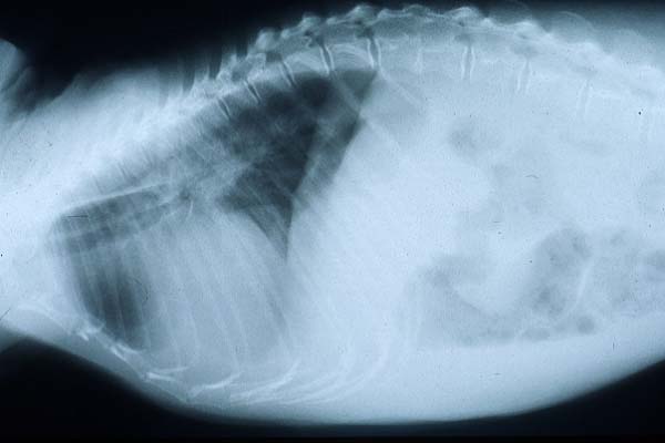
- Case 20-4. Lateral and Dorsal Radiographs. The lateral
view shows diffuse fluid density of the lung space (pulmonary
edema). Both views demonstrate an enlarged cardiac silhouette.
-
- History: This animal arrived at our facility in 1988.
It underwent an ovariohysterectomy in 1993, and had subsequently
been treated for periodontal disease. In June 1997, it was observed
to have ascites when sedated for a routine health check. Abdominocentesis
was performed and the fluid revealed to be a transudate. A CBC
was normal and serum biochemistry profile revealed a mild increase
of BUN and creatinine. Approximately 1 week later, the animal
was noted to be weak and anorexic. It was again sedated and intravenous
fluids were administered. On auscultation, this monkey was noted
to have a gallop rhythm. Thoracic radiographs revealed an enlarged
cardiac silhouette, compatible with generalized cardiomegaly.
An ECG revealed increased P wave amplitude compatible with right
atrial enlargement, and increased PR interval consistent with
1st degree heart block. A repeat serum biochemistry profile indicated
a moderate increase of BUN, creatinine, and bilirubin. Several
days later, this animal was noted to be increasingly weak and
anorexic, and was euthanatized.
-

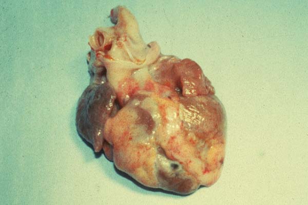
- Case 20-4. Gross Heart. There is multifocal myocardial
pallor.
- Gross Pathology: Approximately 100 ml of ascitic fluid
was present and the liver was firm. The heart appeared slightly
enlarged and weighed 27.7 grams, which was 0.73% of this animal's
body weight (~3.79 kg). [For comparison, the heart weight as
a percent of body weight ranges from 0.35-0.65% in similarly
sized (body weight) rhesus monkeys; our laboratory does not have
similar historical data for cynomolgus macaques].
-
- Laboratory Results: Serum biochemistry- mildly to
moderately increased BUN, creatinine, and bilirubin.
-
- Contributor's Diagnosis and Comments: Severe multifocal
to coalescing myocardial fibrosis with myofiber atrophy and myofiber
karyomegaly
-
- Myocardial fibrosis has been previously reported in western
lowland gorillas, orangutans, and chimpanzees. Generally, these
animals are mature to aged (often in the 2nd and 3rd decades
of life, occasionally older), they often have underlying cardiac
dysfunction, and die during restraint or under anesthesia. [A
review of our archives reveals no record of having ever diagnosed
a similar lesion in any rhesus or cynomolgus monkey from our
studies, though we do not generally use aged primates in safety
studies nor maintain them on site for such a duration].
-
- Myocardial fibrosis also occurs in humans, having been reported
to be associated with hypertension, chemotherapy, and as a feature
of chronic ischemic heart disease. Two types of fibrosis are
noted, the first type being a scarring phenomenon, with the replacement
of lost myocardium by fibrous tissue and having an apparent vascular
relation (ie. anoxia/hypoxia). The second type is interstitial
fibrosis, where a delicate network of collagen fibers encircle
individual myofibers. This latter phenomenon represents an aging
process and is not believed to be a disease-related alteration.
-
- Specific etiologic agents or entities causing myocardial
fibrosis are rarely identified, though potential etiologies would
include viral infection (picornaviruses such as coxsackie B and
encephalomyocarditis viruses), vitamin E/selenium deficit, hypertension,
hypercholesterolemia, and heredity. Frequently, death results
from congestive cardiac failure secondary to the myocardial fibrosis.
-
- AFIP Diagnosis: Heart: Myocardial fibrosis, interstitial
and replacement, multifocal to coalescing, moderate, with myofiber
atrophy, karyomegaly, and mild multifocal lymphocytic inflammation,
cynomolgus monkey (Macaca fascicularis), non-human primate.
Conference Note: The conference participants and the Department
of Cardiovascular Pathology essentially agree with the contributor's
diagnosis and comments. In additional to the myocardial fibrosis
and myofiber atrophy, in many conference participants' slides
there were small, scattered foci of lymphocytic inflammation.
There was also some discussion about the amount of karyomegaly
normally seen in aged macaques. Although the myocardial fibrosis
and atrophy could lead to compensatory myocardial karyomegaly,
without age-matched controls, it is difficult to assess the significance
of this finding.
-
- Contributor: Merck Research Laboratories, Departments
of Safety Assessment and Laboratory Animal Resources, West Point,
PA, 19486.
-
- References:
- 1. Callaway MP, Tyrrell CJ, Williams MP, Marshall AJ: Chemotherapy
induced myocardial fibrosis. Clin Oncol 6:55-56, 1994
- 2. Hansen JF, Alford PL, Keeling ME: Diffuse myocardial fibrosis
and congestive heart failure in an adult male chimpanzee. Vet
Pathol 21:529-531, 1984
- 3. Klima M, Burns TR, Chopra A: Myocardial Fibrosis in the
Elderly. Arch Pathol Lab Med 114:936-942, 1990
- 4. Munson L, Montali RJ: Pathology and diseases of great
apes at the National Zoological Park. Zoo Biol 9:99-105, 1990
- 5. Schulman FY, Farb, A Virmani R, Montali RJ: Fibrosing
cardiomyopathy in captive western lowland gorillas (Gorilla gorilla
gorilla) in the United States: A retrospective study. J Zoo and
Wild Med 26(1):43-51, 1995
-
- J Scot Estep, DVM
Captain, United States Army
Registry of Veterinary Pathology*
Department of Veterinary Pathology
Armed Forces Institute of Pathology
(202)782-2615; DSN: 662-2615
Internet: estep@afip.osd.mil
-
- * The American Veterinary Medical Association and the American
College of Veterinary Pathologists are co-sponsors of the Registry
of Veterinary Pathology. The C.L. Davis Foundation also provides
substantial support for the Registry.
-
- Return to WSC Case Menu