Results
AFIP Wednesday Slide Conference - No. 19
2 February 2000
- Conference Moderator:
LTC Dale G. Dunn
Department of Veterinary Pathology
Armed Forces Institute of Pathology
Washington, DC 20306-6000
-
- NOTE: Click on images for larger views. Use
browser's "Back" button to return to this page.
Return to WSC Case Menu
Case I - 99N185 (AFIP 2694824)
-
- Signalment: 9-week-old, springer spaniel, female,
canine
-
- History: On presentation, the puppy had a large central
corneal opacity associated with cords of tissue running from
the iris collarette to the posterior cornea. The density of the
opacity precluded visualization of the posterior aspect of the
globe.
-
- Gross Pathology: All non-ocular tissues were normal.
The left eye had an asymmetric white opacity of the cornea and
lens.

- Case 19-1. Eye. Pigmented strands of uveal tissue
extend from the iris and ciliary body to the cornea.
-
- Laboratory Results: none.
-
- Histopathology of brain, intestine, kidney, spleen, liver,
heart, pancreas, lungs, ovary, uterus, and thyroid gland were
within normal limits. Both eyes displayed indications of cataracts
along the posterior aspect of the lens.
-
- Histosections of the left eye revealed uveal cords of pigmented
cells, spindle cells, and blood vessels stretching between the
iris and the posterior cornea. Descemet's membrane and the corneal
endothelium were disrupted at the site of adhesion. Radiating
from these sites was often a multicellular, partially pigmented,
variably vascularized membrane of spindle cells, pigmented cells,
blood vessels, and glassy collagen similar to Descemet's membrane.
At different sites along the cornea, there was localized disruption
of Descemet's membrane with endothelial-like cells between it
and the posterior stroma.
-
- Most of the posterior stroma was within normal limits. The
anterior stroma exhibited disorganized lamellae with apparent
edema and some vascularization centrally. A zone of poor epithelial
attachment was present centrally and was associated with membrane
thickening, mineralization, and fragmentation. Some of the fragmented
basement membrane was entrapped in the most superficial stroma,
indicating band keratopathy.
-
- Contributor's Diagnoses and Comments:
- 1) Anterior segment dysgenesis consistent with Peters' anomaly.
2) Superficial corneal edema and vascular invasion.
3) Posterior nuclear cataract.
-
- Peters' anomaly is a developmental defect of the anterior
segment, resulting in cords of uveal tissue adhering to the periphery
of a central corneal leukoma. Histopathologically, Descemet's
membrane and the corneal endothelium are reduced or absent in
the area of the corneal opacification. The corneal stroma in
this area is thin and hypercellular, and Bowman's layer may be
absent. Approximately 80% of reported Peters' anomaly cases are
bilateral. Glaucoma is frequently associated.
-
- Normally corneal endothelium, corneal stroma, iris, and the
iridocorneal angle arise from sequential waves of neural crest
cells. Embryologically, a defect of neural crest migration is
thought to result in Peters' anomaly. The abnormality is thought
to be a heterogeneous defect but it may be associated with Pax6,
which is a transcription factor in the "paired-box containing"
gene family.
-
- AFIP Diagnoses:
- 1. Eye: Incomplete Descemet's membrane and loss of corneal
endothelium, central, focally extensive, with pigmented and vascularized
iridokeratic cords, springer spaniel, canine.
2. Eye, cornea: Superficial edema and vascularization.
3. Eye, lens: Cataract.
4. Eye, retina: Dysplasia, focal (not present in all sections).
-
- Conference Note: Arriving at a consensus on the morphologic
diagnosis for this case became immediately problematic for conference
participants because descriptions of both Peters' anomaly in
the human medical literature and persistent pupillary membrane
(PPM) in the veterinary medical literature are essentially identical.
This conflict prompted a more descriptive approach to the morphologic
diagnosis. However, conference participants did agree that using
the current definitions either designation could apply in this
case. We reviewed this case in consultation with the Department
of Ophthalmic Pathology. They, understandably, favored a diagnosis
of Peters' anomaly, stating that in humans PPM does not attach
to the cornea. However, they conceded that this opinion does
not fully account for the presence of the small blood vessels
(that could be of pupillary membrane origin) evident within the
iridokeratic cords. Reconciling these terminology differences
is beyond the scope of this conference and awaits a review and
revision of the subject in the veterinary medical literature.
-
- Contributor: University of Wisconsin, School of Veterinary
Medicine, Department of Pathobiological Sciences, 2015 Linden
Drive West Madison, WI 53706.
-
- References:
- 1. Churchill AJ, Booth AP, Anwar R, Markham AF: Pax6 is normal
in most cases of Peters' anomaly. Eye 12:299-303, 1998
- 2. Nakanishi J, Brown SJ: The histopathology and ultrastructure
of congenital, central corneal opacity (Peters' anomaly). Am
Journ of Ophthal 72(4):801-812, 1971
- 3. Spencer WH: Ophthalmologic Pathology, 4th ed., pp. 170-173.
WB Saunders, Philadelphia, PA, 1996
- 4. Wilcock BP: The Eye and Ear. In: Pathology of Domestic
Animals, eds. Jubb KVF, Kennedy PC, Palmer N, 4th ed., vol. 1,
pp. 449-450. Academic Pres, San Diego, CA, 1993
- 5. Williams DL: A comparative approach to anterior segment
dysgenesis. Eye 7:607-616, 1993
-
-
- Case II -94N236 (AFIP 2500585)
-
- Signalment: 8-year-old, Siberian husky, spayed-female
canine.
-
- History: An icteric dog with mucosal petechia was
presented. Anemia and thrombocytopenia determined to be autoimmune
were treated with steroids, but the dog developed respiratory
distress and was euthanized. Hemorrhages were noted on the iris.
-
- Gross Pathology: Icterus and pulmonary vascular thrombosis
were found. The eyes were fixed in Bouin's solution.
Contributor's Diagnoses and Comments:
- 1. Iridal hemorrhage
2. Goniodysgenesis, Siberian husky, canine
-
- Glaucoma is a common cause of ocular pain and vision loss
in animals. Primary glaucoma in dogs usually occurs in globes
with an abnormal morphological development of the iridocorneal
angle structures known as goniodysgenesis or mesodermal dysgenesis.
Affected dogs have abnormal angle morphology prior to the development
of glaucoma. Furthermore, only a small percentage of dogs with
goniodysgenesis will develop glaucoma. If one eye becomes affected,
the risk of disease in the other eye is high, but the time of
onset is unpredictable. The eye submitted has the typical features
of goniodysgenesis in a normotensive eye from a commonly affected
breed. Rather than a series of primary pectinate ligaments, this
eye has a solid sheet of uveal tissue connecting the iris base
and the end of Descemet's membrane. The terminus of Descemet's
membrane also shows abnormal thickening and branching, and extends
deep into the ciliary body. The iris hemorrhage was likely due
to thrombocytopenia.
-
- AFIP Diagnoses:
- 1. Eye: Peripheral anterior segment dysgenesis (goniodysgenesis),
Siberian husky, canine.
2. Eye, iris: Hemorrhage, focally extensive.
3. Eye: Anterior uveitis, plasmacytic and lymphocytic, diffuse,
mild.
4. Eye, ora ciliaris retinae: Cystic degeneration.
Conference Note: This case was also reviewed in consultation
with the Department of Ophthalmic Pathology. As discussed in
the conference note for case I of this conference, improper formation
of the anterior segment results in a variety of histologic lesions.
In humans, peripheral anterior segment dysgenesis consists of
a spectrum of defects known by their eponyms. Axenfeld's anomaly
consists of peripheral defects including a prominent Schwalbe's
line, with or without strands, between the iris and cornea. Rieger's
anomaly consists of these peripheral changes complicated by iridal
defects. Rieger's syndrome encompasses these changes and non-ocular
defects.
-
- The Department of Ophthalmic Pathology preferred a diagnosis
of Axenfeld's anomaly for this case. However, strict adherence
to medical pathology terminology is difficult, given the lack
of a Schwalbe's line in nonprimate species. Conference participants
favored the less specific diagnosis of peripheral anterior segment
dysgenesis and considered goniodysgenesis an accurate and reasonable
descriptive synonym for this histologic change. For an excellent
review of anterior segment dysgenesis see reference 5 below.
-
- Contributor: University of Wisconsin-Madison, Department
of Pathological Sciences, School of Veterinary Medicine, 2015
Linden Drive West, Madison, WI 53706-1102.
-
- References:
- 1. Gwin RM: Current Concepts in Small Animal Glaucoma: Recognition
and Treatment. Vet Clin of N Am/ Sm An Pract 10:357-376, 1980
- 2. Martin CL: Scanning Electron Microscope Examination of
Selected Canine Iridocorneal Angle Abnormalities. Scan Elect
Mic Ex 11:300-306, 1975
- 3. Spencer WH: Ophthalmologic Pathology, 4th ed., pp. 170-173.
WB Saunders, Philadelphia, PA, 1996
- 4. Wilcock BP: The Eye and Ear. In: Pathology of Domestic
Animals, eds. Jubb KVF, Kennedy PC, Palmer N, vol. 1, 4th ed.,
pp. 449-450. Academic Pres, San Diego, CA, 1993
- 5. Williams DL: A comparative approach to anterior segment
dysgenesis. Eye; 7:607-616, 1993
-
-
- Case III - Eli Lilly and Co, Rat A, X6,100 and Eli Lilly
and Co, Rat A, X10,200Rat A (AFIP2686560 )
-
- Signalment: F344 rat, female, approximately 2 years
of age
-
- History: The nodule was an incidental finding at necropsy.
After histologic examination, remaining formalin-fixed tissue
was processed for transmission electron microscopy (TEM).
-
- Gross Pathology: At necropsy, the rat had a partially
ulcerated skin nodule, approximately 1.3 x 0.5 cm, on the pinna.
The nodule was firm and white on section.
Contributor's Diagnosis and Comments: Amelanotic melanoma
-
- Histologic sections (limited slides submitted).
Skin (pinna). The dermis is expanded by a densely cellular neoplasm
composed of closely-packed spindle cells with indistinct cell
borders, scant amounts of cytoplasm and oval nuclei, generally
with 1-3 inconspicuous nucleoli. Zero-1 mitotic figures are noted
per 40x field. The neoplastic cells are arranged in interwoven
bundles, and whorls, sometimes forming a "storiform"
pattern. The neoplastic cells abut the epidermis and surround
the auricular cartilage. The overlying epidermis is covered by
serocellular crusts, is attenuated or partially ulcerated in
some areas, and has multifocal areas of hyperplasia with formation
of small rete pegs that extend into the neoplasm.
-
- Immunohistochemical staining (no slides submitted).
Neoplastic cells had minimal to slight diffuse cytoplasmic staining
and more densely-granular, nuclear staining for S-100 protein.
The location, histologic appearance and positive S-100 reactivity
of the neoplasm were suggestive of amelanotic melanoma. This
diagnosis was confirmed by identification of premelanosomes in
the neoplastic cells via TEM. Findings in this case are similar
to those described by Nakashima, et al.
-
- Four stages of melanosomes have been identified by Fitzpatrick
et al (Yoshimoti, 1991). However, in albino rats, normal melanocytes
usually contain only premelanosomes (stage II melanosomes) which
cannot produce melanin. Spontaneous melanomas reported previously
in F344 rats had characteristic premelanosomes, in contrast to
chemically-induced uveal melanomas which also contained stage
III and IV (pigmented) melanosomes (Yoshitomi, 1993). The morphologic
features of the neoplasm presented here are consistent with a
spontaneous, aural, amelanotic melanoma.

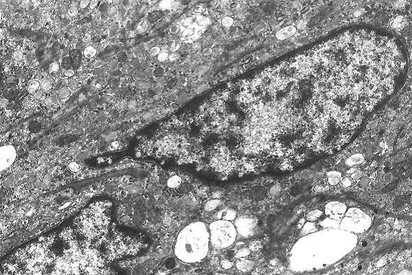
- Case 19-3. Rat A, 6100x
This electron micrograph contains portions of multiple, interdigitating
spindle cells, 2 with visible nuclei. The cells have ovoid nuclei
with dispersed chromatin and abundant cytoplasmic organelles,
and are joined by desmosomes. The most prominent organelles are
multiple, ovoid, approximately 0.3 x1.0 (single membrane-bound
structures, with multiple internal membranous filaments arranged
in parallel to the long axis [premelanosomes (stage II melanosomes)].
Other cytoplasmic organelles are present in low number and include
rough endoplasmic reticulum (RER), mitochondria and multiple
membrane-bound vacuoles with scant contents (most likely dilated
RER).

- Case 19-3. Rat A, 10200x
This electron micrograph illustrates the finer detail of the
premelanosomes as well as the multiple desmosomes between the
neoplastic cells, and absence of a basal lamina.
-
- AFIP Diagnosis: Pinna (per contributor): Spindle cells
with numerous intracytoplasmic premelanosomes and absence of
melanin, Fisher 344 rat, rodent.
-
- Conference Note: This case was chosen for the Wednesday
Slide Conference because it provided the opportunity to review,
describe and interpret an electron micrograph. Conference participants
readily identified the spindle cells as having features suggesting
melanocytic origin. Given signalment alone, they suspected amelanotic
melanoma but few were willing to make a definitive diagnosis
based on the few cells evident in the micrograph. With the additional
history of a mass on the pinna, most were confident of the diagnosis.
- The differential diagnosis of this case includes schwannoma.
Schwann cells in both rats and humans can contain melanosomes,
but they are usually surrounded by a basal lamina. Although only
two cells are present in the electron micrographs, a basal lamina
is not observed in these photos.
-
- Although amelanotic melanomas are considered rare in albino
rats (such as the Fisher 344), they are relatively common in
pigmented varieties. Along with the pinna, other common sites
for melanomas in rats include the uvea, tail, and genitalia.
-
- Contributor: Lilly Research Laboratories, PO Box 708,
Greenfield, IN 46140.
-
- References:
- 1. Nakashima N, Mitsumori K, Maita K, Shirasu Y: Amelanotic
melanocytic tumors of the pinna in six F344 rats. J Vet Med Sci
53(2):291-296, 1991
- 2. Yoshitomi K, Boorman G: Palpebral amelanotic melanomas
in F344 rats. Vet Pathol 30(3):280-286, 1993
- 3. Yoshitomi K, Boorman G: Spontaneous amelanotic melanomas
of the uveal tract in F344 rats. Vet Pathol 28(5):403-409, 1991
-
-
- Case IV - 19361 (H&E); 19361 BH (Brown-Hopps Gram)(AFIP
2681374)
-
- Signalment: 2-month-old male New Zealand white rabbit
(Oryctolagus cuniculus).
-
- History: This rabbit was purchased from a supplier
of laboratory rabbits and acclimated in the recipient animal
facility for a few days. It was in excellent condition and had
no signs of disease. The investigator withheld food for 24 hours
in preparation for surgery. The rabbit was found dead in its
cage the morning of the scheduled surgery.
-
- Gross Pathology: At necropsy, the perineum, thighs
and hocks had tan, watery fecal staining. The serosa of the cecum
had a few scattered petechiae and the wall appeared thickened.
The cecal lumen was » 1.0 to 1.5 cm in diameter and contained
pink, gelatinous mucoid material; the mucosa was
roughened and dark pink. The colon and the rectum contained minimal
fluid feces and had normal appearing mucosae. Touch preparations
were made for Gram stain of contents at three levels: cecum,
mid colon and rectum. The liver, thymus and tracheal mucosa were
reddened.
-
- Laboratory Results:
Aerobic Bacterial Cultures:
Conjunctiva- negative culture
Nasal swab: gamma Streptococcus, Staphylococcus epidermidis,
Corynebacterium sp. (non-pathogenic)
Small intestine: negative culture
Cecum: Enterococcus, Streptococcus viridans, Bacillus sp.
Colon- Enterococcus, Streptococcus viridans, Corynebacterium
sp. (non-pathogenic)
- Anaerobic Bacterial Cultures: None
- Gram stains of contents of large intestine:
Cecum: Mucoid contents contained massive numbers of large C-shaped
and coiled Gram positive rods compatible with Clostridium spiroforme
(see 2 X 2 slides from this case and those of Gram stained cecal
contents from a normal control rabbit from the same supplier).
Colon: Few large Gram positive rods compatible with Clostridium
spiroforme
Rectum: Few large Gram positive rods compatible with Clostridium
spiroforme
Contributor's Diagnosis and Comments: Necrotizing typhlitis,
peracute, severe and diffuse, compatible with Clostridium
spiroforme mediated enterotoxemia, probably provoked by extended
withholding of food.
Additional findings in this case included: (i) mild necrotizing
enteritis in the sacculus rotunda and appendix, (ii) mild acute
mesenteric lymphadenitis, (iii) severe myeloid depletion of bone
marrow and spleen, (iv) severe congestion of viscera, and (v)
moderate neuronal shrinkage and basophilia in Ammon's horn of
the dentate gyrus consistent with anoxia. All of these changes
were thought to be directly or indirectly associated with the
C. spiroforme mediated enterotoxemia.
-
- Specimens of intestine were fixed intact in alcoholic formalin
(10% formalin in 70% ethanol) to limit autolysis of the mucosa
and allow visualization in sections of the undisturbed intestinal
contents-mucosal relationships. The severe necrotizing lesions
in the cecum and similar lesions in the sacculus and appendix,
coupled with the presence of massive numbers of large curved
or coiled Gram positive bacteria typical of C. spiroforme
in the contents of the affected bowel, provided very strong presumptive
evidence of C. spiroforme mediated enterotoxemia as the
central disease process in this case. Definitive proof would
have required culture of C. spiroforme and demonstration
of C. spiroforme enterotoxin in the intestinal contents by biochemical,
immunological, cytotoxicity assay or other methods, not routinely
available in most labs. Enteritides due to aerobic bacteria were
ruled out by negative cultures. There was no morphologic evidence
of Clostridium pilifome infection in the intestine or
liver. Additionally, current health surveillance testing had
shown the stock to be serologically negative for C. piliforme
and rotavirus, negative for Lawsonia intracellularis by
PCR testing and negative for other common nonintestinal pathogens
of rabbits.
-
- C. spiroforme is considered one of the leading causes
of enteropathy in weanling rabbits. Overgrowth of C. spiroforme
and other toxin producing clostridia in the cecum (mainly) of
rabbits has been associated with numerous factors, including
weaning, dietary change and antibiotic administration. The extended
withholding of food and the young age (perhaps limited GI flora?)
of this rabbit probably were major contributing factors to the
massive overgrowth of C. spiroforme in the cecum, resulting
in a fatal case with all of the characteristic features of peracute
C. spiroforme mediated enterotoxemia.
-
- Much is known about the molecular pathogenesis of enterotoxemia
due to C. spiroforme. The toxin is a protein that consists
of two functional domains, A and B. The B domain binds to receptors
on enterocytes and delivers the toxic A moiety into the cytosol.
Toxin A is an ADP (adenosine 5'-diphosphate)-ribosyltransferase
that removes nicotinamide from a ribose of nicotine adenine dinucleotide
and attaches the ribose to Rho, small GTP (guanosine 5'-triphosphate)-binding
proteins known as Rho GTPases involved in the regulation of actin
filament assembly. Ribosylation functionally inactivates Rho
and results in depolymerization of filamentous (F) actin which,
in turn, results in loss of cell polarity and adhesion, leading
to rounding and necrosis of enterocytes. Function of the Rho
GTPases is of critical importance as they regulate a plethora
of other cell functions, including cytokinesis, phagocytic NADPH,
serum- and growth factor-mediated signaling, nuclear signaling
and induction of apoptosis. Based on studies of similar toxins
A and B of Clostridium difficile which have been studied
far more extensively, toxins A and B of C. spiroforme may inactivate
Rho through other chemical processes, and have numerous other
deleterious effects such as inducing secretion and fluid accumulation,
chemotaxis, cytokine and chemokine secretion, and many others
(see Fasano, 1999; Guerrant et al., 1999).
- AFIP Diagnosis: Cecum: Typhlitis, erosive, acute,
diffuse, severe, with loss of glands and numerous luminal coiled
Gram positive bacilli, New Zealand white rabbit (Oryctolagus
cuniculus), lagomorph.
-
- Conference Note: Conference participants agreed with
the contributor that this case probably represents an example
of enterotoxemia resulting from Clostridium spiroforme infection.
While the morphology of this organism is fairly distinctive,
participants reaffirmed the need for bacterial culture and toxin
detection for definitive diagnosis. The contributor has provided
an excellent review of the clinical and pathological findings
as well as the pathogenesis and differential diagnosis for enterotoxemia
caused by C. spiroforme.
-
- Contributor: Department of Comparative Medicine, University
of Alabama at Birmingham, Birmingham, AL 35243-0019.
-
- References:
- 1. Aktories K: Identification of the catalytic site of clostridial
ADP-riboslytransferases. Adv Exper Med Biol 419:53-60, 1997
- 2. Bouquet P, Gill DM: Modulation of cell functions by ADP-ribosylating
bacterial toxins., In: Sourcebook of Bacterial Protein Toxins.
eds. Alouf JE, Freer JH, pp. 23-44. Academic Press: San Diego,
CA, 1991
- 3. Butt MT, Papendick RE, Carbone LG, Quimby FW: A cytotoxicity
assay for Clostridium spiroforme enterotoxin in cecal fluid of
rabbits. Lab Anim Sci 44:52-54, 1994
- 4. Carman RJ, Evans RH: Experimental and spontaneous clostridial
enteropathies of laboratory and free living lagomorphs. Lab Anim
Sci 34:443-452, 1984
- 5. DeLong D, Manning PJ: Bacterial diseases, III. Enterotoxemia.
In: The Biology of the Laboratory Rabbit, eds. Manning PJ, Ringler
DH, Newcomer CE, 2nd ed., pp. 140-143. Academic Press, San Diego,
CA, 1994
- 6. Fasano A: Cellular microbiology: can we learn cell physiology
from microorganisms?: Am J Physiol 276:C765-C776, 1999
- 7. Guerrant RL, Steiner TS, Lima AAM, Bobak DA: How intestinal
bacteria cause disease. J Infect Dis 179(Suppl 2):S331-337, 1999
- 8. Holmes HT, Sonn RJ, Patton NM: Isolation of Clostridium
spiroforme from rabbits. Lab Anim Sci 38:167-168, 1988
- 9. Popoff MR, Milward FW, Bancillon B, Boquet P: Purification
of the Clostridium spiroforme binary toxin and activity of the
toxin on HEp-2 cells. Infect Immun 57:2462-2469, 1989
- 10. Rappuoli R, Pizza M: Structure and evolutionary aspects
of ADP-ribosylating toxins., In: Sourcebook of Bacterial Protein
Toxins. eds. Alouf JE, Freer JH, pp.1-21. Academic Press, San
Diego, CA, 1991
- 11. Sears CL, Kaper JB: Enteric bacterial toxins: Mechanisms
of action and linkage to intestinal secretion. Microbiol Rev.
60:167-215, 1996
- 12. Simpson LL, Stiles BG, Zepeda H, Wilkins TD: Production
by Clostridium spiroforme of an iota like toxin that possesses
mono(ADP-ribosyl)transferase activity: Identification of a novel
class of ADP-ribosyltransferases. Infect Immun 57:256-261, 1989
- 13. Yonushonis WP, Roy MJ, Carman RJ, Sims RE: Diagnosis
of spontaneous Clostridium spiroforme iota enterotoxemia in a
barrier rabbit breeding colony. Lab Anim Sci 37:69-71, 1987
-
- J Scot Estep, DVM
Captain, United States Army
Registry of Veterinary Pathology*
Department of Veterinary Pathology
Armed Forces Institute of Pathology
(202) 782-2615; DSN: 662-2615
Internet: estep@afip.osd.mil
-
- * The American Veterinary Medical Association and the American
College of Veterinary Pathologists are co-sponsors of the Registry
of Veterinary Pathology. The C.L. Davis Foundation also provides
substantial support for the Registry.
- Return to WSC Case Menu