Results
AFIP Wednesday Slide Conference - No. 16
January 12, 2000
- Conference Moderator:
Dr. Steven E. Weisbrode, Diplomate, ACVP
The Ohio State University
Department of Veterinary Biosciences
- Columbus, OH 43210
-
- NOTE: Click on images for larger views. Use
browser's "Back" button to return to this page.
Return to WSC Case Menu
-
- Case I - 99-1848 (AFIP 2679493)
-
- Signalment: One-year-old, male thoroughbred horse.
-
- History: Ataxia of one-month duration. Static narrowing
of cervical spinal canal at C3-4 seen radiographically.
-

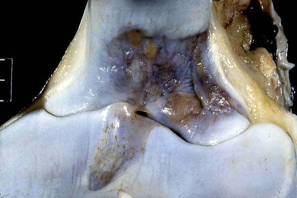
- Case 16-1. Proximal radius. Reddish-brown areas bordered
by pale gray cartilage are the synovial fossae. Indistinct vertical
streaks in the cartilage are small grooves (wear lines) which
appear histologically as areas of cartilage compression and chondrocyte
necrosis.
- Gross Pathology: Mid sagittal diameter of spinal canal
at C3-4 was 13 mm. Radial-ulnar joint had developing synovial
fossae and mild degenerative joint disease (linear "scoring").
-
- Contributor's Diagnoses and Comments: Specimen is
proximal radius. There is a focal depression in articular cartilage
and corresponding subchondral bone. Cartilage in this region
has a thickened superficial fibrous layer. In some areas, chondrocytes
in the tangential layer have a stellate or myxomatous appearance.
Multifocally there is hypocellularity or coagulation necrosis
in the radial layers. A tidemark is more prominent in cartilage
in the depression compared with adjacent cartilage, and subchondral
bone beneath the depressed cartilage is more porous. In areas
corresponding to scoring or wear lines in the non-depressed cartilage,
there is mild degenerative joint disease characterized by cartilage
hypocellularity, reactive chondrocyte clusters and variable fibrillation
of the matrix.
- 1. Normal synovial fossa
2. Mild degenerative joint disease ("wear lines")
-
- The gross and microscopic appearances of the irregular depressed
regions are characteristic of early stages of synovial fossae.
These are grooves of uncertain function found on articular surfaces
of ruminants, horses, pigs and dogs. It has been suggested that
they might act as reservoirs for synovial fluid or might reflect
disuse atrophy in non-weight bearing regions of the joint. Although
in some locations in cattle these fossae begin to develop prenatally,
in most species and locations, they are not apparent at birth
but progressively develop in the first several months of life.
With complete development, the articular cartilage of the fossa
is replaced by fibrous tissue which directly communicates with
the marrow through the relatively porous subchondral bone. The
scoring or wear lines in the gross photo are common in the elbow
joints of horses and are often subclinical. They have been described
as being associated with cases of cervical vertebral stenotic
myelopathy (as in the current case). It is speculated that ataxia
might cause increased turbulence of synovial fluid resulting
in these wear lines.
-
- AFIP Diagnoses:
- 1. Bone, articular cartilage, superficial and tangential:
Chondrocyte necrosis, multifocal, thoroughbred, equine.
2. Bone, articular cartilage: Synovial fossa (normal)
-
- Conference Notes: Histologically the score lines that
are visible adjacent to the fossa are characterized by focal
areas of chondrocyte necrosis. Although grossly there are visible
and generally palpable depressions or scores, histologically
the cartilage is not scored and is only intermittently depressed
over these areas. Physiologically these depressions are caused
by necrosis of chondrocytes resulting in decreased glycosaminoglycans
that in turn results in decreased water binding and subsequent
cell shrinkage. Processing frequently obscures this subtle lesion.
-
- Contributor: Department of Veterinary Biosciences,
The Ohio State University, 1925 Coffey Road, Columbus, OH 43210
-
- References:
- 1. Palmer N: Bones and Joints. In: Pathology of Domestic
Animals, eds. Jubb KVR, Kennedy PC, Palmer N, 4th ed., vol. 1,
p. 144. Academic Press, San Diego, 1993
- 2. Rooney JR, Robertson JL: Foreleg. In: Equine Pathology,
p. 155. Iowa State University Press, Ames, IA, 1996
- 3. Wagner KM, Heje N-I, Aarestrup FM, Ravn BT, and Osterby
J: The morphology of synovial grooves (Fossae synoviales) in
joints of cattle of different age groups. J Vet Med Assoc 40:359,
1993
-
-
- Case II - 99-1413 (AFIP 2693021)
-
- Signalment: 4-year-old Scottish terrier, male, castrated
dog
-
- History: Presented with a 2cm long oral mass of the
left rostral mandible surrounding and caudal to the canine tooth.
The surgeon's report indicated the mass "shelled out".
There has been no recurrence 1.5 mos. after surgery.
-

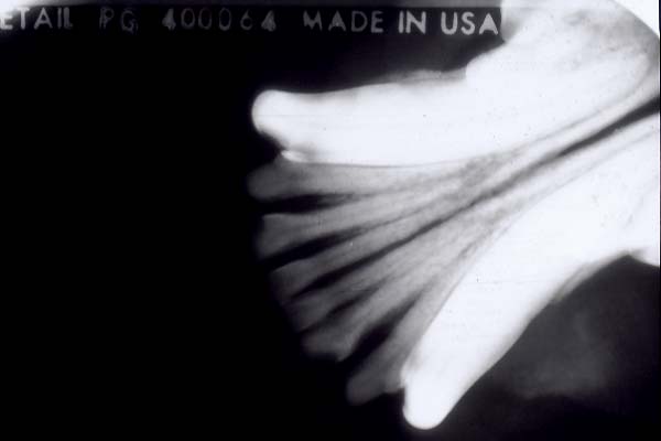
- Case 16-2. Radiograph of mandible. A nodular mass
of low radiodensity extends from the lateral aspect of the mandible
(lower right corner), but does not deform it.
-
- Gross Pathology: Three, white, firm to hard, smooth
multilobulated fragments of the gingival mass measure up to 3
X 1.7 cm.
-
- Laboratory Results: Radiograph of left mandibular
ramus (see 2x2) illustrates a soft tissue mass with subtle mottling;
there is a smooth border along the mandible-tumor interface.
Interpretation by Drs. Gregory Daniel and Kari Anderson: "Left
rostral mandibular soft tissue neoplasia with no evidence of
bony destruction. Primary consideration is given to ossifying
epulis".
-
- Contributor's Diagnosis and Comments: Aggressive osteoblastoma,
left mandible, canine.
-
- The non-decalcified, rounded mass has 3 remarkable elements
consisting of more superficial osteoblasts merging with more
and more mineralized and non-mineralized matrix deeper in the
tumor: The outer portion is more cellular as it is dominated
by epithelioid osteoblasts; these cells surround mineralized
bony fragments which gradually become branching trabeculae with
limited lattice formation deep in the mass; fragments and trabeculae
often have wide osteoid seams. A hyaline matrix (osteoid), sometimes
stippled with mineralization is adjacent to foci of confluent
mineralization. Very occasional deep trabeculae contain chondrocytes
(not in all sections).
-
- The epithelioid component has cells with a variable amount
of lightly eosinophilic to basophilic cytoplasm with discrete
margins in some regions. Nuclei of vary in size, and are oval
to irregular with fine chromatin and indistinct nucleoli. Very
occasional normal mitotic figures are present. The nuclei of
these cells become more elongate and their cytoplasm less conspicuous
as they are surrounded by more and more hyaline matrix (osteoid).
The most discrete epithelioid cells are surrounded by a deeply
basophilic material (mucin). Occasional multinucleated giant
cells (with oval nuclei resembling those of the epithelioid population
but smaller) are present within the trabecular region usually
unassociated with the surfaces of the trabeculae.
-
- The tumor has undergone coagulation necrosis in a region
at the periphery of the basal-most portion. Tumor extends to
the surgical margin. The original diagnosis was parosteal osteogenic
sarcoma reflecting Roy Pool's description (not the Atlas of Tumor
Pathology's description) in order to emphasize an expected less
aggressive behavior. The AFIP fascicle describes osteoma, osteoid
osteoma and osteoblastoma. This tumor somewhat resembles a variant
of osteoblastoma, the aggressive osteoblastoma. This tumor is
not an osteoma which usually has well-formed trabeculae surrounded
by pavemented osteoblasts; trabecular spaces in an osteoma typically
contain fibrovascular tissue, not the epithelioid cells seen
here. Cellular atypia and bony invasion were not present to suggest
the osteoblastic variant of osteogenic sarcoma.
-
- Note: 8 months following the original surgery the
mass recurred and a hemimandibulectomy was performed. A smooth
firm sessile mass (1.5 x 1.2 cm) was present caudal and medial
to the canine and surrounding the first premolar. A similar mass
1.5 x .6 cm mass was located lateral to the canine tooth. Histologically
the mass is very similar to the original biopsy.
-
- AFIP Diagnosis: Gingiva: Benign fibro-osseous neoplasm,
Scottish terrier, canine.
-
- Conference Notes: This unusual lesion was the subject
of a lengthy discussion. While this lesion is moderately cellular,
has multifocal mild nuclear atypia and occasional mitotic figures,
the small size, absence of bone destruction and absence of greater
atypia are more consistent with a benign process. The Departments
of Orthopedic Pathology and Oral Pathology were consulted and
both favor a benign process. The Department of Orthopedic Pathology
noted that "the absence of vascularity, osteoclastic and
blastic activity does not fit an osteoblastoma". Conference
participants and the 2 consulted departments could not definitively
determine whether the lesion was purely peripheral or intraosseous
with perforation. The Department of Oral Pathology favors a variant
of ossifying fibroma if the lesion is central and peripheral
ossifying fibroma if the lesion is peripheral.
- Contributor: Department of Pathology, College of Veterinary
Medicine, University of Tennessee, Knoxville, TN 37901-1071
-
- References:
- 1. Fechner RE, Mills SE: Atlas of Tumor Pathology, Third
Series, Fascicle 8, Tumors of the Bones and Joints, pp. 26-38.
Armed Forces Institute of Pathology, Washington, DC, 1993
- 2. Hoffman S, Jacoway JR, Krolls SO: Atlas of Tumor Pathology,
Second Series, Fascicle 24, Intraossoeous and Parosteal Tumors
of the Jaw, eds Hartmann WH, Sobin LH, pp. 203-216. Armed Forces
Institute of Pathology, Washington, DC, 1985
- 3. Pool RR: Tumors of Bone and Cartilage In: Tumors in Domestic
Animals, ed. Moulton JE, 3rd ed., pp. 159-163;193-194;222-225.
University of California Press, Berkeley, CA, 1990
-
-
- Case III - MP9B (AFIP 2679485)
-
- Signalment: Adult, male, C57BL6 mouse (Mus musculus)
-
- History: Eleven days following experimental, intravenous
inoculation with Mycoplasma pulmonis, the mice exhibited lameness
with reddening and swelling around multiple synovial joints of
the limbs, and edema of the hind feet.
-
- Gross Pathology: Purulent fluid was in multiple joint
spaces. The periarticular tissue of affected joints was swollen
and edematous.
-
- Contributor's Diagnosis and Comments: Tarsus: marked,
suppurative polyarthritis and chronic active periarthritis with
synovial proliferation, osteolysis, and subperiosteal bone proliferation.
-
- Etiology: Mycoplasma pulmonis
-
- The section consists of a decalcified sagittal section through
the tarsus. Affected joints have distended synovial spaces containing
proteinaceous material and degenerate neutrophils. Proliferative
synovial cells lined the thickened synovial membrane, often interrupted
by areas of erosion. The periarticular soft tissue contained
organized layers of histiocytic and neutrophilic inflammation
with scattered focal accumulation of degenerate neutrophils into
microabscesses. Early fibroplasia of the synovia and fascia was
suggestive of burgeoning ankylosis. Articular inflammation frequently
undermined articular cartilage, destroyed subchondral bone, and
entered the metaphyseal medullary cavity. Lytic areas in metaphyses
were filled with histiocytic inflammation and proliferative fibrocartilage,
and also lined by new cancellous bone. Along the periosteal surface
of cortical bone adjacent to the joints, there was prominent
reactive bone proliferation. Chronic, mixed inflammation of tendons,
entheses, and tendon sheaths was also present.
-
- Mycoplasmas are responsible for multiple, naturally occurring
inflammatory diseases including pneumonia, polyserositis, and
arthritis in many species, including rodents. Mycoplasma arthritidis
is the agent typically responsible for mycoplasmal polyarthritis
in the rat and mouse. Experimental polyarthritis similar to that
produced by M. arthritidis can be produced by Mycoplasma
pulmonis in the mouse by intravenous inoculation, although M.
pulmonis is usually responsible for lymphocytic pneumonia
in the rodent. By intravenous exposure, the pathogenesis and
outcome of the arthritic lesion of M. pulmonis is similar
to that of M. arthritidis (Lindsay, 1978). In general,
infected mice fail to clear the mycoplasma organism and develop
chronic proliferative arthritis characterized by periods of remission
and exacerbation (Kono, 1980).
-
- Histologically, an acute phase (1 week post inoculation)
consists of suppurative inflammation and edema of the articular
and periarticular tissues with sporadic tendonitis. Two to three
weeks post inoculation, affected joints display acute and chronic
features of the inflammatory process, and by 4 weeks and onward,
the chronic phase consists of hyperplasia of the synovial membrane,
mononuclear cell infiltration, granuloma formation, pannus, destruction
and proliferation of subchondral cortical bone, and destruction
of articular cartilage (Harwick, 1976). This pattern resembles
the lesions of rheumatoid arthritis in humans, for which this
experimental disease is a suitable model.
-
- AFIP Diagnosis: Tarsus: Synovitis and tenosynovitis,
chronic, suppurative, multifocal, moderate, with pannus and osteophytes,
C57BL6 mouse (Mus musculus), rodent.
-
- Conference Notes: Conference participants favored
Mycoplasma sp. as the most likely etiology of this lesion. Participants
discussed the need to exclude other possible causes including
bacterial and viral infections and autoimmune diseases.
-
- Contributor: Lilly Research Laboratories, PO Box 708,Greenfield,
IN 46140
-
- References:
- 1. Harwick HJ, Mahoney AD, Kalmanson GM, Guze LB: Arthritis
in mice due to infection with Mycoplasma pulmonis. II. Serological
and histological features. J Infect Dis 133(2):103-112, 1976
- 2. Kono M, Tanaka H, Yayoshi M, Araake M, Yochioka M, Imai
M: Mycoplasma pulmonis arthritis in congenitally athymic (nude)
mice. Histologic features. Microbiol Immunol 24(5):381-391, 1980
- 3. Lindsay JR, Cassell GH, Baker HJ: Diseases due to Mycoplasmas
and Rickettsias. In: Pathology of Laboratory Animals, eds. Benirschke
K, Garner FM, Jones TC, vol II, pp. 1507-1513. Springer-Verlag,
New York, 1978
- 4. Palmer N: Bones and Joints. In: Pathology of Domestic
Animals eds. Jubb KVR, Kennedy PC, Palmer N, 4th ed., vol. 1,
p. 144. Academic Press, San Diego, 1993
-
-
- Case IV - 8903/99 (AFIP 2698157)
-
- Signalment: A 42-day-old male Ross broiler chicken.
-
- History: This bird was culled due to lameness and
submitted with other lame broilers to the laboratory for necropsy.
-
- Gross Pathology: On examination of a mid-line frontal
section of the proximal tibiotarsus, a small zone of pale/yellow
tissue was observed in the region of the growth plate.
-
- Laboratory Results: A profuse growth of Staphylococcus
aureus was recovered following bacterial culture of one half
of the affected proximal tibiotarsus.
-
- Contributor's Diagnosis and Comments: Bacterial chondronecrosis
of the physeal cartilage with osteomyelitis due to infection
with S. aureus.
-
- The pale stained physeal chondrocytes and matrix may indicate
the edge of a chondronecrotic lesion that contrasts with the
dark blue, normally stained zone. Note also clumps of bacteria
closely associated with necrotic cartilage and accumulating in
the metaphyseal blood vessels. This condition is a common cause
of lameness in fast-growing broiler chickens. It is most frequently
detected in the proximal femur and proximal tibiotarsus.
-
- AFIP Diagnosis: Tibiotarsus: Osteomyelitis, necrotizing,
heterophilic, diffuse, moderate, with focally extensive physitis
and epiphysitis, necrotizing vasculitis, congestion, hemorrhage,
and numerous colonies of cocci, Ross broiler chicken, avian.
-
- Conference Notes: Bacterial osteomyelitis affecting
the proximal tibiotarsus and the distal femur is an important
cause of lameness in commercial poultry. Staphylococcus aureus
is the most common cause of chondronecrosis and osteomyelitis
in chickens, although Mycoplasma sp, Escherichia coli, Staphylococcus
xylosus, and other bacteria have been isolated from affected
birds. McNamee et al reported an increased frequency of lesions
in association with high nutritional levels and dual infection
by chicken anemia virus and infectious bursal disease virus,
thus providing a model for study of the natural disease.
A possible pathogenesis for localization of infection in the
tibia and femur is deposition of bacteria in the growing ends
of metaphyseal vessels following development of a continuous
bacteremia after a primary infection of the respiratory tract.
Predisposing factors discussed in conference include lack of
anastomoses in metaphyseal vessels, an incomplete endothelial
lining,and locally decreased phagocytic activity.
-
- Contributor: Veterinary Sciences Division, 43 Beltany
Road, Omagh, N. Ireland BT78 5NF
-
- References:
- 1. McNamee PT, McCullagh JJ, Thorp BH, Ball HJ, Graham DA,
McConaghy D, Smyth JA: A longitudinal investigation of leg weakness
in two commercial broiler flocks. Vet Rec 143:131-135, 1988
- 2. McNamee PT, McCullagh JJ, Thorp BH, Ball HJ, Connor T,
McConaghy D, Smyth JA: Development of an experimental model of
bacterial chondronecrosis in broilers following exposure to Staphylococcus
aureus by aerosol, and inoculation with chicken anaemia and infectious
bursal disease viruses. Avi Pathol 28:26-35, 1998
-
- J Scot Estep, DVM
Captain, United States Army
Registry of Veterinary Pathology*
Department of Veterinary Pathology
Armed Forces Institute of Pathology
(202)782-2615; DSN: 662-2615
Internet: estep@afip.osd.mil
-
- * The American Veterinary Medical Association and the American
College of Veterinary Pathologists are co-sponsors of the Registry
of Veterinary Pathology. The C.L. Davis Foundation also provides
substantial support for the Registry.
- Return to WSC Case Menu