Results
AFIP Wednesday Slide Conference - No. 13
December 1, 1999
-
- Conference Moderator:
LTC Jeff Eggers, Diplomate, ACVP
Chief Veterinary Pathology, CID
59th MDW/HSR
1255 Wilford Hall Loop, Bldg 4430
Lackland AFB, TX 78236-5319
-
- NOTE: Click on images for larger views. Use
browser's "Back" button to return to this page.
Return to WSC Case Menu
-
- Case I - UFSM # 1 (AFIP 2559049)
-
- Signalment: 4-year-old zebu cross, female.
-
- History: This animal is from a group of 19 young adult
cows that were in a pasture heavily infested with Tetrapterys
multiglandulosa. The owner was advised of this being a poisonous
plant, but still chose to leave the animals in the pasture. After
40 days he noticed that 4 cows were sick. There was subcutaneous
dependant edema (submaxilar, ventral neck, and brisket), depression,
and the animals tired easily. After 15 days, the cows were removed
from the pasture but even then the clinical signs became progressively
worse and the 4 cows died. Subcutaneous edema extended up to
the ventral abdomen (kodakchrome A); the jugular vein was distended
and pulsating and there was cardiac arrhythmia. After exercise
the cows became breathless.
-
- Gross Pathology: An extensive translucent gelatinous
dependent subcutaneous edema was confirmed at necropsy. Marked
cavitary edema (ascites, hydropericardium, and hydrothorax) and
edema of the mesentery were also noticed. Discolored pale areas
were seen in the myocardium through the epicardium and sharp
white patchy areas and streaks (kodakcrome B) were present on
the cut surface of the myocardium. The liver was slightly swollen
and had a nutmeg appearance particularly on the cut surface.
-
- Contributor's Diagnoses and Comments: Morphologic
diagnosis:
Myocardium, necrosis of myofibers and fibrosis, multifocal, severe.
- Etiologic diagnosis: Toxic cardiomyopathy
- Etiology: Tetrapterys multiglandulosa toxin
-
- Other histopathological changes included centrilobular congestion
and fibrosis (congestive heart failure) of the liver.
-
- The ingestion of plants of the genus Tetrapeterys, family
Malpighiaceae, causes primarily degenerative and fibrotic changes
in the myocardium, and related signs of cardiac failure. The
natural disease occurs only in cattle over 1 year of age in southeastern
Brazil and only in pastures where Tetrapterys spp. occur and
after the cattle have stayed for 1 to 2 months on the pasture;
it was experimentally reproduced in cattle by feeding fresh or
dried sprouts of T. acutifolia and T. multiglandulosa
(3). Most of the cases have subacute (1-5 weeks) or chronic (months)
courses. Most common clinical signs include: edema of the brisket,
prominent pulsating jugular vein and cardiac arrhythmia; in the
terminal stages dyspnea and labored breathing are noticed. The
natural disease occurs throughout the year with no seasonal preference,
has variable morbidity rates, and high mortality. Occasionally
some animals may recover; pregnant cows may abort. The main necropsy
findings include: subcutaneous and cavitary edema; pale areas
in the heart, perceptible through the epicardium; and sharp whitish
areas and streaks across most of the cut surface of the myocardium.
When present, liver lesions are mild and related to congestive
heart failure (nutmeg liver). The main histopathological findings
are degeneration and lysis of myofibers marked massive necrosis
of myocardium and fibrosis. Fibrosis occurs with a diffuse interstitial
distribution or as extensive focal areas. Tetrapterys spp. poisoning
must be differentiated from other primary or secondary cardiomyophies
of cattle (1) such as high altitude (brisket) disease, St. George
disease, and another disease with similar clinical signs (swollen
brisket disease) (2) which occurs in the south of Brazil. These
3 diseases can be differentiated by epidemiological data. The
first two are not reported in Brazil. Brisket disease presents
changes (medial thickening) in the arteries and arterioles of
the lungs and is usually reported on cattle above 2,000m while
Tetrapterys spp. poisoning occur in cattle pasturing at 20-700m
and only where Tetapterys spp. can be found. Other cardiomiopathies
such as those induced by ionophore antibiotic (eg. monensin,
salinomycin, lasalocid) poisoning should be included in the differential
diagnosis.

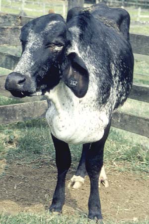
- Case 13-1. Antemortem photo. There is a pendulous
and multinodular enlargement of the brisket area. (Image
provided by Colegio Brasileiro de Patologia Animal, Seropedica,
RJ, Brazil, with permission).

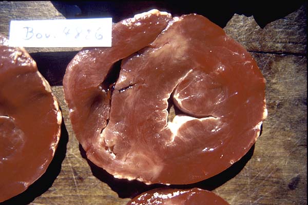
- Case 13-1. Cardiac muscle. There are two focally extensive
pale, white, areas in the left ventricular wall. (Image
provided by Colegio Brasileiro de Patologia Animal, Seropedica,
RJ, Brazil, with permission).
-
- AFIP Diagnoses:
- 1. Heart, cardiomyocytes: Vacuolar degeneration, diffuse,
moderate, with multifocal necrosis, fibrosis, mineralization,
and chronic inflammation, zebu cross, bovine.
2. Heart, cardiomyocytes: Sarcocysts, few.
-
- Conference Note: Although the specific etiology of
this case is not found outside of southern Brazil, lesions of
myocardial degeneration, necrosis and fibrosis occur in a variety
of conditions including Vitamin E/Selenium deficiency, and various
toxicities including those caused by gossypol, monensin and other
ionophore antibiotics, Cassia occidentalis (coffee senna), Eupatorium
rugosum (white snake root), Karwinskia humboldtiana (coyotillo),
fluoroacetate containing plants (Acacia georginea, Gastrobilium
spp, Oxylobium spp.), and others. Other causes of similar lesion
include uremic cardiomyopathy and ischemia. Storage diseases
such as generalized glycogenosis due to deficiency of 1,4-glucosidase
may cause diffuse cardiomyocyte vacuolization.
-
- Contributor: Universidade Federal de Santa Maria,
Departamento de Patologia, 97119-900, Santa Maria, RS, Brazil
-
- References:
- 1. Peixoto PV, Loretti AP, Tokarnia CH: "Doenca do Peito
inchado", Tetrapterys spp. poisoning, brisket disease and
St. George disease: A comparative study. Pesq Vet Bras 15:43-50,
1995
- 2. Robinson WF, Maxie MG: The Cardiovascular System In: Pathology
of Domestic Animals, 4th ed., vol. 3, eds. Jubb KVF, Kennedy
PC, Palmer NC, pp. 27-31. Academic Press, San Diego, CA, 1993
3. Tokarnia CH, Gava A, Peixoto PV, Stolf L, Moaraes, SS: A "doenca
do Peito inchado" (edema da regiao esternal) em bovinos
no estado de Santa Catarina. Pesq. Vet. Bras. 9:73-83, 1989
- 4. Tokarnia CH, Peixoto PV, Doebereiner J, Consorte LB: Gava
bovinos caracterizadas por alteracoes cardiacas. Pesq Vet Bras
9:23-44, 1989
-
-
- Case II - OL 8257 (AFIP 2694777)
-
- Signalment: 48-week-old, male, Charles River CD rat.
-
- History: The rat was part of an experimental study.
It was administered a test compound for four days. On the fourth
day, the rat was found dead and was necropsied.
-
- Gross Pathology: Kidneys were moderately enlarged
and pale with many pinpoint white spots on the capsular surface
and many white streaks on the cut surface, apparently following
nephrons.
Contributor's Diagnoses and Comments:
- 1. Kidney: Severe, diffuse, acute, tubular degeneration and
necrosis with intratubular, birefringent, acicular crystals.
2. Kidney: Mild, diffuse chronic progressive nephropathy ("old
rat nephropathy").
-
- Etiology: xemilofiban
Pathogenesis: Administration of large quantities of xemilofiban,
forming crystalline precipitates in renal tubules and leading
to obstruction of urine flow and tubule degeneration and necrosis.
-
- Note: To preserve the water soluble crystals, a modified
hematoxylin and eosin stain was performed, involving less time
in hematoxylin and eosin and minimal washing steps.
-
- Diffusely throughout the section, proximal tubules are mildly
to moderately dilated. There is marked, diffuse degeneration
and necrosis of proximal tubular epithelial cells with accumulation
of small amounts of cellular debris in dilated tubules. Scattered
tubules are lined by denuded basement membranes, which are occasionally
partly mineralized. In scattered tubules, there are variable
quantities of birefringent, refractile, acicular, 10-50 microns
long, clear to brown crystals. Occasional crystals are present
in the center of small aggregates of inflammatory cells, consisting
of a mixture of macrophages, lymphocytes, plasma cells, and neutrophils.
Most glomerular urinary spaces are mildly to moderately dilated.
In addition, there are scattered regenerative ("basophilic")
tubules and a few scattered small interstitial foci of lymphocytes,
plasma cells, and macrophages. Glomerular tufts have segmental
thickening of glomerular basement membranes and mesangial matrix
with mild to moderate segmental thickening of Bowman's capsule
with mild to moderate hyperplasia and hypertrophy of parietal
cells.
-
- The compound administered to the rat is a fiban known as
xemilofiban, the development of which is currently discontinued.
This compound is an ester prodrug, which is absorbed after oral
administration and is de-esterified to its pharmacologically
active acid.
-
- Infrared spectroscopy, Ramen spectroscopy, time-of-flight
secondary ion mass spectroscopy (TOF-SIMS), and liquid chromatography/mass
spectroscopy (LC/MS) were used to identify the crystals in situ.
These techniques confirmed that the intratubular crystals were
composed of the acid metabolite of xemilofiban.
-
- The fibans are a group of compounds that block the glycoprotein
IIb-IIIa receptor on platelets. This receptor ordinarily binds
fibrinogen, the final step of platelet aggregation, allowing
platelets to aggregate in vivo and in vitro. The fibans reversibly
block the receptor in a concentration-dependent way, causing
a titratable inhibition of platelet aggregation.
Platelets of humans and dogs are highly sensitive to the effects
of fibans. In dog toxicity studies, the limiting toxicological
findings were intramural hemorrhages in the stomach and urinary
bladder and/or gingival bleeding. The fibans evaluated did not
cause obstructive nephropathy in dogs. However, rat platelets
are comparatively insensitive to the fibans. Therefore, higher
dosages are needed to produce toxicity in rats, and compound-related
bleeding does not occur. The principal toxicity of these compounds
in rats, at large multiples of human dosages, was the formation
of crystalline precipitates in kidneys. These precipitates lead
to obstruction of urine flow, varying degrees of renal inflammation,
tubular degeneration, and secondary uremia.
Differential diagnosis for crystalline precipitates in renal
tubules includes uric acid, sulfonamide, acyclovir, methotrexate,
and oxalate crystals. Presence of uric acid crystals in renal
tubules results from the destruction of cells, especially erythrocytes
in newborn animals. Collecting tubules are filled with yellow
crystals and their presence may give a radial pattern to yellowish
streaks. Uric acid crystals may also appear in gout (especially
in birds), where they may be found in the interstitial stroma
with a radial pattern.
Various drugs, such as acyclovir, methotrexate, primadone, and
sulfonamides, can cause acute renal failure through the deposition
of crystalline material in renal tubules. Incidence of intraluminal
sulfonamide crystals was greater when only less-soluble forms
of the drug (sulfapyridine, sulfathiazole, and sulfadiazine)
were available. This problem is currently rare, since newer sulfonamides
are short acting and have greater solubility. The incidence of
sulfonamide crystal precipitates is exacerbated by limited intake
of water and acidic urine. The typical lesion is a plugging of
the lower collecting tubules with masses of fine crystals, resulting
in obstruction. The lesions are due to local toxic and mechanical
effects. Hypersensitivity does not play as important a role in
animals as it does in humans. The crystals of most of the sulfonamides
are elongated, acicular, anisotropic, and yellowish in color.
-
- Oxalate crystals in renal tubules can occur from ingesting
plants with toxic levels of oxalate, ethylene glycol, oxalate
salts, or plants infected with fungi that produce oxalates. Plants
are the usual source of oxalate poison in sheep and cattle. Some
of the common plants that contain oxalates are rhubarb (Rheum
rhaponticum), halogeton (Halogeton glomeratus) and greasewood
(Sacobatus vermiculatus), common sorrel (Rumex acetosa), pigweed
(Amaranthus retroflexus), mangles, and sugar beets. Ethylene
glycol is the usual source of oxalate poisoning in humans, dogs,
and cats. It is metabolized in the liver by alcohol dehydrogenase,
where it is oxidized to glycolic acid, then to glyoxylate, and
finally to oxalate. Calcium oxalate is precipitated in renal
tubules during the process of clearance. The crystals are nearly
transparent, birefringent, irregularly rhomboidal, and 30-40
microns long.
-
- AFIP Diagnoses:
- 1. Kidney: Tubular ectasia, diffuse, moderate, with intratubular
birefringent acicular crystals, multifocal tubular degeneration
and necrosis, and subacute tubulointerstitial nephritis, Charles
River CD rat, rodent.
2. Kidney: Nephropathy, mild, characterized by glomerular and
tubular basement membrane thickening, chronic interstitial nephritis,
and tubular regeneration (chronic nephropathy of rats).
-
- Conference Note: The contributor has provided an excellent
discussion of both the pathogenesis of fiban toxicity and the
differential diagnosis for crystalline precipitates in renal
tubules. Conference participants agreed with the contributor's
morphologic diagnosis. A discussion of the differential diagnosis
and pathogenesis of nephrosis ensued. This was followed by a
brief review of the characteristic features of chronic progressive
nephropathy in rats ("old rat nephropathy"). These
features include interstitial fibrosis, chronic inflammation,
tubular ectasia and proteinosis, glomerular basement membrane
thickening and mesangial proliferation. Participants remarked
that this case provides an excellent example of a more acute
lesion superimposed on more chronic lesions.
-
- Contributor: Searle/Monsanto, 4901 Searle Parkway,
Skokie, IL 60077
-
- References:
- 1. Jones TC, Hunt RD, King NW: Veterinary Pathology, 6th
ed., pp. 1131-1132, pp. 724-726. Williams & Wilkins, Baltimore,
MD, 1997
- 2. Maxie GM: The Urinary System. In: Pathology of Domestic
Animals, eds. Jubb KVF, Kennedy PC, Palmer N, 4th ed., vol. 2,
pp. 490-493. Academic Press, San Diego, CA, 1993
- 3. Levin S, Friedman RM, Cortez E, Hribar J, Nichols M, Schlessinger
S, Fouant M, Khan N: Lesion and identification of crystalline
precipitates of glycoprotein IIb-IIa antagonists in the rat kidney.
Tox Pathol 27:38-43, 1999
- 4. Perazella, Mark MD: Crystal-induced acute renal failure.
Am Journ of Med 106:459-465, 1999
-
-
- Case III - 99T6-20 (AFIP 269553)
-
- Signalment: Young adult, Xenopus laevis (African
clawed frog), female
-
- History: Several frogs had been found dead. Others
appeared to have increased shedding and external mucus production.
Aeromonas hydrophilia was isolated from external lesions. Ova
from frogs are used for calcium channel studies.
-
- Gross Pathology: Skin is rough and reddish over abdomen
and legs. Frogs do not change pigment/coloration in response
to light.
-
- Laboratory Results: Aeromonas hydrophilia pure culture
-
- Contributor's Diagnosis and Comments: Skin: Dermatitis,
subacute, multifocal with nematodes consistent with Pseudocapillaroides
(Capillaria) xenopi.
-
- These laboratory-raised frogs are housed in a state-of-the-art
aquarium system that incorporates filtration and water changes.
They had been at the institution for 8 months. Frogs from the
same tank necropsied 2 months before had dermatitis but no evidence
of nematodiasis. Tank treatment with levamisole (2 times, 2 weeks
apart) resulted in no further evidence of infection.
-
- AFIP Diagnosis: Skin: Hyperplasia, epithelial, diffuse,
moderate, with mild chronic-active dermatitis and many intraepithelial
aphasmid nematodes, African clawed frog (Xenopus laevis),
amphibian.
Conference Note: Conference participants unanimously agreed
with the contributor's identification of Capillaria xenopodis
(Pseudocapillariodes xenopi). It is the only nematode
parasite reported in the epidermis of African clawed frogs. The
key histologic features of this aphasmid parasite include: stichosomes
(large round basophilic structures) thin cuticle, thin to inapparent
musculature, and digestive tract lined by many uninucleate cells.
Only a few affected animals in the population show the clinical
signs of rough skin, anorexia and emaciation. Histologically,
there is epidermal hyperplasia and mild inflammation. This epidermal
hyperplasia significantly limits the normal epidermal functions
of respiration and osmotic balance. Generally animals waste away
for 2-3 months and eventually succumb to bacterial septicemia,
most commonly caused by Aeromonas hydrophila.
-
- Other cutaneous pathogens of frogs include Citrobacter
freundii, Flavobacterium, chytridiomycosis, chromomycosis,
Chlamydia, and Mycobacterium.
-
- Other nematode parasites that localize in epithelium include:
Trichosomoides crasicauda in the urothelium of rats, Gongylonema
sp. in various locations including the esophagus of cattle and
primates, Anatrichosoma sp. in the nasal mucosa of primates,
and Capillaria bohmi in the nasal mucosa of foxes.
-
- There are many other significant species of Capillaria. C.
hepaticum infects the hepatic parenchyma of rodents and rarely
dogs. C. aerophilus infects the nasal passages of many
animals. C. feliscati infects the urinary tract of cats.
C. micronata infects the urinary tract of minks. C.
putorii infects the stomach of cats.
Contributor: Air Force Research Laboratory, 2509 Kennedy
Circle, Brooks AFB, TX 78235-5118
-
- References:
- 1. Crawshaw GJ: Amphibian Medicine. In: Kirk: Current Veterinary
Therapy XI Small Animal Practices, pp. 1219-1230. WB Saunders,
Philadelphia, PA, 1992
- 2. Barker IK, Dreumel AAV, Palmer N: The Alimentary System.
In: Pathology of Domestic Animals, eds. Jubb KVF, Kennedy PC,
Palmer NC, 4th ed., vol. 2, p. 269. Academic Press, San Diego,
CA, 1993
- 3. Dungworth DL: The Respiratory System. In: Pathology of
Domestic Animals, eds. Jubb KVF, Kennedy PC, Palmer NC, 4th ed.,
vol. 2, p. 562. Academic Press, San Diego, CA, 1993
- 3. Kelly R: The Liver and Biliary System In: Pathology of
Domestic Animals, eds. Jubb KVF, Kennedy PC, Palmer NC, 4th ed.,
vol. 2, p. 376. Academic Press, San Diego, CA, 1993
- 4. Maxie MG, Prescott JF: The Urinary System In: Pathology
of Domestic Animals, eds. Jubb KVF, Kennedy PC, Palmer NC, 4th
ed., vol. 2, p. 518. Academic Press, San Diego, CA, 1993
- 5. Ruble G, Berzins IK, Huso DL: Diagnostic Exercise: Anorexia,
Wasting, and Death in South African Clawed Frogs. Lab Ani Sci,
45(5):592-594, 1995
-
-
- Case IV - 99250 (AFIP 2699550)
-
- Signalment: 2-year-old, male New Zealand white rabbit,
Oryctolagus cuniculus.
-
- History: This animal received an intradermal injection
along the dorsal midline of 20.0 mg purified brown recluse spider
toxin (venom) as part of a spider bite therapy study. The submitted
sections of skin were taken 48 hrs post-injection.
-
- Gross Pathology: A 6 cm X 5 cm irregularly shaped,
pale blue to blue-black mottled area of skin was present on the
dorsal midline. Mild erythema and dermal and subcutaneous edema
extended ventrally from the central lesion.
Contributor's Diagnosis and Comments: Haired skin and
subcutis: Necrosis and hemorrhage, focally extensive, severe
with necrotizing vasculitis, fibrin thrombi, collagen degeneration
and edema.
The lesion is typical of "necrotic arachnidism" associated
with the bite of the North American brown recluse spider (Loxosceles
reclusa). Bites from brown recluse spiders are common and
show considerable variation in clinical presentation. The early
lesion is characterized by erythema, pruritus, pain, swelling
and blister formation. In more severe cases, localized necrosis
develops with a central area of deep blue to purple discoloration
surrounded by a red rim of erythema. The lesion spreads in a
gravity-dependent manner and eventually progresses to form a
disfiguring dark black eschar.
-
- Brown recluse venom is cytotoxic and hemolytic. Several enzymes
have been identified in the venom including lipases, hyaluronidase,
alkaline phosphatase, and most importantly sphingomyelinases.
Sphingomyelinases are primarily responsible for both tissue necrosis
and complement-dependent hemolysis. The pathogenesis is thought
to involve intravascular coagulation locally at the site of the
bite followed by necrosis and an influx of PMNs. Endothelial
damage and thrombosis of small capillaries have been demonstrated
ultrastructurally as early as 3 hours after envenomation.
-
- More recent studies have shown Loxosceles venom induces E-selectin
expression on endothelial cells and stimulates endothelial production
of NF-[Kappa]B-dependent proinflammatory chemokines IL-8, GM-CSF,
and GRO [Alpha]. IL-8 is a potent neutrophil agonist, and parenteral
infusion of IL-8 neutralizing antibody has been shown to attenuate,
but not eliminate lesion development experimentally. Leukocyte-derived
inflammatory mediators likely play an important role in the continued
progression and refractory nature of these lesions.
-
- Histologic evaluation of chronic lesions in people reveals
persistent arterial wall necrosis, subcutaneous fat necrosis
and residual scarring of the dermis and subcutis.
-
- In addition to local tissue necrosis, systemic signs are
occasionally reported following brown recluse spider bite, most
commonly headache, joint pain, abdominal pain and vomiting. "Systemic
loxoscelism" refers to a rare, severe systemic reaction
usually occurring several days after envenomation. This reaction
is characterized by massive hemolysis, acute renal failure, thrombocytopenia,
and can progress to disseminated intravascular coagulation, coma
and death, particularly in young children.
-
- AFIP Diagnosis: Haired skin and subcutis: Necrosis
and hemorrhage, focally extensive, severe, with necrotizing vasculitis,
fibrin thrombi, acute inflammation, collagen degeneration and
edema, New Zealand white rabbit, lagomorph.
-
- Conference Note: The differential diagnosis discussed
in conference for this type of lesion included thermal injury,
bacterial septicemia, drug reaction, type three hypersensitivity
(Arthus) reaction to bacterial toxins or drugs, and infectious
agents such as Rickettsia rickettsi and several toxins.
-
- Contributor: Pathobiology, Clinical Research, Wilford
Hall Medical Center, 1255 Wilford Hall, Lackland AFB, TX 78236
-
- References:
- 1. Butz WC, Stacy LD, Heryford NN: Arachnidism in rabbits:
Necrotic lesions due to the brown recluse spider. Arch Path,
9: 97-99. 1971
- 2. Berger RS, Adelstein EH, Anderson PC: Intravascular coagulation:
The cause of necrotic arachnidism. J of Invest Derm, 61:142-150,
1973
- 3. Clowers TA. Wound assessment of the Loxosceles reclusa
spider bite. Journal of Emergency Nursing, 22:283-287, 1996
- 4. Masters EJ. Loxoscelism: Images in clinical medicine.
NEJM, 339(6):379, 1998
- 5. Pucevich MV, McChesney T. Histopathologic analysis of
human bites by the brown recluse spider. Arch Dermatol, 119:
851, 1983
- 6. Rees RS, O'Leary JP, King LE. The pathogenesis of systemic
loxoscelism following brown recluse spider bite. J Surg Res.
35(1), 1983
- 7. Tambourgi DV, et al. Sphingomyelinases in the venom of
the spider Loxoxceles intermedia are responsible for both
dermonecrosis and complement-dependent hemolysis. Biochem and
Biophys Res Commun 251(1):366-73, 1998
- 8. Wilson DC, King LE. Spiders and spider bites. Derm Clini
8(2): 277-286, 1990
- 9. Wasserman GS, Anderson PC. Loxoscelism and necrotic arachnidism.
J. Toxicol Clin Toxicol 21(4&5): 451-472, 1983-84
-
- J Scot Estep, DVM
Captain, VC, USA
Registry of Veterinary Pathology*
Department of Veterinary Pathology
Armed Forces Institute of Pathology
(202)782-2615; DSN: 662-2615
Internet: estep@afip.osd.mil
-
- * The American Veterinary Medical Association and the American
College of Veterinary Pathologists are co-sponsors of the Registry
of Veterinary Pathology. The C.L. Davis Foundation also provides
substantial support for the Registry.
-
- Return to WSC Case Menu