Results
AFIP Wednesday Slide Conference - No. 4
29 September 1999

Conference Moderator:
MAJ Dana P. Scott, Diplomate, ACVP
Department of Veterinary Pathology
Armed Forces Institute of Pathology
Washington, DC 20307
NOTE: Click on images for larger views. Use browser's "Back" button to return to this page.

Return to WSC Case Menu
 
Case I - 99-2195 (AFIP 2676140)
 
Signalment: 8-month-old, spayed, female, golden retriever
 
History: This dog had progressive weakness in hind legs for two weeks.
 
Contributor's Diagnosis and Comments: Spinal nephroblastoma
 
Microscopically there is a nonencapsulated intradural mass composed of many blast-like mesenchymal and epithelial cells that form tubules and juvenile glomerulus-like structures. These findings are diagnostic of a spinal nephroblastoma. These tumors are probably derived from ectopic remnants of metanephros or pronephros.
 
AFIP Diagnosis: Spinal cord: Thoracolumbar spinal tumor of young dogs, golden retriever, canine.

Conference Note: Thoracolumbar spinal tumor of young dogs (also reported as nephroblastoma, embryonal nephroma, embryonal adenosarcoma, renal adenosarcoma, and Wilm's tumor) is an uncommon neoplasm of large breed dogs, and is invariably located between the tenth thoracic and second lumbar vertebra. The histogenesis of this tumor has not been firmly established. While the histomorphology and location are suggestive of ectopic nephroblastoma and a recent publication reported that one of these tumors (as well as this case) stained positively for Wilm's Tumor Gene Product WT1, the Department of Urogenital Pathology of the Armed Forces Institute of Pathology has reviewed many of these tumors (reference #5) as well as this one and do not recognize them as nephroblastomas. WT1 has not been proven specific for renal tissue in dogs; in humans, it is reported to stain many tissues including spinal cord. Therefore, the terminology recommended by the World Health Organization Histological Classification of Tumors of Domestic Animals is preferred.
 
Thoracolumbar spinal tumor of young dogs is an intradural (inside the dura), extramedullary (outside the medulla spinalis or spinal cord) neoplasm. In several sections, this tumor appears to have infiltrated into the spinal cord, but this is interpreted as extension into the Virchow-Robin's space.

Contributor: Pal-Path, Inc., 1277 Record Crossing Road, Dallas, Texas 75235
 
References:
1. Jubb KVF, Huxtable CR: The Nervous System: Neoplastic disease of the Nervous System. In: Pathology of Domestic Animals. 4th ed. vol 1. eds. KVF Jubb, PC Kennedy, and N Palmer. pp 429-439. San Diego, Academic Press, Inc. 1993
2. Koestner A, Bilzer T, Fatzer R, Schulman FY, Summers BA, Van Winkle TJ: Histological Classification of the Tumors of the Nervous System of Domestic Animals. In: World Health Organization, Histological Classification to Tumors of Domestic Animals, ed Schulman FY, 2nd ed., vol. 5, pp. 26-27. The Armed Forces Institute of Pathology, Washington, DC, 1999
3. Macri NP, Alstine WV, Coolman RA: Canine spinal nephroblastoma. J Am Anim Hosp Assoc 33:302-306, 1997
4. Pearson GR, Gregory SP, Charles AK: Immunohistochemical demonstration of Wilms tumor gene product WT1 in canine "neuroepithelioma" providing evidence for its classification as an extrarenal nephroblastoma. J Comp Path 116:321-327, 1997
5. Ribas JL, Bridges CH, Turk JR, Benton CS: An unusual thoracolumbar tumor of young dogs. In: Proceedings of the Twenty-Fourth Annual Meeting of the American College of Veterinary Pathologists, Baltimore Maryland, p. 141, 1989
6. Summers BA, Cummings JF, deLahunta A: Veterinary Neuropathology, pp. 386-390. Mosby, St Louis, MO, 1995
7. Summers BA, deLahunta A, McEntee M, Kuhajda FP: A novel extramedulary spinal cord tumor in young dogs. Acta Neuropathol 75:402-410, 1988
 
 
Case 2- N97-115 (AFIP 2594273)
 
Signalment: Two-year-old, gelding, quarter horse, equine
 
History: This animal had a lack of appetite and was losing weight for about six months. The horse had been treated symptomatically by a referring veterinarian, but the specific abnormality was not diagnosed. Euthanasia was elected and the animal was submitted for necropsy.
 
Gross Pathology: The major gross abnormality observed was an enlarged and firm pancreas.
 
Laboratory Results:
 Test

 Values
 
 Albumin

 11.5 g/dl
 
 Tot. Protein

 5.0 g/dl
 
 Alk. Phos.

 129 IU/L
 
 Calcium

 4.3 mg/dl
 
 Phosphorus

 3.1 mg/dl
 
 Glucose

 87 mg/dl
 
 BUN

 26.9 mg/sl
 
 Potassium

 4.9 mEq/L
 
 
Contributor's Diagnosis and Comments:
Chronic diffuse, eosinophilic/ fibrotic pancreatitis with multifocal granulomas.
 
Etiology: Unknown. Possibilities include toxins, viruses, parasites and immunologic reactions.
Equine multisystemic eosinophilic epitheliotropic disease is a rare disease of horses. Affected horses frequently manifest chronic weight loss as the primary clinical sign. Organs/systems most often affected include gastrointestinal, skin, pancreas, liver, lungs and salivary glands. Characteristic microscopic lesions found in the pancreas include fibrosis, inflammation, ductular epithelial and acinar loss, and multiple, variably sized granulomas.
 
AFIP Diagnosis: Pancreas: Pancreatitis, chronic-active and eosinophilic, diffuse, severe, with marked fibrosis, atrophy and loss of exocrine and endocrine pancreas, ductular hyperplasia and multiple eosinophilic granulomas, quarter horse, equine.
 
Conference Note: Conference participants agreed that the histopathologic changes present within the pancreas are consistent with the lesions described in multisystemic eosinophilic epitheliotropic disease (MEED) of horses. However, without the evidence of multi-system involvement, most were reluctant to conclude that this is a case of MEED.
 
Wilkie, et al., suggested that chronic eosinophilic gastroenteritis and chronic eosinophilic dermatitis of horses are different manifestations of a multisystemic eosinophilic disease. MEED is typically seen in young horses, but ages of affected animals range from 3 to 13 years. Horses generally present with weight loss and pitting edema, and may have diarrhea. The disease characteristically involves multiple epithelial tissues, to varying degrees, including pancreas, salivary gland, gastrointestinal tract, biliary epithelium, bronchial epithelium and skin. The histologic features are infiltration of affected tissues by eosinophils, lymphocytes and plasma cells, and formation of eosinophilic granulomas. This cause of this disease is unknown.
 
Contributor: Tuskegee University, School of Veterinary Medicine, Tuskegee, AL 36088
 
References:
1. Hillyer NH, Mair TS: Multisystemic eosinophilic epitheliotropic disease in a horse: Attempted treatment with hydroxyurea and dexamethasone. Vet Rec 130: 392-395, 1992
2. La Perle KMD Blomme EAG: Multisystemic eosinophilic epitheliotropic disease of horses, Presented at the 25th Annual Southeastern Veterinary Pathology Conference (#21), Tifton, GA, May 1997
3. Wilkie JSN, Yager JA, Nation PN, Clark EG, Townsend HCG, Baird JD: Chronic eosinophilic dermatitis: A manifestation of multisystemic, eosinophilic, epitheliotropic disease in five horses. Vet Pathol 22: 297-205, 1985
 
 
Case III - MO1250-99 (AFIP 2679505)
 
Signalment: Striped skunk (Mephitis mephitis), juvenile, female (1-pound)
 
History: This skunk was observed to have "cold-like symptoms" including serous, mucous, and/or purulent discharge from the nostrils and conjunctivae, and coughing. The skunk was euthanized and presented for necropsy.
 
Gross Pathology: Muco-purulent exudate had accumulated around both eyes including conjunctival sacs. Both lungs were partially consolidated and dark-red. The footpads were white-gray, thick, flaky, and rough with small, irregularly dispersed pink-red pits.
 
Laboratory Results: The brain, lungs, and spleen were positive for canine distemper virus by florescent antibody (FA) testing. The brain was negative for rabies virus by FA testing. Hemolytic E coli was isolated from the lungs.

Contributor's Diagnosis and Comments: Non-haired skin (of footpad): Epidermal hyperplasia with hyperkeratosis and parakeratosis, degeneration, syncytia formation, necrosis and apoptosis (of the stratum basale), and cytoplasmic inclusion bodies.
 
Disease: Canine distemper-induced epidermal hyperplasia ("hard-pad disease")
 
Cause: Canine distemper virus (morbillivirus)
 
Histologically, cytoplasmic inclusion bodies were also observed in epithelia of the bile ducts, endometrium, renal pelvis, esophagus, tongue, and lungs. This case is an example of canine distemper-induced "hard-pad" disease in a skunk. Skunks are mustelids and are susceptible to canine distemper virus infection.
Case 4-3.Gross Image
 
AFIP Diagnosis: Nonhaired skin, footpad: Hyperkeratosis, parakeratotic, diffuse, severe, with epidermal hyperplasia, syncytial cells and epidermal eosinophilic intracytoplasmic inclusion bodies, striped skunk (Mephitis mephitis) mustelid.
 
Conference Note: Canine distemper virus (canine morbillivirus) is a member genus Morbillivirus in the Paramyxoviridae family. This family is composed of large (100-300nm) pleomorphic, double stranded RNA viruses. Other members of the Paramyxoviridae include the genera Paramyxovirus and Pneumovirus. Members of the genus Morbillivirus include rinderpest virus, peste des petits ruminants virus, phocine distemper virus, at least three strains of cetacean morbillivirus and measles virus.
 
Canine morbillivirus infects a wide range of species including wolves, coyotes, ferrets, minks, skunks, raccoons, large cats, badgers, lesser pandas and others. The site of initial infection is respiratory tract epithelium followed by dissemination to and replication within the tonsils and bronchial lymph nodes. A viremia develops and the virus is transported primarily within macrophages to lymphoid tissues, lungs, gastrointestinal tract, skin, eyes and brain.
 
In dogs, clinical manifestations include bronchopneumonia, dermatitis, vomiting and diarrhea, and central nervous system signs including spasms, myoclony, epileptic seizures, ataxia and paresis. Dogs that survive the disease may develop late complications such as demyelinating encephalomyelitis and "hard-pad" disease.

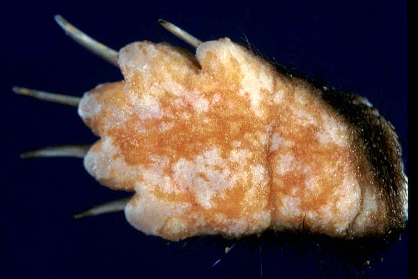
The differential diagnosis for hyperkeratosis in dog includes canine distemper, zinc-responsive dermatosis, generic dog food dermatosis, acrodermatitis of bull terriers, superficial necrolytic dermatitis and contact dermatitis. The presence of intracytoplasmic inclusion bodies within hyperplastic keratinocytes is consistent with canine distemper.

Contributor: Animal Disease Diagnostic Laboratory, ADDL-1175, Purdue University, West Lafayette, IN 47907
 
References:
1. Dungworth DL: The Respiratory System. In: Pathology of Domestic Animals, Eds. Jubb KVF, Kennedy PC, Palmer N, 4th ed., pp. 617-624. Academic Press, San Diego, CA, 1993
2. Fowler ME: Carnivora. In: Zoo and Wild Animal Medicine, Ed. Fowler ME, 2nd ed., 800-807. WB Saunders Co. Philadelphia, PA, 1986
3. Jones TC, Hunt RD, King NW: Veterinary Pathology, 6th ed., pp. 311-315 Iowa State University Press, Ames, IA, 1997
 
 
Case IV - D98-23773 (AFIP 2676032)
 
Signalment: 6-month-old, male raccoon.
 
History: This animal had pruritic scabby skin lesions on the dorsal ear, neck and thorax. The lesions were resistant to antibiotics, steroids, and fungicidal treatment.
 
Contributor's Diagnosis and Comments: Chronic active, eosinophilic and pyogranulomatous dermatitis, multifocal, severe, with furunculosis, fibrosis, ulceration, serocellular crusting, and intralesional embryonated nematode parasite eggs.
 
Microscopically, there are marked multifocal chronic active, eosinophilic, and pyogranulomatous inflammatory infiltrates in the dermis associated with many embryonated nematode parasite eggs, with the morphologic features consistent with a primitive filarial nematode.
 
AFIP Diagnosis: Haired skin: Dermatitis and panniculitis, pyogranulomatous and eosinophilic, multifocal, moderate, with vesico-pustules, necrosis, and numerous larvated nematode eggs, raccoon (Procyon lotor), carnivore.
 
Conference Note: In this case, the presence of 44 x 27 mm, larvated nematode eggs within the subcutis and dermis is consistent with Filaria taxidea. This worm has been reported in skunks, racoons, badgers, and a lesser panda. This parasite is a primitive filarial nematode. The adult female lives in the subcutis and deposits her eggs (via a uterine opening next to the mouth) in the dermis, where they produce a marked inflammatory reaction as they migrate to the surface. It has been proposed that dipteran insects (flies, gnats and mosquitoes) take part in the life cycle by ingesting the eggs from the infected hosts and spreading them to other hosts.

There are six groups of nematodes that can have larvated eggs: Filarids, Rhabditoids, metastrongilids, aphasmids, spirurids and oxyurids.

The diagnostic features of trematodes, cestodes, and nematodes were discussed:
 
Key features of Trematodes, Cestodes, & Nematodes

 Trematodes

 Cestodes

 Nematodes

 Body cavity

 none

 none

 pseudocelom

 Digestive tract

 paired ceca

 none

 single

 Reproduction

 hermaphroditic

 hermaphroditic

 male or female

 Surface

tegument 

 tegument

 cuticle (non-living)

 Eggs

 thick, opaque
(except schistosomes)

 within proglottid

 varied

 Suckers

 oral or ventral

 on scolex

 none
 
Contributor: Pal-Path, Inc., 1277 Record Crossing Road, Dallas, Texas 75235
 
References:
1. Gardiner CH, Loomis MR, Britt JO, Montali RJ: Dermatitis caused by Filaria taxideae in a lesser panda. JAVMA 183(11):1285-1287, 1983
2. O'Toole D, Williams ES, Welch V, Nunamaker CE, Lynn C: Subepidermal vesiculobullous filarial dermatitis in free-ranging American Badgers (Taxidea taxus). Vet Pathol 30:343-351, 1993
3. Saito EK, Little SE: Filarial dermatitis is a striped skunk. J Wild Dis 33(4):873-876, 1997.
 
J Scot Estep, DVM
Captain, VC, USA
Registry of Veterinary Pathology*
Department of Veterinary Pathology
Armed Forces Institute of Pathology
(202)782-2615; DSN: 662-2615
Internet: estep@afip.osd.mil
 
* The American Veterinary Medical Association and the American College of Veterinary Pathologists are co-sponsors of the Registry of Veterinary Pathology. The C.L. Davis Foundation also provides substantial support for the Registry.
 
Return to WSC Case Menu