Signalment: Adult, male, cynomolgous macaque (Macaca fascicularis).
History: This animal developed an acute multifocal to coalescing dermatitis affecting the entire body surface. Affected foci were 0.5 to 1.0 cm in diameter, erythematous, and rarely contained central vesicles (see included gross photos). The animal died during sedation for supportive treatment.
Gross Pathology:
Dermatitis, vesicular, multifocal to coalescing, generalized.
Hepatitis, necrotizing, multifocal, acute, severe.
Splenitis, necrotizing, multifocal, acute, severe.
Laboratory Results: Virology: Simian varicella virus was cultured from samples taken at the time of the gross examination.
Etiology: Simian varicella virus.
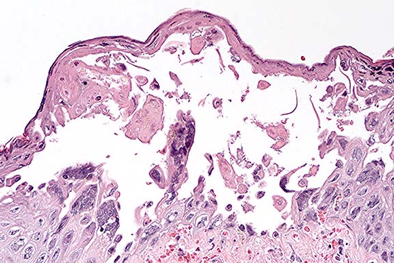
In contrast to the gross skin lesions, the microscopic lesions are relatively mild and include degeneration and necrosis of surface and adnexal epithelium and vesicle formation. Intranuclear viral inclusions are present in almost all affected epithelial foci. A mild variable neutrophilic infiltrate and multifocal subcuticular hemorrhage are present in the dermis of some affected sections. In sections with subcutaneous hemorrhage, small vessels are congested, lined by rounded endothelium and contain occasional fibrin aggregates. Some vessel walls are necrotic and rare endothelial cells contain intranuclear inclusions. Similar vascular lesions are present in the spleen.
Conference Note: Conference participants interpreted the sialoadenitis as a chronic process unrelated to the acute herpesviral infection.
Simian varicella virus (SVV) infection in nonhuman primates causes disease that is similar in many ways to that caused by varicella zoster virus (VZV) in humans. VZV is transmitted by aerosol, disseminates hematogenously, and causes a self-limiting primary infection in immunocompetent humans consisting of widespread vascular skin lesions (chickenpox, or varicella). VZV also infects satellite cells around neurons in the dorsal root ganglia and remains latent there for many years. Recrudescence occurs in elderly or immunosuppressed patients, and causes shingles (zoster), which is characterized by pain and vesicular skin eruptions in the area of distribution of the affected sensory nerve. Acute SVV infection in Old World monkeys causes similar generalized skin lesions. Additionally, viral infection of ganglia by SVV has been documented, though the associated inflammation is much less severe than that seen in shingles.1 The route of ganglionic infection is unclear. A shingles-like recrudescence has not been reported in SVV-infected monkeys.
Both SVV and VZV cause inflammation and necrosis, accompanied by eosinophilic intranuclear inclusion bodies, in multiple organs, including liver and lung. Common clinicopathologic abnormalities seen in both infections include thrombocytopenia and increased aspartate aminotransferase (AST). A major difference is that in humans, varicella is seldom fatal, whereas the mortality rate of SVV infection in nonhuman primates is high.
Contributor: Department of Comparative Medicine and Washington Regional Primate Research Center, Box 357190, Health Sciences, University of Washington, Seattle, WA 98195-7190.
Signalment: Adult, male, Sprague-Dawley rat.
History: This animal was in the control group of a subchronic toxicity study and was necropsied on day 120 of the study.
Gross Pathology: At necropsy, gross morphological changes
were limited to bilaterally enlarged external ears.
Contributor's Diagnosis and Comments: External ear: Inflammation,
pyogranulomatous with cartilaginous proliferation and osseous
metaplasia, severe, compatible with auricular chondropathy of
Crl:CDÒ Rats.
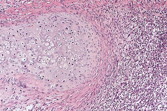
Conference Note: In addition to Sprague-Dawley and fawn hooded rats, a similar auricular chondropathy has been reported in Wistar rats.2 The pathogenesis of this condition remains unclear. Relapsing polychondritis in humans is associated with antibodies to type II collagen, the major component of cartilage matrix.4 Prieur et al described spontaneous auricular lesions in rats which resembled those caused by immunization of rats with type II collagen, and they suggested the use of these rats as an animal model for the study of the role of immunity to type II collagen as a mechanism of disease of cartilage.3 In contrast, a later study with Crl:CD(SD)BR rats demonstrated no immunity to type II collagen in affected rats; the author concluded that the chondritis was likely due to an autoimmune response initiated by a chronic inflammatory process at the insertion site of metal ear tags.4
Contributor: Novartis Pharmaceuticals, 556 Morris Avenue, Summit, NJ 07901-1398
Signalment: 6-week-old, conure, avian.
History: This baby conure was being hand raised and was feeding okay. Unexpectedly, it was found dead.
Gross Pathology: A 3 mm nodular mass was adherent to the endocardial surface of the left atrium.
Contributor's Diagnosis and Comments: Vegetative mural endocarditis of the left atrium caused by a fungus of the Zygomycetes class.
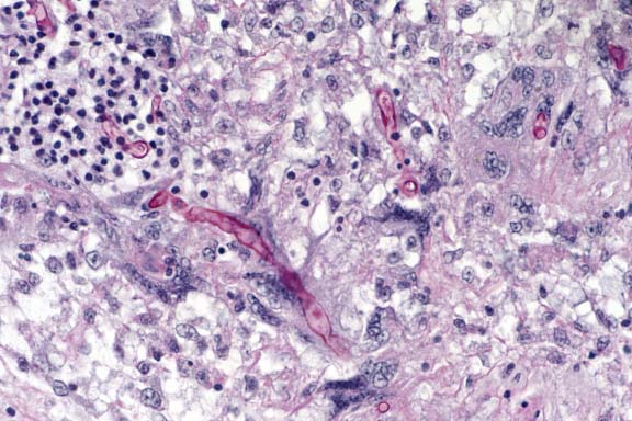
Conference Note: Conference participants discussed the morphologic differences between Aspergillus sp., common pathogens of birds, and Zygomycetes, which are occasionally seen in immunocompromised or debilitated birds. Aspergillus sp. hyphae are typically 3 to 6 mm wide, septate and branched. Branching is dichotomous and often at acute angles. Typical mucoraceous hyphae vary from 5 to 20 mm wide and branch nondichotomously at right angles. The hyphal walls are thin and often become twisted or folded.
The biological behavior of zygomycosis and aspergillosis in birds is similar. Infection of the lungs is usually primary, followed by necrotizing vasculitis, invasion of blood vessels by fungal elements, and subsequent hematogenous dissemination to multiple organs.
Contributor: Iowa State University, College of Veterinary Medicine, Ames, IA 50011.
International Veterinary Pathology Slide Bank:
Laser disc frame #18419
Signalment: 6-year-old, male, rhesus macaque (Macaca mulatta).
History: The animal was found dead. It had a history of decreased appetite during the last few days. 16 months prior, it was on antibiotics for an infection of a catheter site.
Gross Pathology: An indwelling catheter was present from the dorsal cervical subcutaneous tissue into the right jugular vein and into the right atrium. Bilateral, red, firm to meaty circular to irregularly shaped foci, up to 5 mm in diameter, were disseminated over the natural and cut surfaces of the lung. The left epididymis was enlarged focally and contained white purulent fluid.
Laboratory Results: Bacteriology results:
Blood: Staphylococcus aureus
Lung: Staph. aureus, Klebsiella pneumonia
Epididymis: Staph. aureus, Klebsiella pneumonia

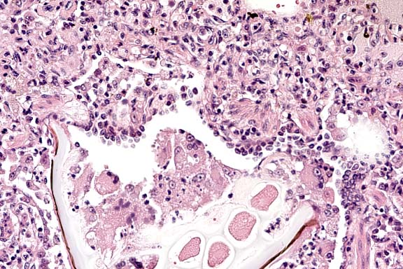
Conference Note: Conference participants discussed the importance of recognizing that two distinct processes are evident. One process is chronic, bronchocentric, and a result of foreign material introduced into the airways. The other process is more acute, angiocentric, and a result of staphylococcal bacteremia.
Staphylococcus aureus and Streptococcus pneumoniae are pathogenic cocci that cause pneumonia in primates. In contrast to the present case, S. pneumoniae usually appears as small diplococci and causes a fibrinohemorrhagic pneumonia, often accompanied by fibrinous pleuritis and pericarditis.
Contributor: North Carolina State University, College of Veterinary Medicine, 4700 Hillsborough Street, Raleigh, NC 27606
International Veterinary Pathology Slide Bank:
Laser disc frame #8494
Terrell W. Blanchard
Major, VC, USA
Registry of Veterinary Pathology*
Department of Veterinary Pathology
Armed Forces Institute of Pathology
(202)782-2615; DSN: 662-2615
Internet: blanchard@email.afip.osd.mil
* The American Veterinary Medical Association and the American College of Veterinary Pathologists are co-sponsors of the Registry of Veterinary Pathology. The C.L. Davis Foundation also provides substantial support for the Registry.