Signalment: 1.5-year-old, castrated male, Angora goat.
History: This goat was one of four goats that died out of a herd of 35. All of the animals were purchased from a local breeder. At the time of purchase, all of the goats had been vaccinated with Covexin 8ä (immunizes against diseases caused by Clostridium chauvoei, Cl. septicum, Cl. novyi type B, Cl. hemolyticum (or Cl. novyi type D), Cl. tetani, and Cl. perfringens types C and D), and Lepto 5ä (immunizes against diseases caused by Leptospira canicola, L. grippotyphosa, L. hardjo, L. icterohemorrhagiae and L. pomona). All were negative for Brucella abortus and Coxiella burnetti, and were infested with lice (later identified as Damalinia crassipes) and coccidia. The animals were treated with a pyrethrin product for the lice and with Amproliumä for the coccidia. The goats were maintained on a diet of sweet feed and pelleted alfalfa hay.
This goat was observed down in the pasture. It was unconscious and vocal, exhibited muscle twitching and weakness, was non-responsive, and had no menace reflex. The clinician reported that the goat preferred to be in lateral recumbency, and displayed opisthotonus when placed in sternal recumbency. Body temperature was 102° F, heart rate was 72 beats per minute, and the respiratory rate was 60 per minute. Treatment was initiated: thiamine 10 mg/kg (500 mg) SQ, every 6 hours for 24 hours; 2 liters of intravenous saline, to which was added 80 ml 23% calcium gluconate and 40 ml (120 mg) dexamethasone sodium phosphate. Five grams of Cefasolä (an antibiotic) was also added to the first liter. Later in the afternoon, the goat was sitting upright in the stall, but was unable to stand. The bilateral loss of menace response was interpreted to be loss of vision, but palpebral and corneal reflexes were still present. The next morning, the goat was unconscious in the stall with no menace or palpebral reflexes. After consultation with the investigator, euthanasia was elected.
Gross Pathology: The animal was well-muscled and had
excessive subcutaneous and cavitary adipose tissue. Throughout
the pelage, there were numerous adult chewing lice and nits (Damalinia
crassipes). Gross diagnoses were:
Lungs: Aspiration pneumonia.
Brain, meninges: Congestion, diffuse, moderate.
Pelage: Pediculosis.
Carcass: Obese.
Contributor's Diagnoses and Comments:
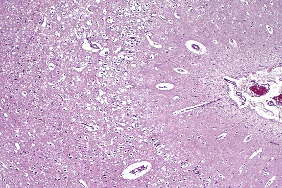
1. Cerebrum, diencephalon, white tracts: Distended myelin sheaths,
multifocal, mild.
2. Cerebrum, lateral and fourth ventricles: Distention (hydrocephalus).
3. Cerebrum, gray matter, level of the mammillary body: Necrosis,
laminar, bilateral, segmental, mild, with spongiosis.
4. Cerebrum, white matter, level of the mammillary body: Myelin
sheaths, distention, bilaterally symmetrical, mild.
5. Cerebellum; brain stem: Myelin sheaths, distention, bilaterally
symmetrical, moderate.
6. Medulla, ventral and lateral funiculi: Myelin sheaths, distention,
bilaterally symmetrical, diffuse, marked.
7. Spinal cord, cervical, thoracic, lumbar and sacral: Distended
myelin sheaths, bilaterally symmetrical, multifocal, marked, with
minimal bilateral neuronal necrosis in the ventral horns.
8. Lung: Congestion, diffuse, mild, with multifocal hemorrhage.
9. Lung, airways: Intra-airway plant material (interpreted to
be an agonal event).
The laminar necrosis and mild malacia of the cortical gray matter are consistent with changes seen in goats affected by thiamine deficiency but are not pathognomonic for the condition. Thiamine deficiency in these goats was diagnosed and treated empirically. Once thiamine deficiency was tentatively diagnosed, all of the remaining goats were given bolus subcutaneous injections of thiamine, and only four goats were lost.
Conference Note: Internal hydrocephalus, which was diagnosed
by the contributor, could not be confirmed histologically in the
sections examined in conference. Some participants preferred "malacia"
over "laminar necrosis" because of the entrenched use
of the term "malacia" to describe this lesion in the
veterinary literature. Others, however, contended that "malacia"
is better reserved as a gross descriptive term, and that "necrosis"
is more suitable for histopathologic diagnosis.
In ruminants, polioencephalomalacia (PEM) is most frequently associated
with high concentrate, low roughage diets, and is found most commonly
in well-nourished cattle, 6 to 18 months of age, that have been
in the feedlot for several weeks. It is also seen in animals on
forage, most commonly 5 to 10 days following a change from poor
to good pasture. A similar condition has been reported in animals
fed a molasses/urea diet. In sheep, a drastic change in management,
such as shearing time, may precipitate outbreaks in which only
yearlings are affected. In goats, the disease may be seen in kids
fed milk-replacer diets, and in older animals on high concentrate
diets.
Deficiencies of thiamine or disturbances in its metabolism are often implicated in PEM. Thiamine deficiency has been produced in most domestic species, as well as mink and foxes in which it is known as Chastek paralysis. In carnivores, the deficiency is induced by a thiamine-splitting enzyme naturally present in many species of fish. In ruminants, PEM develops when high levels of thiaminases are formed in the rumen; these substances destroy, inactivate, or interfere with absorption, phosphorylation and ultimate utilization of thiamine. Ruminal acidosis may allow the preferential growth of thiaminase-producing bacteria, such as Clostridium sporogenes and Bacillus thiaminolyticus. Thiaminases, in the presence of certain substrates, may also produce thiamine antimetabolites or analogs. Thiabendazole and levamisole, commonly used anthelmintics, may react with thiamine in a reaction catalyzed by thiaminase I, and PEM may follow their use. Amprolium, a commonly used coccidiostat in ruminants, is an analog of thiamine, and competitively inhibits thiamine at its active binding sites. High levels of sulfates in the water or feed can lead to thiamine deficiency in sheep and cattle; a variety of inorganic sulfur compounds is capable of cleaving thiamine into inactive constituents.
Thiamine is a necessary cofactor in carbohydrate metabolism. Ruminants are primarily dependent upon ruminal microbial synthesis of thiamine, whereas in carnivores, thiamine is a dietary requirement. Thiamine is converted to cocarboxylase, which is a necessary cofactor for 1) pyruvate dehydrogenase in the conversion of pyruvate to acetyl CoA, 2) a-ketoglutarate dehydrogenase in the conversion of a-ketoglutarate to succinyl CoA, 3) transketolase activity in the hexose monophosphate shunt, and 4) branched-chain a-keto acid dehydrogenase. Transketolase is active in white matter and is important in the metabolism of oligodendrocytes. Nervous tissue is entirely dependent on carbohydrate oxidation for its energy, and is thus extremely sensitive to thiamine deficiency.
Contributor: Armstrong Laboratories, 2509 Kennedy Circle, Brooks AFB, TX 78235
International Veterinary Pathology Slide Bank:
Laser disc frame #16, 791, 2883, 2884, 3277, 3492, 3493, 3526,
3527, 3551, 4077, 4078, 4607, 4608, 4853, 4974, 5104, 5105, 5247,
5668, 7538, 7539, 7783, 9187, 9188, 9351, 10507, 10508, 14219-14222,
14233-14235, 22829, 23175, 23176.
Signalment: 8-week-old, crossbreed, pig.
History: This pig was one of six that were displaying central nervous system signs and twelve which had died out of a group of 150. This pig was in right lateral recumbency and, when aided to stand, circled to the right and tilted its head to the right.
Gross Pathology: Mild nasal turbinate atrophy was noted
in the right nasal cavity. No other gross lesions were seen.
Laboratory Results: Streptococcus suis, Pasteurella multocida,
and Actinomyces pyogenes were isolated from a swab of the turbinates.
Escherichia coli was isolated from the small intestine.
Contributor's Diagnosis and Comments: Severe multifocal necrotic vasculitis, Edema disease.
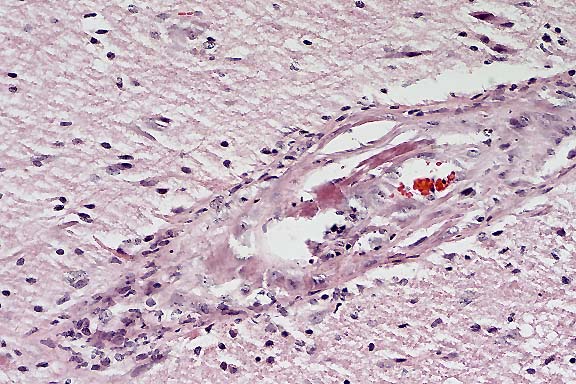
In these sections of brain stem, there is hyaline degeneration and necrosis of the tunica media of small caliber arterioles. Lymphocytes and plasma cells are present in the tunica media and adventitia of affected arterioles. There is adventitial hyperplasia surrounding many vessels. Endothelial cells appear swollen and occasional fibrin thrombi are noted. In some sections, there are focally extensive areas of malacia which contain moderate numbers of mononuclear inflammatory cells or are partially surrounded by a zone of inflammatory cell infiltrates.
Conference Note: Edema disease is a distinct syndrome, usually occurring in pigs shortly after weaning. There are rare reports of the disease in both suckling and mature pigs. Mortality may approach 100%.3 The disease may occur sporadically or as a herd outbreak, and often occurs in association with outbreaks of postweaning E. coli enteritis.
Factors which favor enteric colonization and adhesion of shiga-like toxin II variant (SLT IIv)-producing strains of E. coli are not known. The serogroups involved are part of the normal gut flora of weaned pigs.3 They contain no known pilus adhesins, and do not use an attaching-effacing mechanism, yet they adhere to small intestinal microvilli. Lesions are attributed to the effects of SLT IIv, which binds to the receptor globotetraosylceramide on vascular endothelium. This receptor has a differential distribution in endothelium of various organs, which may account for the apparent organ tropism.
Grossly visible edema may or may not be present at necropsy. When present, it may be subtle and mild. Edema is most consistently found in the mesocolon, gastric submucosa, and in mesenteric lymph nodes. Occasionally, bilaterally symmetrical areas of malacia are visible in the brain stem, and are associated with lesions in cerebral vessels. Inflammation is not a prominent component of the angiopathy, and thrombosis is rare.
Contributor: Veterinary Diagnostic Center, University of Nebraska-Lincoln, Fair St. and East Campus Loop, Lincoln, NE 68583-0907
International Veterinary Pathology Slide Bank:
Laser disc frame #5201, 20467.
Signalment: 1-year-old, 307 kg, male, horse.
History: The yearling was anesthetized for castration. When he recovered from the anesthesia, it became apparent that he was alert, but unable to rise. The surgeon diagnosed a cauda equina syndrome because of the paralysis of the urinary bladder, anal sphincter and tail. A fracture of the vertebral column was suspected and the animal was euthanized.
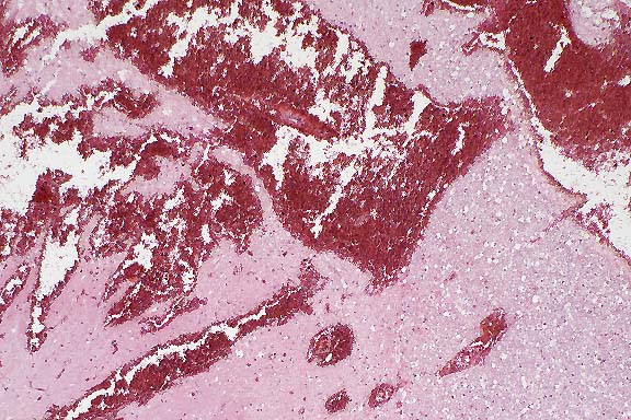
Gross Pathology: The castration wounds were well adapted. There were no alterations of the head or the vertebral column. The spinal cord was removed, and following fixation, was cut into transverse slices at each spinal level. Hemorrhages were observed at the level of T6 and from T13 to the cauda equina. The dorsal horns were especially affected.
Contributor's Diagnosis and Comments: Spinal cord: Severe acute hemorrhages and poliomyelomalacia.
Severe hemorrhages are found in the dorsal horns of the gray matter either as perivascular hemorrhages around congested blood vessels or forming extensive clefts in the neuropil. The neuropil in this area is edematous and contains swollen, degenerated neurons. A few reactive macrophages are present only. The leptomeningeal and spinal veins are severely dilated and congested. The spinal arteries and the spinal canal also contain erythrocytes. Leptomeningeal hemorrhages extend about half the circumference of the dorsal aspect of the spinal cord (in most slides).
Malacia of the spinal cord with extensive hemorrhages has been described as a rare complication following general anesthesia in the horse. The etiology of this so-called "equine post-anesthetic/surgical hematomyelia" is unknown. Factors common to the cases reported in the literature are: 1) The condition occurs in young growing horses which are "heavy for their age" (250-400 kg). 2) All horses were positioned in dorsal recumbency for the surgical procedures. 3) Inhalant anesthesia was used, frequently halothane. 4. Hemorrhages were predominantly localized between T2 and S2.
Conference Note: The etiopathogenesis of this uncommon condition in horses is poorly understood. One author suggests that the pressure of the viscera on the abdominal vessels may produce a condition similar to the supine hypotensive syndrome of human pregnancy, an acute hypotensive state caused by the pressure of one or multiple fetuses on the aorta and/or the posterior vena cava. However, attempts to experimentally reproduce this condition in the bitch were unsuccessful.2
The clinical circumstances and the histological evidence of congestion and ischemia are consistent with the hypothesis that oxygenation of the lower spinal cord was compromised during anesthesia. It is speculated that alterations in blood flow might include both arterial hypotension and stagnant hypoxia resulting from venous compression by the mass of the abdominal viscera in the horse during general anesthesia. Support of this hypothetical explanation would depend on evidence from physiological monitoring during anesthesia. To prevent surgical injuries like this, the clinical precaution of periodic movement of the anesthetized horse during surgery might reduce the risk of prolonged venous compression and the development of this injury.
Differentiation of this condition from other forms of neuropathy and myopathy in the horse is important to the prognosis.
Contributor: Institut fur Pathologie, Tierarztliche Hochschule Hannover, Bunteweg 17, 30559 Hannover, Germany
Signalment: 7-month-old, 400 lb, crossbred female, bovine.
History: This heifer was from a group of calves purchased at a sale barn. Within 3 weeks of purchase, all the calves had labored breathing and were coughing. None of these calves was vaccinated. This heifer was found dead.
Gross Pathology: The trachea and right terminal airways were filled with blood-tinged froth. The left terminal airways contained free and clotted blood. Approximately 40% of the cranioventral lungs were purple and moderately firm. The visceral pleura was dry and granular. The caudal portion of the right cranial lobe was very firm and adhered to the pericardium. The medial retropharyngeal and mediastinal lymph nodes were dark red, enlarged, and exuded a small amount of serous fluid on cut surface.
Laboratory Results:
Microbiology: Hemophilus somnus was isolated from the lungs.
Virology: Results of fluorescent antibody testing for bovine herpesvirus 1, bovine virus diarrhea virus, parainfluenza-3 virus, and bovine respiratory syncytial virus were negative.
Contributor's Diagnoses and Comments: 1. Acute bronchopneumonia, fibrinopurulent, diffuse, severe, with intralesional bacteria (Hemophilus somnus), with multinucleated cells. 2. Bronchiolitis, necrosuppurative, acute.
The principal microscopic lesions are fibrinous bronchopneumonia and necrotizing bronchiolitis, with diffuse alveolar congestion and thickening, and extremely dilated interlobular septa. Medium-sized and small airways are frequently filled with necrotic and inflammatory debris and occasional aggregates of bacteria. Alveoli contain loosely fibrillar to dense multinucleated cells. A few alveoli contain numerous neutrophils, aggregates of bacteria, and cellular debris. Interlobular lymphatics are extremely dilated and contain dense to loosely arranged fibrin thrombi. The remainder of the interlobular space is filled with fibrin intermixed with small numbers of inflammatory cells.
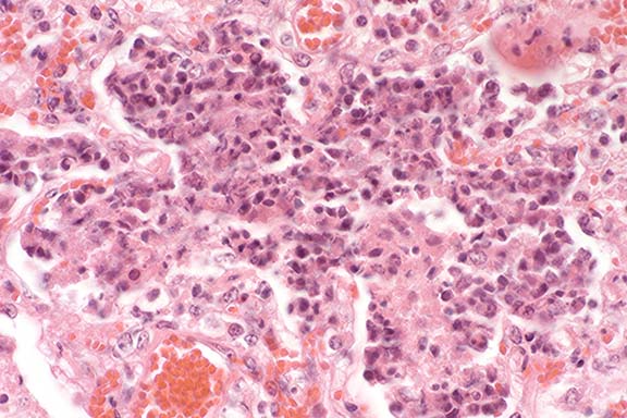
Conference Note: A Gram stain demonstrated small numbers of gram-negative coccobacilli within suppurative foci. Moderate numbers of large, gram-positive, spore-forming bacilli are present in blood vessel lumina. The bacilli are considered to represent postmortem invaders.
Hemophilus somnus is a small, gram-negative, non-motile, pleomorphic coccobacillus. It is part of the normal flora of the male and female bovine genital tract and is regularly isolated from the respiratory tract of healthy animals.
The most common diseases caused by H. somnus are thrombotic (or thromboembolic) meningoencephalitis (TME), pneumonia, and myocarditis. In addition, H. somnus has been associated with the following syndromes in cattle: abortion, metritis/infertility, arthritis, otitis externa, mastitis, vesicular adenitis, orchitis, conjunctivitis, and laryngitis/tracheitis. Usually, clinical disease caused by H. somnus occurs in immature cattle and is associated with environmental stresses such as shipping, mixing of animals from different sources and climatic changes.
Calves are infected by carrier cows, possibly via the respiratory tract, in the first months of life and disseminate the infection in feedlots. The organism multiplies and survives within bovine monocytes. Hemophilus somnus may cause a septicemia that leads to acute death, or may localize in one or several organs causing subacute or chronic, fatal or nonfatal disease. The septicemic phase is brief, and infection is occasionally cleared at this stage without localization. More commonly, septicemia leads to cerebral vasculitis with acute thrombosis, hemorrhage, and necrosis. TME often occurs in calves after respiratory tract infection has developed within the herd. Vasculitis can develop in most organs.
Factors associated with virulence include attachment, serum resistance, growth stimulation by normal flora, interference with phagocyte function, Fc receptors, and toxicity for several bovine cell types. Attachment of organisms facilitates colonization, and vasculitis/thrombosis may develop from bacterial adherence to endothelial cells. Bacterial embolism does not occur, but may be simulated microscopically by intravascular proliferation of bacteria at sites of thrombosis.
Contributor: Department of Pathology & Microbiology, College of Veterinary Medicine, Kansas State University, VCS Building, Room K-221, Manhattan, KS 66506.
International Veterinary Pathology Slide Bank:
Laser disc frame #95, 7760, 12715
Terrell W. Blanchard
Major, VC, USA
Registry of Veterinary Pathology*
Department of Veterinary Pathology
Armed Forces Institute of Pathology
(202)782-2615; DSN: 662-2615
Internet: blanchard@email.afip.osd.mil
* The American Veterinary Medical Association and the American College of Veterinary Pathologists are co-sponsors of the Registry of Veterinary Pathology. The C.L. Davis Foundation also provides substantial support for the Registry.