Signalment: Male transgenic knockout (FVB/N-Trp53tm1Dol) mouse.
History: Euthanized because of a scrotal mass.
Gross Pathology: The scrotum was distended by a 2.0 x 1.0 cm firm pale encapsulated mass that had replaced one testicle.
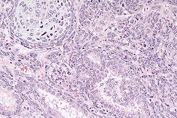
Contributor's Diagnosis and Comments: Testis: Teratoma, malignant.
The mass is partially encapsulated and composed of a variety of haphazardly arranged, well-differentiated tissues from several germ layers, as well as morphologically undifferentiated tissue. Differentiated tissues include nervous tissue, striated muscle, cartilage and less differentiated connective tissue. There also is keratinized squamous epithelium, tissues in nests and forming ducts, tubular structures lined by ciliated respiratory epithelium and intestinal epithelium with goblet cells. Multifocally, there are nests of highly anaplastic "carcinoma" cells.
Teratomas arise from pluripotent stem cells which can differentiate into derivatives of all three primary germ layers (endoderm, mesoderm, and ectoderm). Benign teratomas consist of well-differentiated tissues from the different germ layers while malignant teratomas additionally contain nests of undifferentiated pluripotent stem cells (teratocarcinoma cells). Teratomas and teratocarcinomas typically arise in either the testis or ovary but there are also reports of extragonadal teratoid tumors.
Teratomas are rare in most strains of mice, the exception being inbred strain 129 mice. Selective breeding of strain 129 mice (strain 120/Sv-ter) resulted in one third of the males having spontaneous testicular teratomas. Experimentally, genital ridges from 12-day strain 129 fetuses grafted onto the testes of adult syngeneic mice resulted in an 80% incidence of teratomas at the site. These fetal genital ridges contained pluripotent primordial germ cells. Strain 129 mice also were shown to have increased risk of testicular teratoma when exposed in utero to ethinyl estradiol.
One method of creating transgenic mice is the injection of genetically-modified embryonal stem cells into host blastocysts. These embryonal stem cells have been considered the source of extragonadal teratocarcinomas in the resulting chimeric mice. Whether the gonadal malignant teratoma of the transgenic mouse of the present case originated from injected embryonic stem cells is unknown.
Conference Note: Testicular teratomas are rare in all domestic species except the horse, in which they are the most frequently reported testicular tumor.10 Ovarian teratomas, likewise, are rare, but have been reported in the bitch, sow, mare, and cow.9
Grossly, teratomas vary in color and texture. A cystic or multilocular structure is common. Histologically, teratomas may contain structures derived from all embyronic germ layers, i.e. ectodermal (hair, teeth), neuroectodermal (nervous tissue, melanoblasts), entodermal (salivary gland, respiratory or digestive tissue), and mesodermal (fat, bone, muscle, fibrous connective tissue). Nervous tissue is almost always present.10
International Veterinary Pathology Slide Bank:
Laser disc frame #275, 845, 2737, 5670, 6510, 6511, 8379, 8446,
8447, 8449, 16600, 16601, 16643-45, 20024, 22796.
Signalment: 5.5-year-old, female, Rhesus monkey (Macaca mulatta)
History: This monkey had given birth to a normal baby 6 1/2 months previously and had not been used for any experimental manipulation. In January of 1995, the animal was reported to be weak, dehydrated and anorexic. On physical examination, the monkey was wasted and jaundiced, with a nasal discharge and bilateral rales. Chest X-rays revealed disseminated, circumscribed, variably-sized, solid, radiopaque foci throughout both lung fields. There was marked pallor of the mucous membranes. No palpable lymphadenomegaly was present. The animal was euthanized.
Gross Pathology: Post-mortem evaluation revealed disseminated,
variably- sized nodular foci, generally 1 - 2 cm in diameter,
present throughout the pulmonary parenchyma. These were also noted
in lesser numbers in the liver, spleen and adrenal gland, and
a mass within the uterine lumen and wall caused moderate distension
of this organ. The tissue was generally solid, although somewhat
soft, with frequent central regions of caseation and cavitation.
The lesions were sometimes uniformly hemorrhagic, or associated
with a hemorrhagic rim.
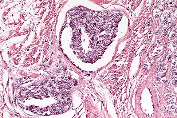
Contributor's Diagnosis and Comments: Choriocarcinoma,
uterus, Rhesus monkey.
Conference Note: This case was studied in consultation
with the Department of Gynecologic and Breast Pathology of the
AFIP. Although careful consideration was given to the contributor's
diagnosis of choriocarcinoma, sections viewed in conference did
not show the biphasic mixture of cytotrophoblasts and synciotrophoblasts
which is characteristic of that tumor. Immunohistochemical stains
performed at the AFIP were focally positive for pancytokeratin
and negative for human chorionic gonadotropin. The discrepancy
between our immuno-histochemical results and those of the contributor
might be related to the time interval between tissue sectioning
and immunostaining at the AFIP. There is extensive invasion of
lymphatic vessels by the neoplastic cells. The possibility of
metastasis to the uterus, or from another part of the uterus,
cannot be excluded.
Contributor: Division of Laboratory Animal Resources, S-1040
BioScience Tower, University of Pittsburgh, Pittsburgh, PA 15261
Signalment: 8-week-old, TCR-a-deficient mouse.
History: This animal was dosed with 10,000 Cryptosporidium parvum oocysts by gavage at 1 week of age, and was euthanized at 8 weeks of age. Infected TCR-a-deficient mice gained weight slower than control mice and developed soft mucoid feces beginning at 4 weeks of age.
Gross Pathology: The distal small intestine and the entire large intestine were thickened.
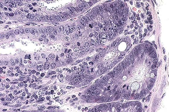
Contributor's Diagnosis and Comments: Cecum: Typhlitis, proliferative, diffuse, moderate, chronic with crypt abscesses and intralesional Cryptosporidium organisms, TCR-a-deficient mouse.
Conference Note: Lymph node was not present in all sections provided to contributors. Plasmacytosis may be another indicator of lack of ab T-cell-mediated suppression of B cells.
Cryptosporidium is an apicomplexan protozoan that infects birds, mammals, fish, amphibians, and reptiles; it is a significant zoonotic agent, especially in immunocompromised humans. Respiratory infection is most significant in birds, whereas the disease in mammals is usually enteric. Cryptosporidial infection has also been reported in the stomach of mice and snakes; the bursa of Fabricius in chickens; and in the biliary and pancreatic ducts and upper respiratory tract of HIV-infected humans, SIV-infected monkeys, and immunosuppressed Arabian foals.
Cryptosporidiosis in mammals is caused by C. muris, C. parvum, or C. wrairi. Cryptosporidium parvum appears to be freely transmissible among numerous host species, whereas C. muris infects the ruminant abomasum and probably the stomach of cats. Cryptosporidium wrairi is a pathogen of guinea pigs. Cryptosporidium meleagridis and C. baileyi infect birds, C. serpentis and C. crotali infect snakes, and C. nasorum infects fish.
Cryptosporidium has a typical coccidian life cycle, with merogony,
gametogony, and sporogony occurring in the brush border of infected
epithelial cells. Sporulated oocysts are either inhaled or ingested,
which is followed by release and invasion of sporozoites in the
respiratory or gastrointestinal tract. Subsequent to invasion
of sporozoites, two types of schizonts develop: type I produce
up to eight merozoites, which recycle to form more type I schizonts
or produce a generation of type II schizonts. The type II schizonts
form four merozoites which invade the host cell and eventually
form gametocytes. Oocysts sporulate within the host to form four
sporozoites. Oocysts are of two forms: 1) thin-walled oocysts,
which excyst within the gut permitting autoinfection and amplification
of the disease and 2) thick-walled oocysts, which excyst and are
passed in the feces to complete the cycle.
In addition to Cryptosporidium, other protozoal genera which sporulate
inside the host include Sarcocystis and Frenkelia.
Contributor: National Animal Disease Center, 2300 Dayton Road, Ames, IA 50010
Signalment: 40-year-old, female, African grey parrot (Psittacus epithacus).
History: This parrot exhibited weakness due to anorexia for several days. Physical examination revealed that the bird was weak, cachectic and had abdominal effusion.
Gross Pathology: There was 15 ml of clear fluid in the thoracic cavity and 25 ml of clear fluid in the abdominal cavity. The aorta and left and right pulmonary arteries were firm, gritty, and had a nodular appearance. The right thyroid was enlarged, 6 mm in diameter. The left thyroid was 3 mm in diameter.
Laboratory Results: Hypoalbuminemia (total protein 1.2 gm/dl, albumin 0.5 gm/dl, and globulin 0.7 gm/dl) was observed in blood chemistry profile.
Contributor's Diagnosis and Comments: Arteriosclerosis, aorta and pulmonary arteries.
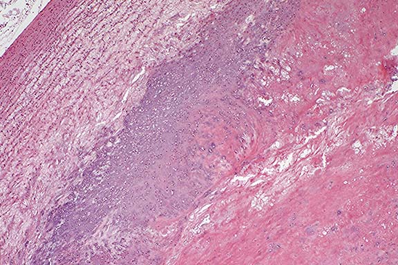
Conference Note: This case was studied in consultation with the Department of Cardiovascular Pathology of the AFIP. The eccentric fibroatheromatous plaque which contains cholesterol clefts resembles human atherosclerosis; however, chondroid metaplasia is unusual in atherosclerosis in humans.
The pathogenesis of atherosclerosis (AS) is an area of current intense research interest. AS and its complications are major causes of morbidity and mortality in humans. Although the distribution of lesions differs from the human, the pig is the only domestic animal species in which AS develops spontaneously.6 It is less commonly seen in dogs; in that species it is almost invariably associated with hypothyroidism or diabetes mellitus.6 A number of animal models of AS have been produced over the last 3 decades, including nonhuman primates, pigs, rabbits, and chickens. Heritable hyperlipidemia of Watanabe rabbits has served as a model of human familial hypercholesterolemia. The apolipoprotein E-deficient (knockout) mouse, recently reported to develop atherosclerotic lesions resembling those in humans, is considered the most promising mouse model.8
The two classic lesions associated with atherosclerosis are the fatty streak and the fibrous plaque. The fatty streak, which often begins in children, may be an early stage in the formation of atherosclerotic lesions. In humans, the fatty streak is characterized by an intimal (subendothelial) accumulation of lipid-laden "foam cells", most of which are macrophages. Smooth muscle cells can be lipid-laden as well. This lesion also contains small amounts of extracellular lipid and variable numbers of lymphocytes. The fatty streak can progress to an intermediate fibrofatty lesion, and ultimately to the more serious fibrous plaque.7
The fibrous plaque (or atheroma) has a characteristic histologic structure. It consists of a subendothelial fibrous cap (composed of proliferating smooth muscle cells, macrophages, lymphocytes, foam cells, and extracellular matrix) which covers a necrotic core consisting of acellular debris, extracellular lipid with cholesterol crystals, and some foam cells.7
Advanced atherosclerotic lesions are associated with 4 classic complications: 1) calcification of the lesion increases the rigidity of the vessel wall; 2) rupture of the plaque may result in thrombosis and embolism; 3) hemorrhage into the plaque may result in additional narrowing of the vessel lumen; and 4) fibrous plaques increase vessel wall fragility and cause loss of elastic tissue, which may result in aneurysmal dilatation and vessel rupture.7
The morphology of atherosclerosis differs significantly between dogs and humans. In canine arteries, lipid accumulates primarily in the media and adventitia, whereas in humans the accumulation is primarily intimal.6
Animal studies have shown that adherence of monocytes and lymphocytes to endothelium is one of the earliest events in AS. In the Watanabe rabbit and in humans, increased expression of VCAM-1 in endothelium overlying early foam cell lesions has been demonstrated. Also, there is local production of chemoattractants specific for monocytes, such as monocyte chemotactic protein-1 (MCP-1) and monocyte colony stimulating factor (M-CSF). Oxidative stress in the vessel wall, resulting from the generation of free radicals and other reactive species, is also believed to play an important role in the pathogenesis of AS.7 In support of this hypothesis, epidemiologic studies in humans have demonstrated an association between increased intake of antioxidant compounds, such as vitamin E and vitamin C, and reduced morbidity and mortality from coronary artery disease.9
Contributor: The Animal Medical Center, 510 East 62nd Street, New York, NY 10021
International Veterinary Pathology Slide Bank:
Laser disc frame #7907, 7908, 9394, 15657, 15669, 18397, 18398,
18403, 18410, 18416, 18480, 18481.
Terrell W. Blanchard
Major, VC, USA
Registry of Veterinary Pathology*
Department of Veterinary Pathology
Armed Forces Institute of Pathology
(202)782-2615; DSN: 662-2615
Internet: blanchard@email.afip.osd.mil
* The American Veterinary Medical Association and the American
College of Veterinary Pathologists are co-sponsors of the Registry
of Veterinary Pathology. The C.L. Davis Foundation also provides
substantial support for the Registry.