Signalment: Approximately 2-month-old, 11 kg, male, Yorkshire cross pig.
History: This pig was to be used on a dermatology research protocol. On arrival from the breeder, the animal was noticed to have a moderate mucopurulent nasal discharge.
Gross Pathology: The lining of the internal nasal mucosa was slightly hyperemic and edematous, and coated with a film of mucopurulent material. A cross section was made at the time of necropsy and was placed in decalcifying solution.
Laboratory Results: Bacterial cultures revealed various nonpathogenic fecal contaminants (E. coli, etc.)
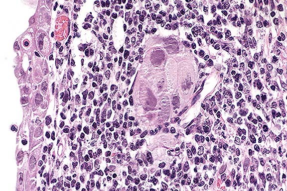
Contributor's Diagnosis and Comments: Rhinitis, subacute, focally extensive, moderate, with intranuclear inclusions and megalocytosis, glandular epithelium of turbinates, consistent with porcine cytomegalovirus (Herpesvirus) aka "inclusion body rhinitis".
Conference Note: Inclusion body rhinitis, caused by porcine herpesvirus 2 (porcine cytomegalovirus) is primarily an acute to subacute disease of suckling piglets characterized by mild to severe necrotizing rhinitis with large basophilic intranuclear inclusions in the epithelium of the mucous glands and ducts in the nasal mucosa. In general, morbidity is high and mortality is low. Infection is usually asymptomatic in older animals; however, in naive herds, disease and occasional deaths may be experienced in 4- to 12-week-old pigs. Infection of pregnant sows may result in delivery of small litters, fetal mummification, stillbirths, neonatal deaths, and runt pigs with rhinitis and/or pneumonia. Subsequent decreased conception rates may occur in affected sows.
Primary viral replication in the nasal mucous glands or the lacrimal or harderian gland is followed by viremia. The site of follow-on viral replication depends on the age of the animal. At an age of 3 weeks or more, when most animals experience infection, the virus disseminates to epithelial sites, particularly the glands of the nasal mucosa, harderian and lacrimal glands, renal tubules, and more rarely the epididymis and mucous glands of the esophagus, hepatocytes, and duodenal epithelium. In the fetal or neonatal pig there is a predilection for reticuloendothelial (RE) cells, particularly capillary endothelium and the sinuses of lymphoid tissues, thus giving rise to generalized lesions. While there is a tendency for replication to predominate in either RE or epithelial cells, they are not mutually exclusive; some individuals have inclusion bodies in both groups of cells. Recovery of virus from pulmonary macrophages of quiescent pigs indicates that the macrophage is a reservoir of infection.
Gross pathologic changes in the nasal passages consist of serous rhinitis with acute congestion of the conchal epithelium in the early stages, progressing to catarrhal or even purulent exudate depending on the type and severity of secondary bacterial infection. Anemia often occurs.
Generalized disease with more extensive and severe lesions is occasionally seen, usually in pigs born alive with congenital infection, but occasionally in older pigs exposed to the virus for the first time. Gross lesions in these pigs include widespread petechiae and edema, including pulmonary edema and congestion, hydrothorax and hydropericardium, multiple firm foci in the ventral lungs, and edema of the throat and tarsal joints. Petechial hemorrhages are seen on the heart, lungs, intestine and particularly the kidneys.
The large basophilic intranuclear inclusion bodies, when correlated with appropriate signs, are diagnostic for inclusion body rhinitis. However, cytomegaloviruses can also produce typical herpetic inclusions without cytomegaly and as such, pseudorabies would have to be eliminated as a cause, particularly in locations such as lung and CNS. The lesions associated with inclusion body rhinitis with a secondary bacterial infection could mimic the early changes associated with atrophic rhinitis. Advanced atrophic rhinitis, with loss of nasal turbinates, should not be difficult to distinguish from inclusion body rhinitis.
Contributor: U.S. Army Medical Research Institute of Chemical Defense, Aberdeen Proving Ground, MD 21010-5425
References
International Veterinary Pathology Slide Bank:
Laser disc frame #15562-15565.
Signalment: Two-year-old, female goat (breed unknown).
History: The doe delivered a kid 8 to 10 days before death. In the 24 hours before death, she had signs of lethargy and was not milking as much, but was still eating normally. Fresh tissues and stomach contents were submitted for examination.
Gross Pathology: No gross lesions were noted in the
organs submitted.
Laboratory Results: Eye fluid had a magnesium content of
1.9 mg/dl and calcium 5.4 mg/dl. A gas chromatograph/mass spectrometric
assay of her stomach contents yielded no significant compounds.
The pH of the stomach contents was 4.7, but the submittor did
not indicate whether these were from the abomasum or rumen. No
significant growth was obtained from aerobic bacterial cultures
of the lung, liver, small intestine, or colon. FA tests on the
tissues were negative for bovine virus diarrhea virus.
Contributor's Diagnosis and Comments: Liver and kidney: Nuclear glycogenosis, hepatocytes and renal tubular epithelial cells, goat.
Intranuclear inclusions were abundant throughout the hepatocytes and the renal tubular epithelial cells. These inclusions were large, acidophilic, and resembled viral inclusions. However, there was no appreciable cellular reaction (necrosis or inflammation) to the presence of the inclusions. Examination of the tissues by transmission electron microscopy revealed no viral particles in the nucleus, but material that was consistent with glycogen.
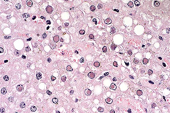
Conference Note: Moderate postmortem autolysis hindered histologic interpretation of both organs. Participants discussed factors that help differentiate antemortem and postmortem changes. Changes that are often associated with autolysis include the absence of inflammation; diffuse changes of a similar magnitude; and increased tissue eosinophilia with loss of cellular and structural detail. This goat had renal changes that were attributed primarily to autolysis; whether acute tubular necrosis was superimposed on the other changes was not definitively determined. Mild chronic interstitial nephritis is a common finding and was not considered to be associated with the nuclear glycogenosis.
A majority of conference participants initially believed that the intranuclear inclusions were most consistent with herpesviral inclusions. However, this was ruled out by the contributor's ultrastructural findings. The periodic acid-Schiff (PAS) reaction, both with and without diastase pretreatment, did not positively stain the nuclei. The differential diagnosis also included lead or bismuth toxicity, both of which may cause intranuclear inclusions in renal tubular epithelial cells. Lead inclusions can also be found in hepatocyte nuclei, and are acid-fast. A Ziehl-Neelsen acid-fast stain performed at the AFIP was negative.
Contributor: University of Kentucky - Livestock Disease Diagnostic Center, 1429 Newtown Pike, Lexington, KY 40511
References:
Signalment: 6-year-old, Pug, intact female, canine.
History: This dog had a history of hypoproteinemia and a chronic cough.
Gross Pathology: Abdominal exploratory revealed moderate thickening of the jejunum. Jejunal mesenteric lymphatics were distended. Multifocal 1-3 mm firm white foci were scattered along the lymphatic vessels.
Laboratory Results: Hypoproteinemia
Contributor's Diagnosis and Comments: Lipogranulomatous lymphangitis and lymphangiectasia, multifocal, chronic, moderately severe, small intestine and mesenteric lymphatics.
Lymphatic structures within the small intestine villi, lamina propria, and muscular tunics are markedly dilated. Dilated lymphatic spaces in muscular tunics frequently contain accumulations of large macrophages. The dilated lacteals result in multifocal marked distortion of mucosal villi and expansion of the lamina propria. There is mild lymphocytic and plasmacytic infiltration throughout the lamina propria and focal elongation of crypts. Mesenteric lymphatics are also dilated. Associated mesenteric adipose connective tissue contains areas of fat necrosis, proliferation of fibroblasts, and accumulations of macrophages, neutrophils and hyperchromatic debris. Special stains for fungal and bacterial organisms were negative.
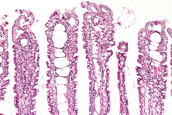
Conference Note: Lymphangiectasia is a common cause of malabsorption and protein-losing enteropathy in dogs. The cause is often presumed to be lymphatic obstruction, such as by neoplastic or inflammatory processes in mesenteric lymph nodes. In many cases, the cause cannot be determined. As the contributor noted, lipogranulomatous inflammation of lymphatics is not common, and the cause is obscure.
Clinical signs of lymphangiectasia result from malabsorption of long-chain fatty acids and loss of plasma proteins and lymph into the gut. Signs include chronic diarrhea, wasting, hypoproteinemia, lymphopenia, relative hypocalcemia, and hypocholesterolemia. Hypoalbuminemia results in peripheral edema, ascites, and hydrothorax, and may have thereby contributed to this dog's chronic cough.
Contributor: Department of Pathobiology, College of Veterinary Medicine, 166 Greene Hall, Auburn University, AL 36849-5519
References:
International Veterinary Pathology Slide Bank:
Laser disc frame #6880, 9474, 10743, 20598, 20608, 20609
Signalment: 4-week-old, layer pullets.
History: There were 30,000 birds in the flock. Birds were inactive, had ruffled feathers, and some had diarrhea with soiling of the vent area. They were undersized for their age, and there was increased mortality.
Gross Pathology: The birds were dehydrated. There were multiple linear hemorrhages within leg muscles. Bursae generally were enlarged, had yellow gelatinous transudate present on the serosal surface and were discolored cream yellow. Plicae were swollen.
Laboratory Results: Bacterial cultures of blood-filtering organs revealed no significant isolations.
Contributor's Diagnosis and Comments: Acute severe necrotizing
bursitis.
Etiology: Birnavirus. Name the Disease: Infectious Bursal Disease
(IBD/Gumboro Disease).
The gross findings of dehydration, multiple linear hemorrhages within leg muscles and the swollen edematous bursae of Fabricius are typical of IBD infection.
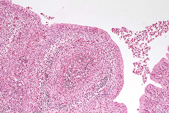
Conference Note: The cloacal bursa (bursa of Fabricius) appears to be the primary target organ of the virus. Gross lesions observed in the bursa follow a sequence of change over time. On day 3 post-infection (PI), the organ begins to increase in size and weight because of edema and hyperemia. By day 4 PI, it is usually twice its normal weight and size; it then begins to become smaller. It continues to atrophy, and by day 8 PI, it is approximately one-third its original weight.1
IBD virus has an affinity for pre-B lymphocytes. Histologic lesions can be found in all lymphoid organs, but are most severe in the cloacal bursa. By 1-2 days PI, there is degeneration and necrosis of lymphocytes in medullae of bursal follicles. Following cell lysis, large amounts of virus are released from the bursa, producing a secondary viremia. In the bursa, lymphoid cells are soon replaced by heterophils, cellular debris, and hyperplastic reticuloendothelial cells. Hemorrhage may occur. As the inflammatory reaction declines, cystic cavities may develop in follicular medullae.
The lesser meal worm (Alphitobius diapernius) is a reservoir host for the virus and may carry the virus for weeks following an outbreak.3
Contributor: Veterinary Laboratory Services, Box 3612, Guelph, Ontario N1H 6R8, Canada
References:
International Veterinary Pathology Slide Bank:
Laser disc frame #4142, 4143, 20493-20496, 21211, 24682
Terrell W. Blanchard
Major, VC, USA
Registry of Veterinary Pathology*
Department of Veterinary Pathology
Armed Forces Institute of Pathology
(202)782-2615; DSN: 662-2615
Internet: blanchard@email.afip.osd.mil
* The American Veterinary Medical Association and the American College of Veterinary Pathologists are co-sponsors of the Registry of Veterinary Pathology. The C.L. Davis Foundation also provides substantial support for the Registry.