Signalment: Adult, male, castrated, bovine.
History: This steer was submitted for slaughter in a USDA-inspected establishment in Texas.
Gross Pathology: The submitting veterinarian noted a
dark, swollen liver with telangiectasis and an enlarged, black
hepatic lymph node with numerous
3-5 mm, yellow, hard granules.
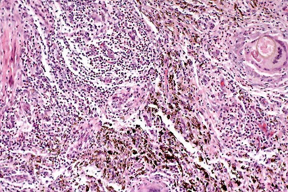
Histology: There is multifocal, nodular to bridging fibrosis of the portal areas with increased numbers of bile ducts and small, congested vessels. Within the fibrous stroma are multiple necrotic foci with large numbers of eosinophils, fibrin, cellular debris, mineral, and numerous oval eggs with a thick, light yellow wall that measure up to 90 x 140 :m in diameter. Many of the eggs are collapsed and defined by a partially disrupted wall that contains mineralized and proteinic globular material. The necrotic foci, eggs, and mineral are surrounded by multinucleated giant cells and epithelioid macrophages that blend into a peripheral zone of fibrovascular tissue with infiltrates of eosinophils, macrophages, plasma cells, lymphocytes and neutrophils. Interspersed with the inflammatory exudate and fibrous stroma is abundant, black granular pigment that is birefringent and yellow-orange with polarized light, consistent with fluke pigment (iron-porphyrin pigment). The surrounding portal areas have mild to moderate multifocal infiltrates of eosinophils, lymphocytes and plasma cells that are often associated with periductal fibrosis and transmigration of the inflammatory cells into the biliary lumina. There is multifocal necrosis and proliferation of plump hyperplastic biliary epithelial cells that surround small numbers of parasite eggs, epithelioid macrophages and multinucleated giant cells in the bile duct lumina.
Contributor's Diagnosis and Comments: Severe multifocal eosinophilic granulomatous cholangiohepatitis with intralesional trematode eggs and pigment.
Etiologic diagnosis: Hepatic fascioliasis, compatible with Fasciola hepatica.
Conference Note: The differential diagnosis of trematodiasis in the bile ducts of cattle should also include Dicrocoelium dendriticum, the lancet fluke. D. dendriticum eggs are much smaller than those of Fasciola hepatica (36-45 x 20-30 :m), and Dicrocoelium usually causes less severe hepatic damage. Also, Eurytrema pancreaticum, which prefers the pancreas, can be found in the bile ducts in severe infections.1,3
Contributor: USDA, FSIS, OPHS-Pathology, Eastern Laboratory, Russell Research Center, P.O. Box 6085, College Station Road, Athens, GA 30604
References:
International Veterinary Pathology Slide Bank:
Laser disc frame #9213, 10689, 16374-6, 22260
Signalment: 12-day-old Angus calf.
History: This was a "poor-doing" calf which developed scours shortly after birth. Treatment with fluorfenical for three days and ceftiofur sodium and electrolytes did not help.
Gross Pathology: The ileum was characterized grossly
by a diffuse fibrinonecrotic membrane replacing the mucosa.
Laboratory Results: A modified acid fast stain for Cryptosporidia
was negative. Fluorescent antibody (FA) examination of intestinal
segments was negative for rotavirus and coronavirus. Electron
microscopic examination of fecal material showed adenoviral particles.
Culture and sensitivity of ileum revealed no significant pathogens.
Virus isolation was positive for bovine adenovirus.
Contributor's Diagnosis and Comments: Ileum: Enteritis, necrotizing and fibrinosuppurative, diffuse, acute, severe with basophilic intranuclear inclusion bodies consistent with bovine adenovirus.

Conference Note: Clinical signs of bovine adenovirus infection include fever, diarrhea which is sometimes bloody, dehydration, and congestion of the mucous membranes of the mouth and muzzle. There may be a serous to mucopurulent ocular and nasal discharge. Gross lesions may be present throughout the gastrointestinal tract. These include ulcers and necrosis in the rumen and abomasum; small intestinal fluid distension, severe multifocal or diffuse necrosis, which may be covered by a pseudodiphtheritic membrane; hemorrhagic necrosis of the colonic mucosa; and marked edema of the mesocolon.5 Other gross lesions include mesenteric lymph node enlargement and edema, multifocal pinpoint red or pale spots in the adrenal cortex, and mottled red areas in the kidneys.1
The true prevalence and significance of enteric bovine adenoviral infection is uncertain. Smyth et al3 believe that adenoviral replication in the vascular endothelium of cattle with enteric disease is underdiagnosed for two reasons: (1) there is no test suitable for diagnosis of this disease in live animals, and (2) rapid autolysis of the intestine often precludes postmortem diagnosis.
Contributor: North Dakota State University, Veterinary Diagnostic Laboratory, Fargo, North Dakota 58105.
International Veterinary Pathology Slide Bank:
Laser disc frame #4351, 20610-11
Signalment: 8-year-old, female, La Mancha goat (Capra aries)
History: This goat was off feed (concentrate) for one
week. It had kids
2½ weeks before presentation. According to the clinician,
the goat had a draining abscess around the face, and respiratory
distress the day before its agonal death.
Gross Pathology: The goat was presented in good nutritional status and with mild postmortem autolysis. The lungs were diffusely firm; however, the cranial half of the lungs were firmer. The tracheobronchial tree contained a copious amount of white frothy fluid. Upon sectioning, a light tan, clear mucus oozed from the airways, and the parenchyma was mottled with innumerable, compact, 1-2 mm white foci. The right mammary gland was firm and congested throughout; no milk was noted in the gland. A whole blood sample taken from the right ventricle of the heart was submitted for serology.
Laboratory Results: Serum was positive for caprine arthritis-encephalitis virus on agar gel immunodiffusion test.
Contributor's Diagnosis and Comments: Lung, pneumonia, interstitial, diffuse, chronic, severe, with alveolar lipoproteinosis (phospholipidosis), caprine arthritis-encephalitis virus.
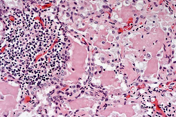
Conference Note: Caprine arthritis-encephalitis (CAE) is a multisystemic lymphoproliferative lentiviral disease of goats that produces persistent chronic-active lesions in the central nervous system, lungs, joints, and mammary glands. This disease leads to a progressive debilitating condition in infected animals. CAE is distributed worldwide.
The major mode of transmission of CAEV to kids under natural conditions is via milk and colostrum. After ingestion, there is a cell-associated viremia with viral replication within blood monocytes and in macrophages of the lung, CNS, synovium, mammary gland, and spleen. Classically, lentiviruses are considered to manifest cell tropism for lymphocytes and macrophages; however, Zink et al4 demonstrated viral RNA in endothelial cells of brain and synovium, as well as in epithelial cells of intestinal crypts, renal tubules, and thyroid follicles. The virus is highly neurotropic, resulting in acute fulminating encephalitis in young kids and chronic encephalomyelitis in those that survive to adulthood. Animals that develop CNS lesions or inapparent infections also develop synovitis, arthritis, and mastitis. The lung lesions are usually found in older animals chronically infected with the virus.
The cachexia which is seen in survivors may be at least partly due to altered metabolism as a result of prolonged elevation of serum TNF-" levels.5 Depressed natural killer cell activity has been observed in infected goats and may contribute to the establishment of a persistent infection.5
The clinical signs and lesions of CAEV infection in goats are similar to those caused by maedi-visna virus (MVV) in sheep. MVV is a lentivirus that is antigenically related to but distinct from CAEV, and is the cause of ovine progressive pneumonia. Experimentally, CAEV can infect sheep, and MVV can infect goats.
Some slides examined in conference contained sections of a nematode parasite in the lung. Participants considered this an incidental finding.
Contributor: Department of Veterinary Pathology, Louisiana State University, School of Veterinary Medicine, Baton Rouge, LA 70803
International Veterinary Pathology Slide Bank:
Laser disc frame #3303, 9706, 20445-7, 20834, 22912
Signalment: 14-month-old, male, quarter horse, equine.
History: Acute onset (24 hours) of neurologic signs including depression, dementia, maniacal behavior, and ataxia leading to recumbency. The horse was unvaccinated. Euthanasia was elected due to progressing signs, poor prognosis, and the possibility of rabies virus infection.
Gross Pathology: No significant gross lesions.
Laboratory Results: A complete blood count and chemistry profile revealed a leukocytosis characterized by a neutrophilia with a mild shift to immaturity. Mild elevations in AST and CK were present in the chemistry profile. Cerebrospinal fluid examination revealed a severe neutrophilic pleocytosis [489 nucleated cells per :l (normal <8 per :l)] and increased protein [172 mg/dl (normal <70 mg/dl)]. The brain was removed and portions were submitted to the state diagnostic laboratory for an IFA test for rabies; negative results were received the following day.
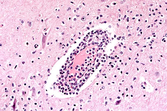
Etiologic diagnosis: Togaviral encephalitis.
Etiology: Eastern equine encephalomyelitis virus.
One section of cerebrum is present with a diffuse, mild to moderate perivascular accumulation of moderate numbers of neutrophils, lymphocytes, plasma cells, and macrophages (perivascular cuffing) with large numbers of neutrophils extending into the surrounding gray matter. Many neurons are in various stages of degeneration and necrosis and some of these are surrounded by dense aggregates of neutrophils and glial cells. There are widely scattered neutrophilic foci throughout the neuropil in the gray matter. The white matter is mildly infiltrated with similar cells, but the majority of the inflammation is contained within the gray matter. The meninges are diffusely infiltrated with small numbers of lymphocytes, plasma cells, neutrophils, and macrophages. Immunohistochemical staining with antibodies against eastern equine encephalomyelitis virus is positive, with the majority of staining in neurons.
Eastern equine enchephalomyelitis (EEE) virus is a single-stranded RNA virus in the family Togaviridae, genus Alphavirus, that causes acute and often fatal illness in horses, ratites, dogs, pigs, cattle, and humans. EEE, western equine encephalomyelitis (WEE), and Venezuelan equine encephalomyelitis (VEE) are caused by related but distinct alphaviruses. EEE and VEE are lethal in approximately 90% of cases, whereas WEE is less virulent with approximately 40% mortality in the horse. In endemically infected areas, EEE and WEE are maintained by a wild bird-mosquito (reservoir-vector) cycle, particularly in swampy or tropical areas. Avian reservoirs maintain sufficient viremia to permit infection of mosquitoes. The infection of domestic animals and humans occurs with the movement of virus from swampy areas carried by reservoirs, vectors, or both. Culiseta and Culex spp. of mosquitoes are most important in maintaining endemic infections.
Episodes of togaviral encephalomyelitis occur in North, Central, and South America, predominantly involving the United States, the Caribbean islands, and the northern regions of South America. This case was diagnosed at the College of Veterinary Medicine at North Carolina State University in July 1995. In 1995, more than 16 cases of EEE were reported in horses in North Carolina. In 1996, more than 11 cases of EEE were reported in horses in North Carolina. In addition, one confirmed canine and one confirmed human case occurred in southeastern North Carolina in the fall of 1996 following Hurricane Fran. Counties in eastern and southeastern North Carolina were ravaged by the hurricane and heavy rainfall. The human case of EEE was that of an adult male who worked as an insurance claims adjuster immediately after the hurricane. The canine case was reported in the same county as the human case.
Neurologic disease in the horse primarily reflects damage to cerebrocortical tissue where the viral infection intensifies. Following natural inoculation, the virus initially infects several tissues, including bone marrow, lymphoreticular tissue, muscle, and connective tissue. A second viremia results in hematogenous infection of the CNS. The exact route of viral entry into the brain is unknown, but viremia with replication in vascular endothelium and subsequent CNS invasion has been proposed. The clinical course is often brief (1 to 2 days), with ataxia and paresis eventually leading to recumbency. Some affected horses survive the infection, but are lethargic due to cerebrocortical injury and are commonly referred to as "dummies". Analysis of cerebrospinal fluid (CSF) usually reveals leukocytosis, characterized initially as neutrophilic, as well as increased protein and xanthochromia. Both a severe neutrophilic pleocytosis and increased protein were present in this case. Occasionally, a prominent eosinophilic pleocytosis is present in CSF.
Gross lesions of EEE are uncommon, but may include edema and multifocal congestion and hemorrhage throughout the brain due to vascular lesions. Microscopic lesions predominate in the gray matter, particularly in the cerebral cortices, thalamus, and hypothalamus. The encephalitic reaction is primarily neutrophilic, with a gradual progression to nonsuppurative inflammation. In fulminating cases where the predominant inflammatory cell is the neutrophil, a characteristic feature is necrosis and fragmentation of the leukocytes. Necrotic neutrophils will occasionally surround degenerate or necrotic neurons. Vascular necrosis and thrombosis are present in some cases. Vasculitis was considered in this case due to the number of inflammatory cells present within vessel walls. However, vasculitis was not diagnosed due to the absence of both fibrin exudation and fibrinoid necrosis of the vessel walls. Due to the extensive neutrophilic infiltration of the surrounding neuropil, the presence of inflammatory cells within vessel walls is most likely due to leukocyte transmigration.
Conference Note: Epornitics in ratites, gallanines, and wild birds may result in widespread morbidity and some mortality, but the lesions in birds are usually limited to the viscera. Encephalitis is not a feature of equine arboviral infection in avians.
Conference participants discussed a differential diagnosis for acute central nervous system disease in the horse, which includes rabies, listeriosis, moldy corn poisoning (leukoencephalomalacia, fumonisin B1 toxicity), lead poisoning, pseudorabies, Borna disease, equine protozoal encephalomyelitis, nigropallidal encephalomalacia (yellow star thistle toxicity), botulism, organophosphate toxicity, equine herpesvirus 1, equine infectious anemia, and others.
Contributor: North Carolina State University, College of Veterinary Medicine, 4700 Hillsborough Street, Raleigh, NC 27606
International Veterinary Pathology Slide Bank:
Laser disc frame #606, 607, 14176-7
Terrell W. Blanchard
Major, VC, USA
Registry of Veterinary Pathology*
Department of Veterinary Pathology
Armed Forces Institute of Pathology
(202)782-2615; DSN: 662-2615
Internet: blanchard@email.afip.osd.mil
* The American Veterinary Medical Association and the American College of Veterinary Pathologists are co-sponsors of the Registry of Veterinary Pathology. The C. L. Davis Foundation also provides substantial support for the Registry.