Results
AFIP Wednesday Slide Conference - No. 10
3 December 1997
- Conference Moderator: Michael A. Eckhaus
NCRR LSS SSB, Bldg. 28A, Rm 117
28 Library Drive, MSC 5210
Bethesda, MD 20892-5210
Return to WSC Case Menu.
Case I - RB97-2341 (AFIP 2593330)
Signalment: Adult, female, pregnant rabbit.
History: This doe was part of an atherosclerosis study
involving embryo transfer. This naturally impregnated doe was
due to deliver on 4/07/97. She delivered one underdeveloped stillborn
kit on 4/03/97 but failed to expel the others. She was lethargic
and depressed and continued to be so on 4/04/97. The animal was
euthanized and submitted for necropsy.
Gross Pathology: The pubic symphysis was partially dilated,
and a serosanguineous discharge was present within the vaginal
canal. Both uterine horns were distended, with the right horn
containing three partially macerated fetuses and the left horn
containing two fetuses in similar stages of degeneration. The
uterine wall was extremely friable and hemorrhagic, with fibrin
strands on the serosal surface of the uterus. A total of seven
placentas was noted, with four present on the right side and three
on the left side. Both the placentas and the wall of the uterus
appeared necrotic. Mild atherosclerosis was present involving
the proximal aorta. The heart appeared grossly normal. The lungs
were diffusely moderately congested. The liver was yellowish-brown
with increased friability, consistent with lipidosis. The kidneys,
spleen, pancreas, and GI tract appeared normal, with a moderate
amount of ingesta present in the stomach.
Laboratory Results: Bacterial cultures were obtained
from the surface and lumen of the uterus. Staphylococcus aureus
was cultured from an intrauterine site.
Contributor's Diagnosis and Comments: Uterus: endometritis,
necrotizing, Staphylococcus aureus, diffuse, severe, acute. Placenta:
ischemic necrosis, diffuse. Fetuses: maceration.
Etiology: Staphylococcus aureus
This rabbit had severe necrotizing metritis due to bacterial
infection of the uterus with Staphylococcus aureus. Numerous bacterial
colonies consistent with Staph. were present in the affected uterine
mucosa, and culture of the uterus yielded pure growth of Staphylococcus
aureus.
Staphylococcosis is characterized by fatal septicemia or suppurative
inflammation in an organ or body site caused by Staphylococcus
aureus and is a common disease in both domestic and wild rabbits.
The disease is usually sporadic and of little economic importance
for commercial rabbitries. Most often, suppurative lesions are
seen with staphylococcal infections, often leading to chronic
abscessation in affected sites. The acute, septicemic form occurs
mostly in neonatal kits and can have lesions ranging from few
and nonspecific to multifocal and suppurative in various organs,
including the lung, kidney, spleen, heart, and liver.
Metritis and fetal death associated with staphylococcal infection
in rabbits are more rare and infrequent findings, but have been
reported in some commercial rabbitries causing serious economic
losses. Pyometra and metritis associated with Pasteurella multocida
are more commonly seen, along with uterine adenocarcinoma, as
the major uterine abnormalities in rabbits. Other bacterial agents
reported infrequently to infect the uterus in rabbits include
Chlamydia, Listeria monocytogenes, Moraxella bovis, Brucella melitensis,
and Salmonella. In the staphylococcal outbreaks, metritis and
abortions were reported, along with mastitis, ulcerative pododermatitis,
interdigital abscesses, lung abscesses and pyothorax. Young rabbits
showed no lesions prior to their death.
- Staphylococcus aureus, a gram-positive, 1 µm, non-motile
and non-spore- forming coccus, is the species most commonly isolated
from affected rabbits and is also the most prevalent bacterial
pathogen affecting humans. It can be spread by direct contact
or by aerosol and can reside in the nasal sinuses or lungs without
showing any apparent disease. Staphylococci produce many extracellular
proteins that add to their pathogenicity: surface proteins that
promote adherence, enzymes that degrade host compounds, and toxins
that damage host cells. Staphylococci induce platelet aggregation,
and S. aureus specifically produces coagulase, both properties
which promote thrombosis of small vessels, as seen in the uterine
submucosa of this rabbit. There is a high correlation between
those Staphylococci that produce coagulase and those that produce
toxins. Since the uterine infection was so acute and severe,
it was likely that a staphylococcal toxin was also involved,
most likely alpha-toxin which selectively damages the endothelial
layer of blood vessels by forming hexameric pores within the
plasma membrane.
-
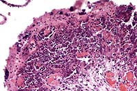
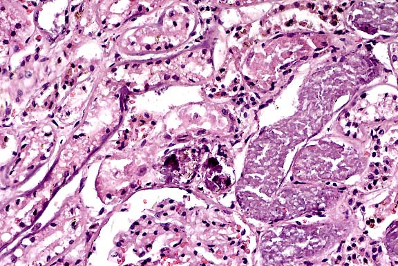
Case 10-1. Uterus. Diffuse caseous necrosis of the endometrium
with bacterial colonies on the surface and dense fibrin thrombi
in the lamina propria. 20X
AFIP Diagnosis: Uterus: Endometritis, necrotizing, acute,
diffuse, severe, with fibrin thrombi, edema, numerous cocci,
and transmural lymphoplasmacytic, neutrophilic and histiocytic
inflammation, rabbit, lagomorph.
Conference Note: A gram-stained section viewed in conference
confirmed that the cocci are gram-positive. Participants also
viewed additional histologic sections from this case, including
sections of placenta and fetal lung. The placenta was diffusely
necrotic, and there were numerous colonies of cocci within the
placenta and mesometrium, and within several fetal bronchioles.
The latter finding suggests that there was periparturient fetal
distress, with aspiration of placental fluid.
In addition to coagulase and -toxin, other significant soluble
factors produced by staphylococci include hyaluronidase, which
depolymerizes hyaluronic acid of connective ground substances;
staphylokinase, which activates serum proteases to induce fibrinolysis;
and -toxin, which lyses erythrocytes.1
Contributor: Veterinary Resources Program, National
Center for Research Resources, National Institutes of Health,
Bethesda, MD 20892-5230
References:
- 1. Cheville NF (ed): Ultrastructural Pathology, Iowa State
Univ Press, pp. 672-75, 1994.
- 2. Hollman A, Girvan G: Staphylococcosis in a commercial
rabbitry. Vet Rec 119: 187, 1986.
- 3. Carolan MG: Staphylococcosis in rabbits. Vet Rec 119:
412, 1986.
- 4. Hobbs BA, Parker RF: Uterine torsion associated with either
hydrometra or endometritis in two rabbits. Laboratory Animal
Science, 40(5), pp. 535-6, 1990.
- 5. Johnson JH, Wolf AM: Ovarian abscesses and pyometra in
a domestic rabbit. J Am Vet Med Assoc, 203(5), pp. 667-9, 1993.
- 6. Laher L, Thorin-Trescases N, Ding A, Laporte R, Osol G:
Alpha-toxin perfusion: a new method for selective impairment
of endothelial function in isolated vessels or intact vascular
beds. Can. J. Physiol. Pharmacol. 73: 1669-73, 1995.
- 7. Percy DH, Barthold SW (eds): Pathology of Laboratory Rodents
and Rabbits, Iowa State Univ Press, pp. 192-3, 1993.
- 8. Weisbroth SH, Flatt RE, Krauss AL (eds): The Biology of
the Laboratory Rabbit, Academic Press, pp. 227-8, 1974.
International Veterinary Pathology Slide Bank:
Laser disc frame #8424, 8497, 13886
Case II - 4833-96 (AFIP 2592284)
Signalment: Adult, male, Cocker Spaniel dog.
History: The dog was treated for heartworms with Caparsolate®
4 days before death. He began vomiting after the last injection
and vomited blood 2 days later. He became very lethargic and continued
to vomit blood until death.
Gross Pathology: The stomach had red-black blotchy hyperemia
on the serosa and mucosa with a moderate amount of dark red liquid,
but no ingesta, in the lumen. Some dark red fluid was in the anterior
intestine but the intestinal mucosa and serosa were not hyperemic.
The kidneys had moderately pale, stippled cortices. The heart
and lungs did not have any heartworms. Other organs were grossly
normal.
Laboratory Results:
- Parvovirus F.A. test -- negative
- Lung culture -- no bacterial growth
Salmonella screen on intestine -- negative
Liver arsenic (wet weight) -- 3.1 ppm
Contributor's Diagnoses and Comments: Necrohemorrhagic
gastritis, severe. Renal tubular necrosis, moderate. Renal tubular
basement membrane mineral- ization, mild.
The arsenic level of 3.1 ppm was considered positive. Caparsolate®
(sodium thiacetarsamide) is hepatotoxic and nephrotoxic and one
side effect is vomiting, although the severe gastric lesions seen
here are not specifically mentioned in the product information.
Acute arsenic poisoning can cause severe hemorrhagic gastritis
or gastroenteritis regardless of the route of exposure. The toxic
range of arsenic is variable, but 7-10 ppm in the liver or kidney
is considered probably toxic, and normal is less than 0.5 ppm.
Arsenic is rapidly cleared from the body (half-life is 1.5 days)
so the level was probably much higher when the Caparsolate®
injections were being given.
- The apparent false diagnosis of heartworm infection was not
explained, but we occasionally get a dog for necropsy without
heartworms that was positive by in-clinic occult heartworm immunoassay
testing.
-
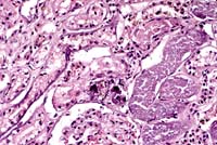
- Case 10-2a. Kidney. Coagulation necrosis of cortical
tubules, focal mineralized lumina debris, and tubular basement
membrane mineralization. 20X

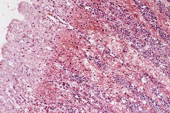
- Case 10-2b. Stomach. Diffuse hemorrhage and necrosis
throughout the mucosa. 10X
- AFIP Diagnoses:
- 1. Stomach: Necrosis and hemorrhage, mucosal and submucosal,
acute, diffuse, with multifocal neutrophilic gastritis, Cocker
Spaniel, canine.
- 2. Kidney: Degeneration and necrosis, tubular, acute, diffuse,
with medullary proteinaceous casts and mineralization of tubular
epithelium and glomerular and tubular basement membranes.
Conference Note: Several conference participants observed
an amphophilic to basophilic granular material, which they interpreted
as mineral, within the gastric mucosa. Results of a Von Kossa
stain performed at the AFIP were equivocal.
Participants considered a differential diagnosis for processes
which might lead to the combination of lesions seen in this dog,
specifically renal tubular and gastric necrosis; gastric hemorrhage;
and mineralization of renal epithelial cells and basement membranes.
Gastric necrosis and mineralization can be secondary to uremia,
so causes of acute renal damage were also considered. The possibility
that this dog had preexisting hepatic or renal disease cannot
be excluded although significant chronic changes are not evident
in the examined sections of kidney. According to the contributor
(personal communication), autolysis of the liver hindered histologic
evaluation of that organ, but hepatic necrosis was not observed.
It is unknown whether or not this dog had hepatic and renal function
tests prior to heartworm treatment. This dog lived in a small
agricultural community, but there was no history of exposure to
arsenic other than via the heartworm treatment, or to any other
known toxins.
Contributor: Arkansas Livestock and Poultry Commission
Laboratory, #1 Natural Resources Drive, Little Rock, AR 72205
References:
- 1. Hatch RC: Poisons causing abdominal distress or liver
or kidney damage. In: Veterinary Pharmacology and Therapeutics,
Booth NH, McDonald LE, eds., Iowa State Press, Ames, IA, 1988,
pp. 1102-1107.
- 2. Kanzler K, managing editor: Veterinary Pharmaceuticals
and Biologicals, 9th ed., Veterinary Medicine Publishing Co.,
Lenexa, Kansas, 1995/1996, pp. 619-620.
- 3. Duncan JR, Prasse KW, Mahaffey EA: Veterinary Laboratory
Medicine: Clinical Pathology, 3rd ed., Iowa State University
Press, Ames, IA, 1994, pp. 190-192.
- 4. Cotran RS, Kumar V, Robbins SL: Robbins Pathologic Basis
of Disease, 5th ed., W.B. Saunders Company, 1994, pp. 980-981.
International Veterinary Pathology Slide Bank:
Laser disc frame #4739, 20585-87
Case III - B96-2171 (AFIP 2595763)
Signalment: 3-month-old, male, F344 rat.
History: This animal was given an experimental material
orally 24 hours before sacrifice.
Gross Pathology: The liver was mottled and pale. There
were no other gross findings.
Laboratory Results: AST: 26,760 (rat normal 75-118)
ALT: 15,360 (rat normal 29-65)
Contributor's Diagnosis and Comments: Moderate to severe,
acute, multifocal to coalescing centrilobular hepatic necrosis.
Acute coagulative necrosis of the centrilobular region (Zone
3 of the liver acinus) is present throughout this section of rat
liver. The necrosis of hepatocytes is most severe in zone 3, with
complete dissolution of cytoplasmic components, karyorrhexis and
karyolysis, and breakdown of sinusoidal endothelium with blood-filled
"lakes" in the immediate vicinity of central veins.
Phagocytosis by large cells assumed to be Kupffer cells is evident
in the centrilobular region, and by Kupffer cells and intact hepatocytes
at the periphery of the necrotic zones. Large numbers of neutrophils
and lesser numbers of other mononuclear cells are abundant in
or at the periphery of the necrotic zone (often approximating
the Zone 2-3 boundary). Increased homogeneity of the hepatocytic
cytoplasm with increased finely granular basophilia exists in
the midzonal to centrilobular regions (Zone 1-2 of the liver acinus).
Mitotic figures are rare throughout the section.
This case of acute hepatic necrosis occurred 24 hours following
administration of coumarin (200 mg/kg). Coumarin and 3,4-dihydrocoumarin
(a related derivative) have widespread use in perfumes, cosmetics,
and other products as a fragrance enhancer. These chemicals were
nominated by the Food and Drug Administration and the National
Cancer Institute for carcinogenicity testing by the National Toxicology
Program (NTP, 1993)
Coumarin toxicity in the liver is characteristic of the spectrum
of lesions observed following acute high-dose administration of
many hepatotoxicants. While no evidence of a regenerative response
is yet apparent, cytoplasmic alterations evident in the midzonal
region are highly suggestive of a chemical toxicity where the
liver might respond with a subsequent regenerative wave of hepatocellular
replication. These early cytoplasmic alterations are also consistent
with studies illustrating the induction of microsomal enzymes
by coumarin, an effect also characteristic of many hepatic toxicants
and chemical carcinogens. Under chronic administration, coumarin
has been shown to induce minor increases in renal tubular cell
adenoma in male F344 rats, but did not induce hepatic tumors in
male or female rats. In these two-year rat studies, the acute
hepatocellular necrosis and cytoplasmic alterations observed at
this dose have been shown to persist with continued exposure,
and were shown to progress to bridging hepatic fibrosis with bile
duct proliferation but not neoplasia.
- The role of cell proliferation in the induction of chemical
carcinogenesis has received considerable attention in toxicologic
pathology. The hepatotoxicity demonstrated by coumarin in F344
rats is not an infrequent finding in industrial toxicology. Particularly,
this form of "sustained" and "regenerative"
cell proliferation has often been implicated in the hepatocarcinogenicity
of tested ingredients. The case with coumarin in the rat illustrates,
however, that continued marked hepatocellular damage and regenerative
repair do not always result in a hepatocarcinogenic response.
-
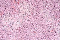
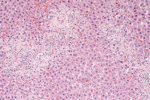
- Case 10-3. Liver. Centrilobular necrosis bridging
to adjacent central vein regions.Periportal and mid-zonal hepatocytes
are not affected. Note the portal triad (toward bottom). 10X
- AFIP Diagnosis: Liver: Necrosis, centrilobular, bridging,
diffuse, with neutrophilic inflammation, F344 rat, rodent.
Conference Note: Coumarin derivatives more familiar
to conference participants are those which have been synthesized
for use as anticoagulant rodenticides, such as warfarin, fumarin,
pindone, diphacinone, brodifacoum, and bromadiolone. Moldy sweet
clover poisoning of cattle was one of the first documented coagulopathies
in animals. The sweet clover plant (Melilotus alba)contains coumarin,
which is converted to dicumarol when the plant spoils in hay or
silage. Ingestion of dicumarol or any of the above compounds results
in the inhibition of vitamin K epoxide reductase, which causes
the production of nonfunctional forms of the vitamin K-dependent
coagulation factors II (prothrombin), VII (proconvertin), IX (Christmas
factor), and X (Stuart-Prower factor). Factor VII has the shortest
half-life, and thus is the first to be depleted, followed by factors
IX and X. Prothrombin, with a half-life of about 40 hours, is
the last to disappear. Depletion of these factors prolongs both
the PT and the APTT, but does not interfere with platelet numbers
or function, nor does it decrease fibrinogen concentration.
Contributor: The Proctor & Gamble Company, Miami
Valley Laboratories, P.O. Box 398707, Cincinnati, Ohio 45239-8707
References:
- 1. National Toxicology Program Technical Report (NTP TR 422).
1993. Toxicology and carcinogenesis studies of coumarin (CAS
No. 91-64-5) in F344/N rats and B6C3F1 mice. US Department of
Health and Human Services, National Institutes of Health (NIH
Publication No. 93-3153).
- 2. Valli VEO: The hematopoietic system. In: Pathology of
Domestic Animals, 4th edition, Jubb KVF, Kennedy PC, Palmer N,
eds., Academic Press, 1993, vol. 3, p. 264.
- 3. Duncan JR, Prasse KW, Mahaffey EA: Veterinary Laboratory
Medicine: Clinical Pathology, 3rd ed., Iowa State University
Press, Ames, IA, 1994, pp. 82-86.
International Veterinary Pathology Slide Bank:
Laser disc frame #16318
Case IV - Mü 2028/96 (AFIP 2600442)
- Signalment: 16-year-old, female, lion (Panthera leo).
History: This animal was euthanized. The lesion was
an incidental finding at necropsy.
Gross Pathology: Bilateral, multiple, thin-walled cysts,
up to 2 cm in diameter, were visible in the ovarian parenchyma.
Contributor's Diagnosis and Comments: Cystic rete ovarii.
Multiple cysts of variable diameter (up to 2 cm) displacing
the ovarian parenchyma are visible. The cyst lining varies from
single to several layers of flattened to cuboidal epithelium.
Some of the cysts are lined by cuboidal ciliated epithelium. Several
cysts are filled with proteinaceous fluid. Occasionally folds
which protrude into the cyst lumen and contain a blood vessel
in the apical part can be observed.
In cats, as in other mammals, the rete ovarii consists of three
parts: an intraovarian rete system, a connecting rete system,
and an extraovarian system. The intraovarian rete is located in
the ovarian medulla and is lined by cuboidal epithelium. At the
tubular extremity of the ovary, the intraovarian system becomes
a reticular formation of dilated tubules lined by ciliated columnar
epithelium- the connecting rete. The extraovarian rete consists
of tubules composed of ciliated columnar cells extending from
the connecting rete and ending blindly in the periovarian tissue.
Cysts of the rete system have been described in different species
including cats. The epithelial lining of the cyst walls can vary
from single to several layers of cuboidal or flattened to columnar
and ciliated epithelial cells.
- In this case the pathological findings in the ovaries were
incidental findings at necropsy and did not result in any clinical
abnormality. The animal was not part of a breeding program and
therefore no attempts were made to breed her.
-
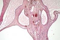
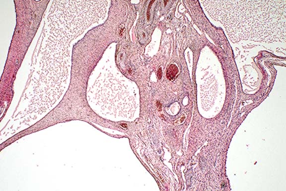
- Case 10-4. Ovary. Multiple lined by very attenuated
epithelium sometimes contain floccular eosinophilic material.
A single ovarian follicle is in the collagenous stroma (center).
2X
- AFIP Diagnosis: Ovary: Cystic rete ovarii, lion (Panthera
leo), feline.
Conference Note: Cysts of the ovarian rete are only
one of sixteen different types of cysts occurring in and around
the ovary of domestic animals.3 The incidence and relative significance
of these is variable among animal species. In the cow and sow,
follicular cysts are most frequent, and are important causes of
infertility. These occur less frequently in the bitch and queen.
Cats with follicular cysts may show signs of hyperestrogenism.
On the other hand, cystic rete ovarii are most common in the bitch
and queen, and seldom if ever cause clinical signs.
Histologic differentiation of the various types of cysts can
be difficult. Important features include the type(s) of epithelium
lining the cyst, presence or absence of smooth muscle in the wall
of the cyst, and anatomic location.
Contributor: Institute of Animal Pathology, Zoo Animal
Pathology, University of Berne, Laenggassstrasse 122, CH 3012
Berne, Switzerland
References:
- 1. Gelberg HB, McEntee K, Heath EH: Feline cystic rete ovarii.
Vet. Pathol. 21:304-307, 1984.
- 2. Wenzel JGW, Odend'hal S: The mammalian rete ovarii: a
literature review. Cornell Vet: 75:411-425, 1985.
- 3. McEntee K: Reproductive Pathology of Domestic Mammals,
Academic Press, Inc., 1990, pp. 52-68.
International Veterinary Pathology Slide Bank:
Laser disc frame #1019, 2704, 5145, 11889
Terrell W. Blanchard
Major, VC, USA
Registry of Veterinary Pathology*
Department of Veterinary Pathology
Armed Forces Institute of Pathology
(202)782-2615; DSN: 662-2615
Internet: blanchard@email.afip.osd.mil
* The American Veterinary Medical Association and the American
College of Veterinary Pathologists are co-sponsors of the Registry
of Veterinary Pathology. The C.L. Davis Foundation also provides
substantial support for the Registry.
Return to WSC Case Menu.