
Signalment: 8-year-old, female, European Shorthair cat (Felis domesticus)
History: Presented with acute vomiting and diarrhea. Results of a clinical examination two weeks later showed poor general condition, and reduced motility and proprioception of the hind legs. The animal was then euthanized.
Gross Pathology: There was enlargement and loss of structure of the jejunal lymph nodes, splenomegaly, and distinct lobulation of the liver.
Laboratory Results: There was a marked leukocytosis (>200,000/µL), with a predominance of medium-sized to large mononuclear cells with obvious azurophilic granulation in blood smears (Pappenheim's stain). A serological test for FeLV antibodies was negative.
Contributor's Diagnosis and Comments: Diagnosis: Large granular lymphoma, extranodal, peripheral blood involvement.
Specimens of lymph nodes (including jejunal lymph nodes), sternal and femoral bone marrow, stomach, jejunum, spleen, liver, kidney, lung, heart, spinal cord and brain were examined histologically.
Jejunal lymph node: High-grade infiltration of the sinuses and the paracortex by medium-sized to large monomorphic, slightly basophilic lymphoid tumor cells. Focal invasion of the capsule and cortical area with partial effacement of the lymph node architecture. Single megakaryocytes can be seen within the sinuses. Within areas of dense tumor cell infiltration there are 3 mitoses per high power filed. The nuclear/cytoplasmic ratio of the neoplastic cells is moderate, numerous nuclei are indented. A marked azurophilic granulation of the cytoplasm near the indentations of the nuclei can be seen in Giemsa-stained plastic sections, whereas this is hardly noticeable in H&E-stained plastic sections and invisible in paraffin section (both H&E- and Giemsa-stained.). The granules react positively for Chloroacetate- esterase and acid phosphatase (plastic sections). The neoplastic cells are labeled by an antibody specific for the epsilon chain of the human CD3 complex (paraffin sections). Tumor cell infiltrations were seen within the tunica muscularis of the jejunum, in the propria mucosae of the gastric fundus, in the lung, in the myocardium, in the kidneys, in the epidural space, in the pachymeninx and in the white substance of the spinal cord.
Ultrastructural findings: Maximum diameter of the tumor cells approximately 10 micrometers. The nuclei are either round or oval or irregularly shaped and indented, with coarse heterochromatin and a small nucleolus. Numerous mitochondria and poorly developed rER and Golgi complex. Numerous membrane- bound electron-dense bodies of up to 1.4 micrometer in diameter and small numbers of multivesicular bodies in the cytoplasm. Scarce matrix granules present in the cytoplasm of some cells.
AFIP Diagnosis: Lymph node: Granulated round cell tumor of cats, European Shorthair Cat, feline.
Conference Note: Much of the conference discussion centered around the difficulty of classifying feline discrete, round cell neoplasms of presumed lymphoid origin. The signalment and history in this case are consistent with the typical clinical syndrome usually associated with large granular lymphoma in cats, i.e. 6-9 years of age, FeLV negative, with involvement of gastrointestinal organs as well as the thymus. This contrasts with feline lymphosarcoma associated with infection with FeLV, in which cases involving the gastrointestinal tract seldom involve thymus, and vice versa.
The cell (or cells) of origin of granulated round cell tumors (GRCT) of cats remains controversial. One report suggests that GRCT are unique among feline neoplasms, and that the morphologic, histochemical, and ultrastructural features are most consistent with globule leukocytes or possibly cells in transition between mucosal mast cells and globule leukocytes.5 Kariya, et al., suggest that feline globule leukocytes are intraepithelial large granular lymphocytes, since they were able to demonstrate immunoreactivity for perforin, a potent mediator of cytotoxicity which is found in granules of cytotoxic T lymphocytes and natural killer (NK) cells.4 Neoplasms composed of cells with morphologic features of globule leukocytes have been reported in several other species, including horses, rats, and man. In humans, immunophenotypic results indicate that granular lymphocytes include NK cells and several subsets of T lymphocytes.4
Contributor: Institute of Veterinary Pathology, University of Munich, Veterinaerstrasse 13, D80539 Munich, Germany
References:
1. Cheney CM, Rojko JL, Kociba GJ, Wellman ML, DiBartola SP, Rezanka LJ, Forman L, Mathes LE: A feline large granular lymphoma and its derived cell line. In Vitro Cell. Dev. Biol. 26:455-463, May 1990.
2. Emile JF, Boulland M-A, Haioun C, Kanavaros P, Petrella T, Delfau-Larue M-H, Bensussan A, Farcet J-P, Gaulard P: CD5 CD56+ T-cell receptor silent peripheral T-cell lymphomas are natural killer cell lymphomas. Blood 87(4):1466-1473, 15 Feb 1996.
3. Jaffe ES: Classification of natural killer (NK) cell and NK-like T-cell malignancies. Blood 87(4): 1207-1210, 15 /feb 1996.
4. Kariya K, Konno A, Ishida T: Perforin-like immunoreactivity in four cases of lymphoma of large granular lymphocytes in the cat. Vet Pathol 34(2):156-159, 1997.
5. McEntee MF, Horton S, Blue J, Meuten DJ: Granulated round cell tumor of cats. Vet Pathol 30:195-203, 1993.
6. Prasthofer EF, Barton JC, Zarcone D, Grossi CE: Ultrastructural morphology of granular lymphocytes (GL) from patients with immunophenotypically homogenous expansions of GL populations (GLE). J. Submicrosc. Cytol 19(2):345-354, 1987.
International Veterinary Pathology Slide Bank:
Laser disc frame # 14281-85, 14459-61, 23336.
Signalment: Five-month-old female Domestic Shorthair cat
History: This cat presented with a 2-3 day history of an uncoordinated and stiff gait. On presentation she was recumbent, mentally dull, and lacked conscious proprioception in all four limbs. Vertical nystagmus could be elicited with manipulation of her head.
Gross Pathology: A smooth, pink to white solid mass was located over the rostral brain stem and displaced the cerebellum caudally. Bilateral hydrocephalus was noted. In addition, the lungs were slightly dull and failed to collapse completely.
Laboratory Results: None submitted.
Contributor's Diagnosis and Comments: Brainstem/cerebellum: Medulloblastoma. Lung (sections not provided): Pneumonia, granulomatous, locally extensive, chronic, mild, with intralesional nematodes (Aelurostrongylus abstrusus).
Medulloblastomas in domestic animals are rare but most frequently reported in calves and puppies. They occur almost exclusively in the cerebellum and cause the expected clinical signs of ataxia and tremors. Spread by subpial extension and dissemination within the central nervous system via cerebrospinal fluid are features of these fast growing and highly malignant tumors.
Although rare in animals, medulloblastomas are the most common embryonal tumors of children and comprise 20% of all pediatric brain tumors. The histogenesis of these tumors is somewhat controversial and the target of active investigation. Originally, the neoplasm was thought to arise from a specific cell of the external granular layer of the cerebellum; however, more current theories revolve around the hypothesis that a population of multipotent progenitor cells gives rise to a number of different tumor types. These tumors are collectively called primitive neuroectodermal tumors, or PNETs, and include neuroblastomas, pineoblastomas, and retinoblastomas.
Two separate cytologic characteristics have been examined in an effort to better understand the origin of medulloblastomas. The first involves the spatial and temporal expression of cell markers (particularly neurofilaments) with regard to the level of cellular differentiation. It appears that differentiation along neuronal or glial lineages is not as common in medulloblastomas of domestic animals as compared to humans. Neuronal phenotype has been confirmed in many human tumors with the use of immunohistochemistry and electron microscopy to evaluate neuronal cytoskeletal components, beta 3 tubulin, MAP-2 [microtubule-associated protein], phosphorylated neurofilament proteins, and synaptophysin. Up to 50% of tumors are positive for glial fibrillary acidic protein (GFAP), but alternative explanations for its presence, such as a stromal induction phenomenon or the induction of perivascular glial cells by the tumor cells, have been proposed. Nestin, a class VI intermediate filament, has been identified in most medulloblastomas. Nestin is found in other embryonal tumors of the central nervous system and is not expressed in mature differentiated neuroectodermal cells or tumors.
The second cellular feature that has been examined is the relationship between certain growth factors and receptor expression by neoplastic cells. Platelet-derived growth factor (PDGF) has been shown to have specific cellular and spatial distribution within the developing central nervous system. Several isoforms have been identified and each acts on unique cellular receptors to partially regulate differentiation. In human medulloblastomas, the expression of PDGF receptors has been shown to correlate with the contrasting phenotypic features of cerebral and cerebellar PNETs. Another family of growth factors, the neurotrophins, includes nerve growth factor (NGF). Different types of NGF receptors with tyrosine kinase activity are associated with changes in patient survival time and freedom from progression of disease.
Because several associations have been made between medulloblastomas and multiple-malignancy syndromes in people, large numbers of specimens have been karyotyped. The most frequent chromosomal abnormality is the loss of the entire short (p) arm of chromosome 17, resulting in isochromosome 17q, but non- random partial deletions also occur. Isochromosome 17q is frequently detected in leukemias, lymphosarcomas, and some gastrointestinal tumors. Although the tumor suppressor gene p53 is located on the short arm of chromosome 17, mutations resulting in decreased expression of p53 do not appear to be common in medulloblastomas. Recent studies present the possibility of a novel tumor suppressor gene at a site distal to p53 on chromosome 17. In support of this idea, the preferential loss of these DNA sequences is reported in several other tumor types.

AFIP Diagnosis: Brain stem: Primitive neuroectodermal tumor, Domestic Shorthair cat, feline.
Conference Note: A majority of conference participants preferred the diagnosis of PNET because cerebellar origin could not be conclusively demonstrated in the sections viewed in conference. This case was also reviewed by members of the Department of Neuropathology of the AFIP. They agree with the interpretation of PNET and add that, in view of the fragments of cerebellum (not present in all slides) and the clinical history of cerebellar signs, the findings are consistent with medulloblastoma.
Contributor: Department of Pathology, Angell Memorial Animal Hospital, 350 South Huntington Ave, Boston, MA 02130.
References:
1. Smits A, et al: Coexpression of platelet-derived growth factor and receptors on medulloblastomas and other primitive neuroectodermal tumors is consistent with an immature stem cell and neuronal derivation. Lab Invest 74: 188-198, 1996.
2. Rutka J, Hoffman H: Medulloblastoma: A historical perspective and overview. J Neurooncol 29: 1-7, 1996.
3. Provias JP, Becker LE: Cellular and molecular pathology of medulloblastoma. J neurooncol 29: 35-43, 1996.
4. Goumnerova L: Growth factor receptors and medulloblastoma. J Neurooncol 29: 85-89, 1996.
5. Cogen PH, McDonald JD: Tumor suppressor genes and medulloblastoma. J Neurooncol 29: 103-112, 1996.
6. Iijima M, Nakazato Y: Pale islands in medulloblastoma consist of differentiated cells with low growth potential. Pathology International 47: 25-30, 1997.
7. Moore KD, et al: In vitro properties of a newly established medulloblastoma cell line, MCD-1. Molecular and Chemical Neuropathology 29: 107-126, 1996.
8. Summers BA, Cummings JF, de Lahunta A: Veterinary Neuropathology, Mosby- Year Book, Inc., pp.375-391, 1995.
International Veterinary Pathology Slide Bank:
Laser disc frame #1920-21, 5587, 14236.
Signalment: 8-year-old male meerkat (Suricata suricatta)
History: This animal was in a zoo collection and noted to have episodes of weakness and abnormal behavior. The clinical diagnosis was syncope from either cardiomyopathy or epilepsy and it presented 3 days before death in a semi- comatose state with dyspnea. There was no response to supportive care, and no other diagnostic tests were performed.
Gross Pathology: The lungs were swollen, dark red and heavy with serosanguinous fluid. Portions sunk in formalin. All other visceral organs appeared normal. Upon opening the calvarium, there were bilateral grayish yellow masses measuring up to 25mm adherent to both cerebral cortices. The right mass was smaller and distinctly indented the frontal cortex, while remaining entrapped in the dura. The left mass was not easily removed from the parenchyma and a third 10mm mass was found at the cerebello-pontine angle, mid-line between the occipital lobes. The texture of all masses was similar, firm and granular with softer foci of possible necrosis.
Laboratory Results: None
Contributor's Diagnosis and Comments: Multiple meningiomas; etiology - unknown.
No primary cardiac pathology was found but there was severe subacute hemorrhagic alveolitis, attributed to respiratory distress syndrome, possibly the consequence of a brain-heart syndrome. The myocardium was edematous with subendocardial tattered and hypereosinophilic fibers. Other microscopic findings included severe global glomerulonephritis and sclerosis with hemoglobinuric nephrosis, acute periacinar hepatic necrosis, a pulmonary alveolar adenoma and a Hurthle cell adenoma.
All three meningeal masses have similar histology, composed of well- circumscribed proliferations of elongated spindle cells with fine cytoplasmic extensions and fibrillary processes, forming empty clefts with intervening hemorrhage. There is a slightly lobulated pattern with more plump spindle cells aggregated in a pseudolobular pattern. Little mineralization is detected but there are many accumulations of hemosiderin-laden macrophages. The masses consistently embed into and deform or atrophy the cortical gyri and, in one area, expand into a sulcus with focal infiltrations via Virchow-Robins spaces.
Meningiomas are mesodermal tumors from the leptomeninges and commonly divided by their morphological pattern and the degree of atypia. Multiple tumors are reported and not often indicative of malignant behavior, in which invasion and metastasis remain the distinguishing criteria. The tumors in this meerkat all appear to classify as the fibroblastic variants, but this is not widely followed in the veterinary literature and rarely carries any prognostic significance. These are judged multicentric in origin (as in domestic cats) and show no sign of malignant cytology or behavior. The theory that meningiomas can derive from previous cranial trauma has usually been discounted and no other etiologic factors are known.


AFIP Diagnosis: Meninges: Cholesterol granuloma, meerkat (Suricata suricatta), viverrid.
Conference Note: Although careful consideration was given to the diagnosis of meningioma, conference participants unanimously preferred the above diagnosis. The lesion is composed of numerous cholesterol clefts, admixed with pigment-laden foamy cells (histiocytes), hemorrhage, and multiple foci of mineralization. Arachnoidal cap cell hyperplasia was observed in some sections. Members of the Department of Neuropathology of the AFIP reviewed this case and believe this lesion represents a reactive process, not a meningioma.
Cholesterol granuloma, or cholesteatosis, in the choroid plexus occurs in 15- 20% of old horses. We are unaware of reports of this condition in meerkats. In horses, cholesterol granulomas are most frequently found in the fourth ventricle; however, those present in the lateral ventricles are more important because they may attain large size and cause hydrocephalus by obstructing the foramina of Monro.
The pathogenesis of cholesteatosis is obscure, but appears to be related to chronic or intermittent congestion and edema, with congestive hemorrhages in the choroid plexuses. During an edematous episode, the interstitial tissues are infiltrated with macrophages containing lipid and hemosiderin. The crystals of cholesterol are deposited in the tissue spaces and apparently act as foreign bodies to stimulate a low-grade productive inflammation.
Contributor: Los Angeles County - DHS, Veterinary Public Health, 3015 Roxanne Avenue, Long Beach, CA 90808
References:
1. Rubenstein LJ. Tumors of the Central Nervous System. In: Atlas of
Tumor Pathology, 2nd series, fascicle 6. Washington, D.C., Armed Forces
Institute of Pathology, 1972, pp. 169-190.
2. Jubb KVF, Huxtable CR. The Nervous System. In: Jubb KVF, Kennedy PC,
Palmer N (eds.): Pathology of Domestic Animals, 4th ed., v. 1. San Diego,
Academic Press, 1993, pp. 334-335, 430-432.
3. Cordy DR. Tumors of the Nervous System and Eye. In: Moulton JE (ed.): Tumors of Domestic Animals, 3rd ed., Berkley, U of California Press, 1990, pp. 650-652.
4. Summers BA, Cummings JF, de Lahunta A: Veterinary Neuropathology,
Mosby- Year Book, Inc., pp.52-3, 1995.
International Veterinary Pathology Slide Bank:
Laser disc frame #1222, 1316, 2660, 7427, 11048, 14569, 16954-55
Signalment: 6-month-old male llama.
History: Dermatitis beginning soon after birth. Crusts and alopecia on face, muzzle, ventral neck, ventral thorax and the perianal region. Treated with antibiotics and antifungal with some improvement, then became worse. Animal has not grown well and was euthanatized.
Gross Pathology: Crusts and alopecia on skin.
Laboratory Results: None
Contributor's Diagnosis and Comments: Chronic hyperplastic and pustular dermatitis with marked hyperkeratosis, consistent with generalized munge.
This is a chronic hyperplastic perivascular dermatitis but the most striking change is the alternating layers of purulent inflammation and hyperkeratosis forming a thick crust on the surface. There is intracellular edema of the superfical epidermis with neutrophilic exocytosis and pustule formation.
The condition in this llama is consistent with a disease called generalized munge or idiopathic necrolytic/neutrophilic/hyperkeratotic dermatosis. The disease occurs most often in young llamas and may be localized to the nose and perioral region or become generalized as in this case. The histology is similar to superficial necrolytic dermatitis of humans and dogs, but there is no underlying liver or pancreatic disease. The cause is unknown, but like the canine disease, it may be a manifestation of deficiencies of zinc, amino acids and fatty acids.
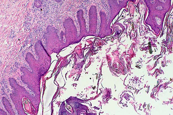
Conference Note: In some of the sections examined in conference, there were one or more cross-sections of an ectoparasite within the hyperkeratotic crust. The organism has jointed appendages with prominent setae, striated muscle, and mouth parts. This case was reviewed by Chris Gardiner, PhD, veterinary parasitology consultant to the AFIP. He confirmed that the organisms are mites but was unable to provide a more specific classification.
In some species of animals, lesions of acariasis can be similar to those
seen in this llama. Although the contributor's diagnosis cannot be excluded,
mange is favored over munge.
Contributor: Department of Biomedical Sciences & Pathobiology,
College of Veteirnary Medicine, Virginia Tech, Blacksburg, VA 24061-0442.
References:
1. Rosychuk RAW. Llama dermatology. In: Veterinary Clinics of North America, Food Animal Practice 7:234-239, 1994.
2. Yager JA, Scott DW. The skin and appendages. In: Pathology of Domestic
Animals, Jubb KVF, Kennedy PC, and Palmer N (eds.), Academic Press, Inc.,
vol. 1, pp. 681-691.
International Veterinary Pathology Slide Bank:
Laser disc frame # - none.
.
Terrell W. Blanchard
Major, VC, USA
Registry of Veterinary Pathology*
Department of Veterinary Pathology
Armed Forces Institute of Pathology
(202)782-2615; DSN: 662-2615
Internet: blanchard@email.afip.osd.mil
* The American Veterinary Medical Association and the American College of Veterinary Pathologists are co-sponsors of the Registry of Veterinary Pathology. The C.L. Davis Foundation also provides substantial support for the Registry.
Return to WSC Case Menu.