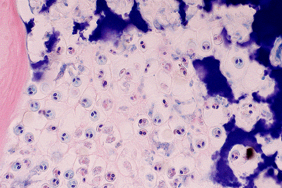
 M. cerebralis trophozoites
within the cartilage of the head in a rainbow trout. Note the
characteristic two prominent polar bodies within each parasite.
(40X, HE, 97K)
M. cerebralis trophozoites
within the cartilage of the head in a rainbow trout. Note the
characteristic two prominent polar bodies within each parasite.
(40X, HE, 97K)
Signalment: Heads from twenty-two, 1-year-old, male and female, rainbow trout (Oncorhynchus mykiss) harvested from Willow Creek, MT (southwest of Bozeman) were submitted for evaluation.
History: Numbers of wild rainbow trout in select rivers and streams in Montana have decreased steadily since 1991. For example, in the 56-mile stretch of the Madison River, between Quake Lake and Ennis Lake, the population has declined from about 3,300 fish per mile in 1991 to about 300 per mile in 1994.1 The cause is unknown, but the Montana Department of Fish, Wildlife, and Parks suspects whirling disease, caused by the myxosporidian protozoa, Myxobolus cerebralis.1
Gross Pathology: No gross lesions were observed.
Laboratory Results: Enzymatic digests performed on head samples showed numerous, approximately 20 æm diameter, acid-fast, spherical spores containing 2 piriform polar bodies and an ovoid sporoplasm, consistent with M. cerebralis.
Contributor's Diagnosis and Comments: Chondritis, granulomatous, multifocal, moderate, chronic, with intralesional myxosporidian trophozoites and spores, consistent with Myxobolus cerebralis.
Granulomatous chondritis with intralesional trophozoites and mature spores are common in juvenile fish infected with M. cerebralis. In older fish, islands of retained, hyperbasophilic cartilage with entrapped spores are within bone; associated inflammation is rare.
The life cycle of M. cerebralis involves a tubificid oligochaete in which numerous motile actinospores (Triactinomyxon gyrosalmo) are produced.2 Actinospores are ingested by trout or directly penetrate the mucous membranes and migrate to bone and cartilage. Asexual reproduction results in numerous trophozoites followed by spore formation within cartilage.3
AFIP Diagnosis: Head, cartilage and bone: Chondritis, granulomatous, multifocal, moderate, with osteitis, perichondritis, and numerous myxosporidian organisms, rainbow trout (Oncorhynchus mykiss), piscine, etiology consistent with Myxobolus cerebralis.
Conference Note: Myxobolus cerebralis utilizes a tubificid worm as an intermediate host. The worm ingests spores of M. cerebralis which then develop into actinosporea in the gut epithelium of the worm. Actinosporea have been shown to infect fish via the water or by ingestion of the worm; presumably the actinosporea penetrate the gut and migrate to cartilaginous tissues where they develop into the spores of M. cerebralis. Although actinosporea have been classified as belonging to the genus Triactinomyxon, it seems that the organisms which comprise this genus may be intermediate forms of myxosporidian parasites. In short, taxonomists have yet to determine how these parasites will be classified.
All species of trout, salmon, and grayling are susceptible to M. cerebralis, the organism responsible for "whirling disease". Clinical signs of whirling disease range from inapparent infection to erratic swimming and death. The abnormal swimming posture and tail-chasing behavior is caused by damage and deformity of the tail and spine secondary to the granulomatous chondritis induced by M. cerebralis. Diagnosis is dependant on demonstrating the protozoa in tissue sections. Identification of the spores may be enhanced by use of Giemsa or acid-fast stains; the spores of M. cerebralis will stain with either.
Vitamin C deficiency is also a common cause of scoliosis in salmonid fish and must be differentiated from infection with M. cerebralis.
Contributor: Washington State University
Department of Veterinary, Microbiology and Pathology
College of Veterinary Medicine, WSU
Pullman, Washington 99164-7040
References:
Signalment: Foal, Equine, 3 - 4 hours old.
History: Bloody diarrhea before death.
Contributor's Diagnosis and Comments: Marked to severe, diffuse, subacute, necrotizing enteritis with emphysema and intralesional bacilli.
This lesion is most consistent with necrotizing enteritis due to clostridial infection. Cultures were not done by the referring veterinarian but the morphology of the bacilli is consistent with Clostridium sp. Some clostridial species induce production of gas within tissues as they become established (gas gangrene) which has been attributed to a composite effect of toxins secreted by the organism.
AFIP Diagnosis: Small intestine: Enteritis, necrotizing, peracute to acute, diffuse, severe, with edema, hemorrhage, emphysema, and myriad mucosal adherent bacilli, breed unspecified, equine, etiology consistent with Clostridium spp.
Conference Note: Clostridial enterotoxemia is usually caused by Clostridium perfringens, a gram-positive, anaerobic, commensal bacillus. Clostridium perfringens has been subdivided into 5 substrains, A through E, depending on the combination of the four major exotoxins (alpha, beta, epsilon and iota toxin) produced. Alpha toxin is a lecithinase which degrades cell membranes. Beta toxin appears to paralyze the gut. Epsilon toxin is a proenzyme that is activated by enzymatic digestion by trypsin. Iota toxin is also a proenzyme that increases capillary permeability. Additionally, strains A, C, and D produce an enterotoxin. The enterotoxin is released only when the bacillus is lysed (thus, it is not an exotoxin) and is associated with the induction of diarrhea.
The majority of veterinary cases of clostridial enteritis in North America are caused by Clostridium perfringens Type C. The toxins produced by C. perfringens type C (alpha and beta toxin) produce a lesion in the intestine that is similar in appearance to autolysis. The villar mucosa is denuded of epithelium, often leaving only shrunken pegs of the lamina propria in place of the villi. The submucosa is expanded by edema and hemorrhage, and may contain neutrophils. There is often emphysema of the intestinal wall due to bacterial gas production.
Clostridium difficile is another cause of acute necrotizing enteritis in neonatal foals. C. difficile produces two toxins, A and B. Toxin A is an enterotoxin and toxin B is a cytotoxin that may act synergistically with Toxin A. The histologic lesion of C. difficile is similar to that of C. perfringens; culture and/or toxin identification is required to differentiate them.
Contributor: Pal-Path, Inc.
1277 Record Crossing Road
Dallas, Texas 75235
References:
Signalment: Juvenile (approximately 90-days-old), Pacific white shrimp, Penaeus vannamei.
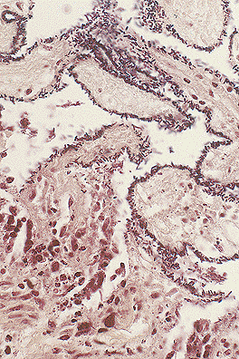
 Necrosis of the cuticular epithelium
in a Pacific white-sided shrimp.(40X, HE, 81K)
Necrosis of the cuticular epithelium
in a Pacific white-sided shrimp.(40X, HE, 81K)
History: Shrimp were lethargic; sea gull activity over the pond; few dead shrimp on bottom of the pond.
Gross Pathology: Tail region of the shrimp was discolored red.
Contributor's Diagnosis and Comments: Acute multifocal cuticular epithelial necrosis, Taura Syndrome Virus.
Taura Syndrome was named after an area in Ecuador where it was first recognized in June 1992. 0.1-5.0 gram shrimp in the nursery phase are primarily affected. Affected shrimp have expanded chromatophores which result in the red discoloration. The causative agent has been tentatively identified as a nodavirus, similar to RNA viruses of insects.
AFIP Diagnosis: Cuticular epithelium: Necrosis, multifocal, Pacific white shrimp (Penaeus vannamei), crustacean.
Conference Note: Taura syndrome virus has been tentatively classified as either a picornavirus or a nodavirus. Taura virus has a worldwide distribution and has substantially impacted farmed shrimp production; pond mortality rates have approached 90% in some outbreaks. Taura syndrome virus primarily affects shrimp of 0.1 to 5.0 gm body weight, that is, approximately 15-40 days after stocking postlarvae.
Histologically, infected shrimp develop multifocal necrosis of the cuticular epithelium. Infected epithelial cells are reported to develop intracytoplasmic inclusion bodies of 1-20 æm in diameter; however, inclusion bodies were not evident in the sections examined during the conference. Affected shrimp have expanded chromatophores which impart a red or blue hue, depending on the dominant chromatophore, to the shell and tail. Infected shrimp often die in molt or have soft shells.
Studies of the structure, genetic sequence, pathogenesis and host range of Taura syndrome virus are in progress.
Contributor: Sanofi Research Division, Post Office Box 5000, 1250 South Collegeville Road, Collegeville, Pennsylvania 19426-0900
References:
1. Chamberlain, GW. Taura Syndrome and China Collapse caused by
new shrimp virus. World Aquaculture, 25 (3), September 1994, pages
22-25.
2. Hasson KW, Lightner DV, Poulos BT, Redman RM, White BL, Brock
JA, and Bonami JR: Taura syndrome in Penaeus vannamei: demonstration
of a viral etiology. Aqua Culture Pathology, 23:115-126, 1995.
Signalment: Six-week-old male broiler chicken, Gallus domesticus.
History: Ten chickens were presented with a history of sudden death. 7% mortality was recorded over a 48 hour period.
Gross Pathology: The cloacal bursae were enlarged 2 to 3 fold and were covered with yellow gelatinous exudate. The bursal lumina contained catarrhal exudate. One bird had moderate intrabursal hemorrhage. Four birds had enlarged spleens with prominent capsular ecchymoses and two birds had blood-tinged ascites. All birds had petechial and ecchymotic hemorrhages in the fascia of the crural musculature.
Laboratory Results: Bacteriological culture of liver, spleen and small intestine did not reveal the presence of pathologic bacteria. Infectious bursal disease (I.B.D.) virus antigen was detected in bursae of all birds by immunofluorescence.
Contributor's Diagnosis and Comments: Cloacal bursa: Lymphoid necrosis, acute, diffuse, severe with involvement of cortical and medullary areas and pronounced interfollicular edema consistent with infectious bursal (Gumboro) disease, broiler chicken, avian.
The degeneration and necrosis of lymphocytes within medullary areas of follicles have resulted in cavity formation. These cavities contain cellular debris. Follicles and inter-follicular areas are infiltrated with heterophils. Reticulo-endothelial cells within bursae are relatively intact.
Infectious bursal disease is a highly contagious birnavirus infection of poultry with a tropism for the bursa of Fabricius. The disease occurs in two forms: clinical disease in which infection of chickens up to 20 weeks of age results in bursal destruction and high mortality (up to 50%), and subclinical disease in which infection occurs in the first 1-2 weeks of life resulting in lymphoid depletion of the bursa and consequent immunosuppression. Mortalities and economic loss result from secondary infections due to the immunosuppression rather the primary viral infection in the subclinical form of the disease.
AFIP Diagnosis: Bursa of Fabricius: Necrosis, lymphoid, diffuse, with lymphoid depletion and heterophilic and histiocytic inflammation, chicken (Gallus domesticus), avian.
Conference Note: Infectious bursal disease virus (IBDV) is a birnavirus that infects chickens and turkeys, although only chickens develop clinical disease. Infectious bursal disease (IBD) is highly contagious and persistent in the environment, surviving up to 122 days in poultry houses. Morbidity often approaches 100% and mortality can be up to 30%; mortality peaks within 12 days of the start of an outbreak. Infectious bursal disease virus has been isolated from mosquitos and a mealworm; however, the role of these insects in transmission of the natural disease is not known.
Clinical signs consist of diarrhea, anorexia, depression, trembling, prostration, dehydration, and death. Common gross lesions include hemorrhages within the thigh and pectoral muscles. The cloacal bursa is enlarged and edematous, with areas of hemorrhage and necrosis; it is often covered with a gelatinous, yellow transudate. The spleen may also be enlarged.
Histologic lesions are seen primarily within lymphoid organs including the cloacal bursa, spleen, thymus, harderian gland, and cecal tonsil. Lesions consist primarily of lymphoid degeneration and necrosis. In the cloacal bursa, there is lymphoid necrosis and infiltration by heterophils and macrophages. As the inflammation and necrosis decline, cystic cavities filled with necrotic debris are left in the medullary region of the follicles. These cystic spaces are delineated by the reticuloepithelial cells of the bursa which are apparently unaffected by the virus. In chickens less than 2 weeks old, infection is clinically inapparent but destruction of the cloacal bursa results in immunosuppression, primarily affecting the humoral immune system. The cellular immune system is affected to a lesser degree and in a transient fashion. Immunosuppression results in production losses due to increased infection rates with other pathogens and vaccination failures.
Marek's disease also causes bursal atrophy and lymphoid necrosis. The presence of lymphoid infiltrates in peripheral nerves, skin and other organs differentiates Marek's disease from infectious bursal disease.
Contributor: Department of Agriculture for Northern Ireland
Veterinary Sciences Division
Stoney Road, Stormont
BELFAST, BT4 3SD, Northern Ireland
References:
International Veterinary Pathology Slide Bank:
Laser disc frame #4142-3, 20493-6, and 21211.
Dana P. Scott
Captain, VC, USA
Registry of Veterinary Pathology*
Department of Veterinary Pathology
Armed Forces Institute of Pathology
(202)782-2615; DSN: 662-2615
Internet: Scott@email.afip.osd.mil
* The American Veterinary Medical Association and the American College of Veterinary Pathologists are co-sponsors of the Registry of Veterinary Pathology. The C.L. Davis Foundation also provides substantial support for the Registry.
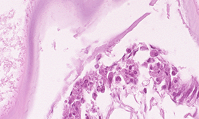
 Classic lesion of clostridial
enteritis, with numerous robust gram-positive bacilli lining necrotic
villar remnants. (40X, Brown-Hopp's, 101K)
Classic lesion of clostridial
enteritis, with numerous robust gram-positive bacilli lining necrotic
villar remnants. (40X, Brown-Hopp's, 101K)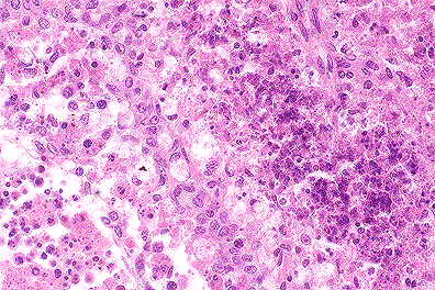 Lymphoid necrosis in the bursa
of a chicken infected with Infectious Bursal Disease (Gumboro's
disease) (40X, HE, 107K)
Lymphoid necrosis in the bursa
of a chicken infected with Infectious Bursal Disease (Gumboro's
disease) (40X, HE, 107K)