 Bacterial embolus within glomerulus
of a rhesus macaque. (40X, HE, 115K)
Bacterial embolus within glomerulus
of a rhesus macaque. (40X, HE, 115K)
Bacterial embolus within glomerulus
of a rhesus macaque. (40X, HE, 115K)
History: This 13-year-old female rhesus macaque was inoculated with a pathogenic molecular clone of simian immunodeficiency virus, SIVmac239. Approximately 120 weeks post infection the animal was subjected to an experimental procedure followed by sequential full thickness excisional skin biopsies at multiple time points. The animal was euthanized after an acute onset of severe anorexia and depression approximately 128 weeks post-viral inoculation.
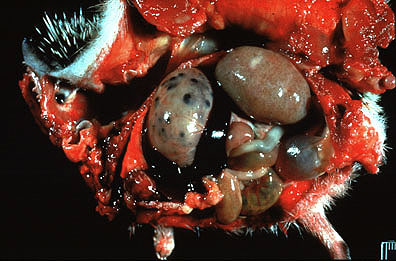
Gross Pathology: At necropsy there were coalescing foci of cutaneous edema and ulceration within the middorsal thorax corresponding to sites of previous biopsy. Adhered to ulcerated areas was a dried serosanguinous crust. The heart contained multiple irregular foci of myocardial hemorrhage and scattered, pale-tan, often slightly raised pinpoint foci visible from the epicardial surface. Adhered to the left A/V valve was a firm, tan to red-brown, granular mass, approximately 1.5 cm in diameter, which protruded into the left atrium. The kidneys contained scattered tan pinpoint foci similar to those seen within the myocardium. In addition, there were several soft, tan, wedge shaped foci compatible with acute renal infarcts. The lungs were pale pink, had delicate fibrinous adhesions between cranial and caudal lung lobes, and contained scattered pale gray, 2-5mm subpleural bullae consistent with lung mite (Pneumonyssus simicola) infestation. The liver had moderately blunted margins, a prominent reticular pattern and contained rare 1-2 cm diameter depressed foci on capsular surfaces. There were scattered petechiae within the mucosa of the small intestine and focal mural hemorrhage near the tip of the cecum. The spleen was soft and markedly enlarged (66 gm, normal range 0.9-11.5 gm). Peripheral and visceral lymph nodes were moderately enlarged. The cerebrum and cerebellum contained rare petechiae.
Laboratory Results: Blood sample from day of necropsy. ref.
range BUN 118 mg/dl (9-23)
CREAT 4.7 mg/dl (0.7-1.3)
PHOS 17.9 mg/dl (0.9-8.0)
CA 6.7 mg/dl (7.6-10.0)
TP 6.8 g/dl (6.0-7.8)
ALB 2.1 g/dl (3.3-4.7)
GLOB 4.7 g/dl (2.1-3.7)
A\G 0.4 (0.86-1.94)
AP 286 U/L (0-74)
ALT 88 U/L (0-59)
AST 281U/L (0-46)
LDH 1849U/L (0-785)
CPK 8746U/L (0-1596)
T-LYMPHOCYTES
CD4 22.1/uL*
CD8 330.75/uL
CD4:CD8 0.07
*CD4<200/uL is indicative of immunosuppression
Bacterial cultures of heart blood yielded abundant Staphylococcus aureus.
Contributor's Diagnosis and Comments:
Sections of kidney reveal widely disseminated microabscesses which multifocally distort and replace normal architecture with 100-500 micron diameter foci composed of lytic cellular debris, degenerate neutrophils, histiocytes and lymphocytes. Abscesses are most often centered upon thrombosed vessels and often contain colonies of large cocci, accompanied by acute hemorrhage (serum, fibrin, and RBC's). Several infarcts extend from thrombosed vessels within the corticomedullary junction and radiate towards the capsular surface. Infarcts appear to be of mixed chronicity with varying degrees of coagulative necrosis, interstitial neutrophil and mononuclear cell infiltration, fibrosis, tubular epithelial regeneration, and parenchymal contracture. Tubules in severely affected areas are often dilated and contain neutrophils within a hyaline proteinaceous material. Glomerular changes include coagulative necrosis with capsular epithelial regeneration/hyperplasia, multifocal capillary thromboses with occasional bacterial colonization, multifocal mesangial cell proliferation, multifocal fibrinous synechiae, and rare glomerular sclerosis. Additional changes include perivascular nodular lymphocytic hyperplasia, mild inner medullary interstitial fibrosis, mild multifocal medullary interstitial mineral deposition, and mild nonsuppurative interstitial nephritis.
Thromboses and microabscesses with associated bacterial colonies were also observed in sections of skin, brain, heart, thyroid, liver, ovary, and submucosa of small and large intestine. Large cocci were adhered to an expansile mitral valve vegetation composed of serum, fibrin, and scattered degenerate leukocytes. Bacteria were not found in sections of lung or lymph nodes. Other lesions included SIV arteriopathy and mild multifocal histiocytic colitis with associated acid fast organisms characteristic of Mycobacterium avium. Arteriopathy appeared to be confined to the lung although similar changes in renal vessels could not be ruled out as the architecture of many such vessels were distorted or effaced by microabscesses. Staphylococcus aureus was isolated from cultures of heart blood collected at necropsy. Infected cutaneous biopsy sites were the presumed portal of entry as coccoid bacteria were abundant in histologic sections from these lesions. Cultures of the wound were not attempted.
Septicemia is an uncommon complication of HIV and SIV infection. However, bacterial infection occurs more frequently in immunosuppressed rhesus macaques infected with type D retrovirus. In these animals, functional deficits in polymorphonuclear leukocytes (PMN) which may predispose to bacterial infections have been documented (4). To our knowledge there is no evidence of similar functional PMN deficits in HIV or SIV infected individuals. In this case, we hypothesize that predisposition towards bacteremia and sepsis was a sequel to chronic percutaneous exposure to bacteria though sequential skin biopsies, and perhaps the presence of a pre-existing sterile mitral valve thrombus, a common lesion in terminally ill, SIV-infected macaques seen at the NERPRC. Relationships between sterile A/V valve thrombi and SIV arteriopathy is currently unclear. SIV arteriopathy occurs in approximately 20% of animals that die with simian AIDS. The lesion is seen in large and medium sized arteries, most commonly in the lung and kidney and is characterized by intimal and medial thickening mediated by proliferation of smooth muscle, collagen, and less commonly macrophage infiltration. Ultrastructural findings indicate primary endothelial cell injury followed by successive thrombosis and repair (2). Attempts to localize virus in these lesions (SIV, CMV) have been unsuccessful. Similarity of these lesions to arteriopathy associated with malignant catarrahal fever (5) and recent isolation of previously undetected gamma herpes-like sequences in Kaposi's sarcoma lesions of AIDS patients (3) makes an argument for a viral etiology particularly compelling; however, proliferative vasculopathies have been observed secondary to a number of distinct mediators of endothelial injury including intravascular parasitism (dirofiliariasis, elaeophorosis), balloon catheter-mediated trauma, and multiple effectors of pulmonary hypertension (1,6,7).
AFIP Diagnosis:
Conference Note: Simian immunodeficiency virus (SIV) belongs to the family Retroviridae, subfamily lentivirinae, which includes human immunodeficiency virus, bovine immunodeficiency virus, feline immunodeficiency virus, equine infectious anemia virus, caprine arthritis-encephalitis virus, and the ovine lentiviruses. There are at least 10 serotypes of SIV based on species infected: SIVsmm from the sooty mangabey is thought to be the progenitor of all other serotypes. SIV uses the CD4 molecule as its cellular receptor and therefore infects CD4+ cells (macrophages and CD4+T lymphocytes). Depletion of CD4+ T cells leads to severe immune dysfunction and death from opportunistic infection or lymphoma. SIV infections must be differentiated from other causes of immunosuppression, the most common of which is infection with type D retrovirus (simian retrovirus).
Lesions thought to be caused by SIV include cutaneous rashes, lymphoid hyperplasia, lymphoid depletion, arteritis, pneumonia, encephalitis, giant cell disease, and glomerulosclerosis. Many participants at the conference believed that multifocally, glomeruli were hypercellular and that this proliferative response may have been directly related to the SIV infection. However, the Department of Nephropathology of the AFIP does not believe that the lesions are consistent with the segmental sclerosis seen with retroviral infection. In their opinion, the lesions probably represent a post-infection glomerulonephritis.
Conference attendees also discussed a differential diagnosis for tubulonecrosis in primates that included SV40, ascending pyleonephritis, and nephrotoxins.
Contributor: New England Regional Primate Research Center, One Pine Hill Drive, Box 9102, Southborough, MA., 01772.
References:
 Cholangiocarcinoma in a Siamese
cat (40X, HE, 129K)
Cholangiocarcinoma in a Siamese
cat (40X, HE, 129K)
Signalment: 11-year-old castrated male Siamese
History: Weight loss, anorexia, alopecia, focal cutaneous erythema which resolved.
Gross Pathology: Emaciated. Multiple circular lesions .5 to 2.5 cm. Grayish white with dark centers in the liver. Peritoneal carcinomatosis and metastasis to lungs.
Laboratory Results: No significant findings.
Contributor's Diagnosis and Comments: Hepatic neuroendocrine carcinoma (hepatic carcinoid).
Primary hepatic neuroendocrine carcinomas are rare, both in the cat and the dog. These tumors can be distinguished from other hepatic neoplasms by their distinct morphologic features.
AFIP Diagnosis: Liver: Cholangiocarcinoma, Siamese, feline.
Conference Note: Neoplasia of hepatic origin is rare in the cat. Hepatocellular carcinoma is the most frequently reported hepatic neoplasm, followed by cholangiocarcinoma. In the cat, cholangioadenomas and cholangiocarcinomas have been associated with cholelithiasis, bile duct hyperplasia, and Clonorchis infection. There have been two reports of hepatic neuroendocrine carcinomas in the cat, one intrahepatic and the other in the extrahepatic bile duct. Neuroendocrine carcinoma of the liver has also been reported in dogs and in humans. Neuroendocrine carcinoma occurs more commonly in the intestines, pancreas, and lung.
This neoplasm is unencapsulated and infiltrative. The neoplastic cells are arranged in nests, packets, and cords, as well as rare tubules. The neoplastic cells have variably distinct cell borders with a moderate amount of eosinophilic, granular cytoplasm. The nuclei are centrally located and round to oval, with stippled chromatin and 1-3 prominent nucleoli; some nuclei are vesicular.
Neuroendocrine carcinomas contain argyrophilic cytoplasmic granules, and immunohistochemical stains for neuron specific enolase, chromogranin, Leu-7, and synaptophysin are often positive. Ultrastructurally, neoplastic neuroendocrine cells contain "dense core" granules.
Immunohistochemically, this tumor is negative for synaptophysin and neuron specific enolase and strongly positive for keratin. The Churukian-Schenk method failed to demonstrate argyrophil granules. This case was also reviewed by the Department of Hepatic Pathology of the AFIP. They noted that the relatively large cells, prominent nucleoli, frequent mitoses, strongly positive immunohistochemical staining for keratin, negative immunohistochemical stains for neuroendocrine differentiation and absence of argyrophilic granules strongly support adenocarcinoma consistent with cholangiocarcinoma.
Contributor: The Animal Medical Center, 510 East 62nd Street, New York, NY 10021.
References:
 Renomegaly in a hedgehog with
PKD (36K)
Renomegaly in a hedgehog with
PKD (36K)
 Markedly ectatic renal tubules
in a hedgehog with PKD (10X, HE, 85K)
Markedly ectatic renal tubules
in a hedgehog with PKD (10X, HE, 85K)
Signalment: 1-year-old male African hedgehog (Atelerix albiventris)
History: An approximately 1-year-old African hedgehog was admitted to the University of Tennessee College of Veterinary medicine with a one day history of right hind limb lameness. The lameness quickly progressed to flaccid paralysis of both hind limbs, depression and death. The hedgehog was the only one affected of 15 inbred animals owned by a breeder.
Gross Pathology: Both kidneys were 3.5 X normal size (2 X 2 X 3.5 cm) and pale but had retained a reniform shape. Their capsule was thin, tightly adherent and generally translucent allowing one to see many, confluent, 1-2 mm diameter clear, fluid-filled cysts. On cut section the cortex and medulla had a cystic appearance.
The cardiac ventricular myocardium had gritty white streaks from the base to the apex; the gastric mucosa was covered by a thin, black, multifocally adherent film; deep hemorrhagic ulcers were visible from the serosa.
Laboratory Results: None submitted.
Contributor's Diagnosis and Comments: Polycystic kidney, African Hedgehog.
The hedgehog died of renal failure due to polycystic kidney disease (PKD). Histologically, the renal parenchyma consisted of many dilated tubules lined by a variably squamous to cuboidal to columnar epithelium with rare papillary projections. The cysts were distended by proteinaceous fluid and contained occasional macrophages and degenerate neutrophils. Glomeruli were markedly decreased in number and often atrophied; their capsules had capsular basement membrane thickening with epithelial crescent formation. The attenuated interstitium consisted of fibrous connective tissue. The staining of tubular epithelium in a select subpopulation of the cysts by lectins specific for distal tubule segments suggested the cysts had arisen from proximal and distal segments of the nephron, collecting tubules and collecting ducts. (Lectin staining performed by Dr. James Thomas Kirby.) These findings suggest this case is analogous to the autosomal dominant form of PKD in humans. Attempted ultrasonographic renal examination of the remaining hedgehogs was unsuccessful since the unsedated animals could not be properly positioned.
The myocardium had many large areas of necrosis with extensive mineralization and a mild eosinophilic infiltrate; a segment of an artery and arterioles were degenerate and mineralized. Skeletal muscle from the right thigh and tongue contained randomly scattered, individually mineralized myofibers and a mild eosinophilic infiltrate; degenerative changes in the thigh skeletal musculature may explain, in part, the presenting signs of hind limb paresis and paralysis.
PKD is characterized by progressive enlargement of the kidneys due to numerous expanding cysts and ultimately leads to renal failure. In humans, PKD is heritable as at least 2 genetically distinguishable diseases - infantile or autosomal recessive PKD (ARPKD) and adult or autosomal dominant PKD (ADPKD). Renal cysts also occur in renal dysplasia and neoplasia and are also seen with hemodialysis, outflow obstruction, hypokalemia and exposure to nephrotoxic cystogenic agents such as diphenylthiazole. In veterinary medicine, PKD has been recognized in many species but no cases have been reported in disease surveys of the African hedgehog. Syndromes resembling both the dominant and recessive forms of human PKD have been described in related domestic cats, and Persian cats appear to be disproportionally affected. PKD has been reported rarely in adult horses, ferrets and related mink. Animal models of heritable forms of PKD occur in mice, rats, rabbits, domestic cats and springboks.
Both heritable forms of human PKD are characterized by bilateral cystic renal disease with enlarged kidneys that retain a reniform shape. In ADPKD, the parenchyma is extensively replaced by cysts that originate from all segments of the nephron, collecting tubules and ducts. In humans with ADPKD, there is an association with cysts in other organs, most commonly the liver, cardiac valvular anomalies, intracranial aneurysms and colonic diverticula, none of which were present in this case. In ARPKD, the cysts arise only from dilated collecting tubules and ducts; on cut section the enlarged kidney of affected newborns has radially arranged thin-walled channels extending from the pelvis to the cortex. In almost all cases there is also biliary dysgenesis and hepatic fibrosis.
Theories concerning the pathogenesis of ADPKD include: 1) prolonged increase in intratubular pressure secondary to partial outflow obstruction from hyperplastic cells or micropolyps; 2) structurally altered tubular basement membranes leading to decreased tubular wall compliance; 3) radial tubular epithelial hyperplasia in response to either physical, chemical or growth factor/hormone/cytokine stimuli; and 4) altered epithelial cell metabolism and transport causing intratubular fluid accumulation due to increased transepithelial secretion of solutes and water. All four mechanisms are likely involved and could result from a primary defect in tubular cell metabolism and tubular cell injury.
AFIP Diagnosis: Kidney, tubules and collecting ducts: Ectasia and cysts, diffuse, with glomerular and tubular atrophy and interstitial fibrosis, African hedgehog (Atelerix albiventris), insectavore.
Conference Note: Polycystic kidney disease (PKD) occurs in pigs, lambs, calves, dogs, cats, and horses. A genetic etiology is proposed in some species, but not in others. As the contributor noted, renal cysts may develop due to obstructive lesions (secondary to interstitial fibrosis, for example), exposure to chemicals (such as diphenylamine, 5,6,7,8- tetrahydrocarbazole-3-acetic acid, alloxan, and diphenylthiazole), renal dysplasia, or they may form as the result of an inherited syndrome. In this case, a single animal was affected; whether or not related hedgehogs were affected is unknown. Additional studies would be needed to classify this case and determine its relationaship to PKD in other species.
Glomerular crescents are often found in cases of severe glomerulonephritis. They are formed by proliferating parietal epithelial cells accompanied by infiltrating macrophages and neutrophils. The stimulus for crescent formation is the presence of fibrin in the urinary space. No crescents were observed in the section examined during the conference.
Contributor: University of Tennessee, College of Veterinary Medicine, Department of Pathology, 2407 River Drive, Knoxville, TN 37996-4500.
References:
 Dermatophilus congolensis
in the epidermis of a lamb.(40X, HE, 128K)
Dermatophilus congolensis
in the epidermis of a lamb.(40X, HE, 128K)
 Eosinophilic intracytoplasmic
inclusion bodies of ovine parapoxvirus. (40X, HE, 90K)
Eosinophilic intracytoplasmic
inclusion bodies of ovine parapoxvirus. (40X, HE, 90K)
Signalment: 8-month-old, black faced mountain cross, male lamb.
History: Skin sections were submitted from an 8-month-old male black faced mountain cross lamb which was euthanized due to the presence of chronic hard crusting lesions on the haired skin of the nose, ears, distal limbs and scrotum and a concurrent chronic ill-thrift.
Gross Pathology: Described above.
Laboratory Results: Not applicable. Contributor's Diagnosis and Comments: Skin: Dermatitis with folliculitis, pustular,
chronic-active, diffuse, moderate to severe, Dermatophilus congolensis and orf virus.
The surface of the lesion is composed of alternating layers of predominantly parakeratotic hyperkeratosis with dense bands of degenerating neutrophils. Epidermal changes are qualitatively similar in both follicular and interfollicular epidermis but are more marked in the follicular canals. The changes include pseudoepitheliomatous acanthosis, marked vacuolar degeneration of keratinocytes, neutrophil exocytosis and the formation of several, varying sized superficial pustules. Numerous gram positive filamentous hyphae with a characteristic railroad track appearance are present within the superficial crust particularly within the hair follicle ostia. Superficial foci of keratinocyte reticular degeneration which exhibit positive immunostaining for orf virus antigen are present in some sections.
Superficial dermal vessels are congested and many are plugged with neutrophils. The dermis in the immediate subepidermal area is edematous and diffusely infiltrated by large numbers of neutrophils, lymphocytes, plasma cells and macrophages. Deeper in the dermis the inflammatory cell aggregates and the edema are largely confined to angiocentric and periadnexal locations.
The main source of infection in dermatophilosis is infected animals including healthy carriers. The two most important predisposing factors in the development of lesions are excessive wetting and skin damage. Excessive moisture activates latent zoospores and can also interfere with epidermal defense mechanisms. Once the zoospores have germinated the branching filaments extend through the damaged epidermis particularly in the hair follicles. An intense inflammatory response is stimulated and this inhibits deeper penetration by the bacteria. The underlying damaged epidermis rapidly regenerates and premature keratinization is stimulated. Bacteria from reservoirs in the hair follicles invade the newly regenerated epidermis and another burst of acute inflammation is stimulated. These cyclic events result in the formation of the characteristic palisading crust.
Concurrent infections with Dermatophilus and orf virus have been described. When they occur concurrently, it is not possible to ascertain which was the primary agent. Most cases of dermatophilosis spontaneously regress within four weeks if the animals are kept dry, and most cases of orf also resolve within four weeks. Chronic dermatophilosis has been associated with a wide range of conditions which produce a reduction in the host's immune response. Cases of persistent orf have been described, and it has been postulated that they occur due to the inability of the infected animal to mount a successful immune response. Therefore, it is probable that the affected animal in this case had diminished immune function.
AFIP Diagnosis: Haired skin: Dermatitis, proliferative, chronic-active, diffuse, severe, with parakeratosis, corneal microabscesses, folliculitis, ballooning degeneration, numerous filaments of paired cocci, and eosinophilic intracytoplasmic inclusions, etiologies consistent with Dermatophilus congolensis and ovine parapoxvirus, black-faced mountain cross, ovine.
Conference Note: Ovine parapoxvirus causes a disease in sheep and goats variously known as contagious ecthyma, contagious pustular dermatitis, and orf. It has a worldwide distribution, a morbidity rate which approaches 90% and a mortality rate of about 1%. Afflicted animals are often hesitant to graze or nurse and can lose body condition. Lesions are usually confined to the lips, oral cavity, muzzle, coronary band, and interdigital cleft; however, systemic infections have been described. Grossly, the lesions manifest as raised, red foci with a surrounding area of hyperemia. The most characteristic feature of the gross lesion is a layer of thick brown-grey crust, which may be elevated 2-4 mm above the skin surface.
Dermatophilus congolensis is is a gram-positive, facultative anaerobe of the order Actinomycetales, and grows in distinctive parallel rows of coccoid cells (resembling a railroad track). Dermatophilus congolensis does not infect intact, healthy epithelium, it requires epithelial damage, from trauma or prolonged wetting, to establish an infection. Dermatophilus infects a number of species including horses, cattle, goats, sheep, and, rarely, pigs, cats, dogs, and humans. Common sites of infection are the back and distal limbs. Grossly, the lesions consist of coalescing crusts, often penetrated by hair. If crusts are removed from active lesions, the underlying epidermal erosions and ulcers often bleed.
Contributor: Department of Veterinary Pathology, Faculty of Veterinary medicine, University College Dublin, Ballsbridge, Dublin 4, Ireland.
References:
International Veterinary Pathology Slide Bank: Laser disc frame #357, 358, 4964, 12862-3
Dana P. Scott
Captain, VC, USA
Registry of Veterinary Pathology*
Department of Veterinary Pathology
Armed Forces Institute of Pathology
(202)782-2615; DSN: 662-2615
Internet: Scott@email.afip.osd.mil
* The American Veterinary Medical Association and the American College of Veterinary Pathologists are co-sponsors of the Registry of Veterinary Pathology. The C.L. Davis Foundation also provides substantial support for the Registry.
 Alternating bands of keratin
and inflammatory cells characteristic of dermatophilosis (10X,
HE, 110K)
Alternating bands of keratin
and inflammatory cells characteristic of dermatophilosis (10X,
HE, 110K)